대한임상신경생리학회지 9(2):93~96,2007 ISSN 1229-6414
Copyright 2007 by the Korean Society for Clinical Neurophysiology 93
조직구괴사림프절염(Kikuchi병)과 동반된 무균뇌수막염과 한쪽 전정신경병증
대구가톨릭대학교 의과대학 신경과학교실
김경집 도진국 이동국
Kyung Jib Kim, M.D., Jin Kuk Do, M.D., Dong Kuck Lee, M.D.
Department of Neurology, School of Medicine, Catholic University of Daegu, Korea
A 34-year-old man presented with a severe headache, fever, and cervical lymphadenopathy followed by generalized tonic-clonic seizure. Evaluations showed splenomegaly, elevated liver enzymes, and 380 white blood cells/mm3 in the cerebrospinal fluid. Two weeks after admission, he developed sudden vertigo. Examination revealed spontaneous horizontal-torsional nystagmus to the right and bithermal caloric tests documented left canal paresis. A cervical lymph node biopsy disclosed subacute necrotizing lymphadenitis. We report a case of aseptic meningitis and unilateral vestibulopathy associated with histiocytic necrotizing lymphadenitis (Kikuchi’s disease).
Key Words: Kikuchi’s disease, Meningitis, Vestibulopathy
INTRODUCTION
Necrotizing histiocytic lymphadenitis, or Kikuchi’s disease (KD), is an uncommon disorder that frequently affects Asian young women. It is characterized by fever, leukopenia, and cervical lymphadenopathy.
Occasionally patients developed extranodal presen- tations.1 KD usually runs a benign course and resolves within several weeks to months and it can be mistaken
for lymphoma or systemic lupus erythematosus. On occasion, myalgia, a malar or butterfly rash, hepato- megaly, or splenomegaly have been noted.2 The diag- nosis of KD can be confirmed by characteristic his- topathologic findings in the involved lymph nodes.
Previously, a few cases with meningitis have been described in KD, but unilateral vestibulopathy in association with aseptic meningitis have not been reported.
CASE REPORT
A previously healthy 34-year-old man presented with high fever, headache, and cervical lymphaden- opathy for 3 weeks. On admission, body temperature was 39.8℃. Examination showed multiple, firm, mo-
김경집․도진국․이동국
J Korean Society for Clinical Neurophysiology / Volume 9 / December, 2007 94
Figure 1. Multiple, enlarged, enhancing lymph nodes at level II on both sides of the neck (arrows).
Figure 3. Finding of caloric tests. Monothermal caloric tests show decreased response in the left ear and right beating nystagmus is shown on videonystagmography (4 channel; ICS medical, USA).
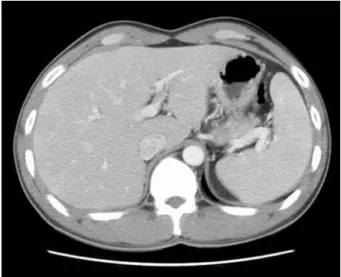
Figure 2. Splenomegaly on abdominal CT.
bile, and non-tender cervical lymph nodes (1-2 cm in size) bilaterally. Other findings were unremarkable.
Neck computed tomography (CT) showed multiple,
enlarged, and enhancing lymph nodes on the anterior neck bilaterally (Fig. 1). Laboratory evaluation reve- aled a white blood cell count of 3,800/mm3, an erythrocyte sedimentation rate (ESR) of 54 mm/h, serum C-reactive protein (CRP) concentration of 90.4 mg/dl, and raised serum aspartate aminotransferase (AST), alanine aminotransferase (ALT), lactate dehy- drogenase (LDH), and γ-glutamyl transferase (GGT) levels. Antiviral titers were not elevated in paired sera for cytomegalovirus or varicella-zoster, rubella, Epstein-Barr (EBV), influenza, parainfluenza, mumps, or rubeola virus.
Ten days after admission, he developed a gener- alized clonic seizure lasting about 10 minutes followed by postictal confusion. A lumbar puncture showed elevated opening pressure of 18 cm H2O with 380 white blood cells/mm3, protein of 238 mg/dl, and glucose of 74 mg/dl. Cultures for bacteria and fungi, and poly- merase chain reactions for enterovirus, EBV, type I and II herpes simplex virus, and Mycobacterium tuberculosis were negative in the cerebrospinal fluid (CSF). Brain CT and magnetic resonance imaging (MRI) were normal. Abdominal CT disclosed splenomegaly (Fig. 2).
The patient was treated with intravenous antibiotics and antiepileptic drugs, which result in rapid impro- vement of the symptoms.
조직구괴사림프절염(Kikuchi병)과 동반된 무균뇌수막염과 한쪽 전정신경병증
J Korean Society for Clinical Neurophysiology / Volume 9 / December, 2007 95 Figure 4. Biopsy finding. The cervical lymph node biopsy shows a necrotic area with nuclear debris (H&E,
×100).
Two weeks after admission, he developed sudden vertigo with nausea and vomiting. Videonystagmo- graphy showed horizontal-torsional nystagmus to the right without fixation in the primary position, which markedly increased in rightward gaze and decreased in the leftward gaze. The nystagmus was completely suppressed by visual fixation. Monothermal caloric stimulation demonstrated a canal paresis in the left ear (Fig. 3). Other neurological and neurotological examinations were normal.
A cervical lymph node biopsy 20 days after admi- ssion, disclosed histopathologic features characteristic of KD, including a broad zone of necrosis accompanied by a histiocytic infiltration (Fig. 4).
DISCUSSION
Kikuchi’s disease is an idiopathic illness that typically causes cervical lymphadenopathy frequently associated with a fever; it generally resolves spon- taneously over 1 to 4 months, and recurrences are unusual.1,2 Since it is mostly self-limiting and may respond to corticosteroids, the disease can frequently be misdiagnosed, leading to inappropriate treatment.3 In fact, many patients with KD who present with fever and meningitis may be assessed as having an infectious disease by physicians who are not aware of the association of KD with meningitis.
Our patient showed high fever, headache, and cervical lymphadenopathy followed by seizure and sudden vertigo. CSF study disclosed aseptic meningitis and caloric test revealed a left canal paresis. A cervical lymph node biopsy documented characteristic histopathologic features of KD. Previously, a few cases of KD associated with aseptic meningitis, encephalitis, or cerebellar ataxia have been described in Asia, but peripheral vestibulopathy associated with meningitis has not been reported in KD.4-6 Meningitis and peripheral vestibulopathy are generally related to systemic viral infections, such as herpes zoster, mumps, or EBV, in which meningitis occurs during the course of an established infection.7
The CSF findings of KD are similar to those in
benign aseptic meningitis occurring in isolation with a presumed viral etiology. Other infections, such as tuberculosis and cat scratch disease should be considered in the differential diagnosis of KD.8 With the increasing prevalence of human immunodeficiency virus infection, atypical pathogens should also be searched according to individual clinical manifesta- tions.
Although CSF cultures and titers for viruses, Mycoplasma pneumoniae, and Coccidioides immitis were negative, the KD-associated meningitis and unilateral peripheral vestibulopathy might have ori- ginated from a viral infection or post-viral immune reaction.
REFERENCES
1. Kuo TT. Kikuchi’s disease (histiocytic necrotizing lympha- denitis). A clinicopathologic study of 79 cases with an analysis of histologic subtypes, immunohistology and DNA ploidy. Am J Surg Pathol 1995;19:798-809.
2. Dorfman RF, Berry GJ. Kikuchi’s histiocytic necrotizing lymphadenitis: an analysis of 108 cases with emphasis on differential diagnosis. Semin Diagn Pathol 1988;5:329-345.
3. Bosch X, Guilabert A, Miquel R, Campo E. Enigmatic Kikuchi-Fujimoto disease. Am J Clin Pathol 2004;122:141- 152.
4. Debley JS, Rozansky DJ, Miller ML, Katz BZ, Greene ME.
Histiocytic necrotizing lymphadenitis with autoimmune phenomena and meningitis in a 14-year-old girl. Pediatrics
김경집․도진국․이동국
J Korean Society for Clinical Neurophysiology / Volume 9 / December, 2007 96
1996;98:130-133.
5. Sato Y, Kuno H, Oizumi K. Histiocytic necrotizing lymph- adenitis (Kikuchi’s disease) with aseptic meningitis. J Neurol Sci 1999;163:187-191.
6. Yi HA, Lee H, Kang YN, Cho YW, Lim JG, Yi SD. A case of viral encephalitis as a presenting symptom of histiocytic necrotizing lymphadenitis (Kikuchi’s disease). J Korean Neurol Assoc 2005;23:135-137.
7. Hollingsworth HC, Peiper SC, Weiss LM, Raffeld M, Jaffe ES. An investigation of the viral pathogenesis of Kikuchi- Fujimoto disease. Lack of evidence for Epstein-Barr virus or human herpesvirus type 6 as the causative agent. Arch Pathol Lab Med 1994;118:134-140.
8. Soman R, Ashar U, Shukla A, Pachauri R, Bhaduri A.
Kikuchi-Fujimoto disease with unusual feature. J Assoc Physicians India 2003;51:314-315.