난자의 회수시기, BSA, Cysteine 및 Myoinositol이 개 난자의 체외성숙에 미치는 영향
김상근1*․ 이봉구2․ 이규승3
Effects of Ovarian Estrus Stage of Oocytes and Supplementation of Medium with BSA, Cysteine and Myoinositol on in vitro
Maturation of Canine Oocytes
Sang-Keun Kim1* ․ Bong-Ku Lee2 ․ Kyu-Seung Lee3
ABSTRACT
This study was carried out to investigate the effects of ovarian estrus stage of oocytes and supplementation of medium with BSA, cysteine and myoinositol on in vitro maturation of canine oocytes. The in vitro maturation(IVM) rate to metaphase II (M II) stage of oocytes recovered from different stage of the reproductive cycle (inactive, follicular and luteal stage) cultured in TCM-199 media were 0.0±0.0%, 10.0±4.1% and 5.7±1.6%, respectively. The IVM rate of oocytes collected from follicular stage was significantly higher in inactive or luteal stage of oocytes. The IVM rates of oocytes recovered from different stage of the reproductive cycle cultured in TCM-199 media with 5% BSA and 0.1 mM cysteine were 0.0±0.0%, 15.8±4.7%, 5.6±1.5%,
1
충남대학교 수의과대학 수의학과(Dept. of Veterinary Medicine, College of Veterinary Medicine, Chungnam National University, Daejeon 305-764, Korea)
2
중부대학교 한방보건대학 애완동물자원학과(Dept. of Animal & Animal Resource Science, Joongbu University, Chungnam, Gumsan 312-702, Korea)
3
충남대학교 농업생명과학대학 동물자원과학부(Division of Animal Science, College of Agriculture and Life Science and Resources, Chungnam National University, Daejeon 305-764, Korea)
* 교신저자 : 김상근(E-mail: [email protected], Tel: 042-821-6754)
respectively. The IVM rates of oocytes recovered from different stage of the reproductive cycle cultured in TCM-199 media with 5% BSA and 10 mM myoinositol were 0.0±0.0%, 18.4±4.6% and 5.7±1.9%, respectively. The IVM rate of follicular stage oocytes was significantly higher in oocytes collected from follicular stage and with cultured 5% BSA and 0.1 mM cysteine or 5% BSA and 10 mM myoinositol compared to other experimental group.
Key words : Ovarian estrus stages, In vitro maturation, BSA, Cysteine, Myoinositol
I. 서 론
국내 애완견은 주로 고단백 및 고지방사료로 사육하면서 운동량이 부족함에 따라 번식장애와 질병으로 이어지고 있다. 불임견의 치료와 복제 개를 생산하기 위해서는 체외성숙 난자를 대량으 로 확보하는 것이 중요한데 개 난자의 확보는 현 실적으로 대단히 어려운 실정이다. 현재까지 보 고 된 개 난자의 체외성숙율은 타 동물에 비해 극히 저조하여 이의 개선이 절실히 필요하다고 하겠다(Gunzel, 1986; Quan 등, 2004).
최근에는 개 난자를 이용하여 12~37%의 체외 성숙율(Hewitt와 England, 1997; Otoi 등, 2000;
Songsasen 등, 2002; Rodrigues 등, 2004)과 배반 포로의 체외발생율(Otoi 등, 2005)을 보고하였는 데. 이는 배양조건의 개선에 기인한 결과로 사료 된다(Holst와 Phemister, 1971; Tsutsui, 1975).
한편, Otoi 등(2004)은 난포기에 채취한 개 난자 를 72시간 배양했을 때 41%의 체외성숙율을 얻 었다고 보고하였다. 이러한 결과는 고양이 난자 의 체외성숙율 40~50%와 소 난자의 체외성숙율 40~60%에 비해 낮은 실정이다(Gomez 등, 2003;
Hansel, 2003). Yamada 등(1992)은 gonadotropin 을 첨가한 배양액에서 72시간 배양했을 때 39%
의 체외성숙율을 얻었으며, 배양 96시간에 ICSI
(intracytoplasmic sperm injection)에 의해 수정 시켰을 때 4세포기의 체외발생율은 13.3%였다고 보고하였다. Hewitt와 England(1999)는 개 난자를 이용하여 48시간 배양 후 GVBD 및 MII기의 발 생율은 각각 33.0%~49.0%와 2.0%~6.0%였다고 하였다.
그러나 불임개체가 많은 소형 개의 불임치료와 복제 개를 생산하기 위해서는 많은 체외성숙 난 자를 얻는 것이 필수적이나 난소확보의 어려움과 배양기술 상 해결되어야 할 난제가 많아서 계속 적인 많은 연구가 필요한 것으로 사료된다.
이에, 본 연구는 번식주기 별 난자의 채취시기, 배양액에 BSA, cysteine, myoinositol 첨가가 개 난자의 체외성숙에 미치는 영향을 조사하였다.
II. 재료 및 방법
1. 난포란의 회수와 체외성숙
개 난소를 적출하여 30~35
oC의 생리식염수에
침지한 후 1시간 이내에 실험실로 옮겨 난소주위
의 조직을 정리하고 배양액으로 2~3회 세척하였
다. 난소를 외과용 blade로 얇게 자른 후 m-PBS
에 부유시켜 난자를 회수하였다. 번식주기 별(휴
지기, 발정기, 황체기)로 구분하여 채취한 난소로
부터 난자를 회수하였다.
2. 난자의 성숙배양
회수한 난자는 5% BSA(bovine serum albumin, Sigma, U.S.A.), 0.1 mM cysteine(Sigma, U.S.A.) 10 mM of myoinositol(Sigma, U.S.A.)이 첨가된 TCM-199(Whittaker, U.S.A.) 배양액 소적 내에 10개의 난자를 주입하고 mineral oil(Squibb, U.S.A.)로 피복한 다음 CO
2배양기내(5% CO
2, 95% air, 37.0
oC)에서 48~72시간 성숙배양을 실 시하였다.
3. 체외성숙율의 판정
성숙 배양한 난자는 0.2%(w/v) hyaluronidase (Sigma, U.S.A.)를 1~2분간 처리에 의해 난구세 포를 제거하고, 나화된 난자는 3.7% formaldehyde triton X-100(Sigma, U.S.A.)에 10분간 고정하고 m-PBS로 세척 후 slideglass에 올린 다음 1.0 uM Hoechst 33342(Sigma, U.S.A.)로 염색 후 세포 및 핵 분열상을 관찰하여 난자의 체외성숙 을 판정하였다.
4. 통계학적 분석
반복실험을 통하여 얻어진 결과는 SAS 8.12 program에 의해 분산분석과 평균치를 구하였으 며, 처리간의 차이를 평가하기 위하여 Duncan의 다중검증을 실시하였다.
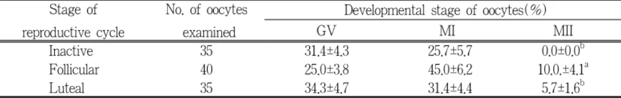
Table 1. Meiotic status of oocytes recovered from different ovarian estrus stage cultured in TCM-199 media
Stage of reproductive cycle
No. of oocytes examined
Developmental stage of oocytes(%)
GV MI MII
Inactive 35 31.4±4.3 25.7±5.7 0.0±0.0
bFollicular 40 25.0±3.8 45.0±6.2 10.0.±4.1
aLuteal 35 34.3±4.7 31.4±4.4 5.7±1.6
ba,b
: Values within column with different superscript differ(p<0.05)
III. 결과 및 고찰
1. 번식주기별 회수 난자의 체외성숙율
번식주기별로 구분하여 채취한 난소로부터 회 수한 난자를 TCM-199 배양액에서 각각 48~72 시간 배양했을 때 체외성숙율은 Table 1과 같다.
휴지기, 난포기, 황체기로 구분한 난소로부터 채취한 난포란을 TCM-199 배양액에서 배양했을 때 체외성숙율은 각각 0.0±0.0%, 10.0±4.1%와 5.7±1.6%로서 난포기에 채취한 난자가 휴지기와 황체기에 비해 높은 체외성숙율을 나타냈다. 이 러한 결과는 개 난자를 이용하여 48시간 SOF+
3% BSA와 SOF+4% BSA액에서 배양했을 때 GVBD 및 MII기의 체외성숙율은 각각 45.0%, 6.0%와 36.0%, 7.0%였다고 한 Hewitt와 England (1999)의 결과에 비해 낮은 성적이었다. 한편, SOF 배양액에 3%의 BSA를 첨가하여 개 난자를 배양했을 때 약간 높은 체외성숙율을 나타냈다고 보고하였다(Hewitt와 England, 1999; Bolamba 등, 1998).
2. BSA와 cysteine 첨가 시 체외성숙율
번식주기별로 채취한 난소로부터 회수한 난자
를 5% BSA와 0.1 mM cysteine을 첨가한
TCM-199 배양액에서 배양했을 때 체외성숙율은
Table 2와 같다.
Table 2. Meiotic status of oocytes recovered from different ovarian estrus stage cultured in TCM-199 media supplemented with 5% BSA + 0.1 mM of cysteine
Stage of reproductive cycle
No. of oocytes examined
Developmental stage of oocytes(%)
GV MI MII
Inactive 34 44.1±7.3 20.6±5.1 0.0±0.0
bFollicular 38 18.4±3.8 47.4±5.8 15.8±4.7
aLuteal 36 41.7±5.2 38.9±6.7 5.6±1.5
ba,b