Influence of Reproductive Status, Serum Type and Estradiol-17β Supplementation on the in vitro Maturation of Canine Oocytes
Fibrianto Yuda Heru
1․ Minkyu Kim
2*1ABSTRACT
Supplementation of serum and estrogen in in vitro maturation(IVM) medium was shown to improve embryo development and quality in several species. This study investigates the effect of ovarian estrus stage on canine oocyte quality and supplementation of medium with canine serum or estrogen on IVM of canine oocytes. As results, in experimental 1, IVM oocytes collected from follicular stage ovaries to MII stages(10.2±1.5%) was higher (p<0.05) with 10% canine estrus stage serum than control(1.3±1.6%), anoestrus stage serum(4.0±1.6%), luteal stage serum (2.7±1.7%) and 10% FBS(1.3±1.6). In experimental 2, 10% canine estrus stage serum supplementation has highest maturation rate to MII stages(10.0±1.8%) and there were significant differences(P<0.05) with another treatment in follicular stages group. In order to investigate the synergic effect of estrous serum and estrogen supplementation, different estrous stage groups of oocytes were cultured with 2 ug/ml estrogen plus various concentrations of different reproductive stage serum and FBS(experimental 3). As results, the rate of maturation to metaphase II(MII) stage was significantly higher(p<0.05) in oocytes from the follicular stage supplemented with estrogen and 10% canine estrus stage serum(11.5%) compared to the other groups(6.0–8.8%).
The present study was demonstrated that canine serum and the estrus cycle of the bitch affect the meiotic competence of oocytes. Hormonal influences within the follicle may be one of the
Received Feb. 14, 2008; Accepted Jun. 5, 2008
1 Faculty of veterinary physiology, Gadgah mada University, Karangmalang Yogyakarta 55281, Indonesia.
2 Department of Animal Science and Biotechnology, College of Agriculture and Life Sciences, Chungngam National University, Daejeon 305-764, Korea
* Correspondence: Min-Kyu Kim(E-mail: [email protected], Tel: +82-42-821-5773, Fax: +82-42-825-9754)
factors responsible for the greater proportion of maturation of oocyte to MII from bitches at the follicular phase.
Key words : Canine oocytes, In vitro maturation, Estrus serum, Estradiol 17-β
Ⅰ. INTRODUCTION
In canine, estrus cycle of bitch is unique that made further complicate the development of research methods to improve in vitro maturation(IVM) in this species. In vitro maturation of oocyte study has been limited to a small number of studies and the rate of maturation of oocytes to metaphase II still low(<40%)(Ottoi et al., 2002). Successful maturation is a prerequisite for the techniques required for assisted reproduction of endangered canine species. The difficulty in obtaining high rates of in vitro matured bitch oocytes is probably due to the peculiar reproductive process of this speces including the hormonal environment and meiotic resumption and progression. In most mammals the oocytes is primarily exposed to high concentration of estrogen before ovulation and in the bitch, ovulated oocytes in the germinal vesicle stage and than completed meiotic maturation take place in oviduct and reach metaphase II by 3-5 days after LH peak (Tsuitsui, 1989).
A few studies have compared the development competence of oocytes from various stages of the estrus cycle (Yamada et. al., 1993; Hewitt and England, 1997; 1999a; Ottoi et al., 2001;
Rodrigues and Rodrigues, 2003), but the result varies among them.
Serum or sources of protein addition in culture medium have been shown to improve the in vitro survival rate of mouse oocyte and to prevent hardening of the zona pelucida. In canine serum supplementation has been used by several researchers(Mahi and Yanagimachi, 1976; Yamada et al., 1993; Hewitt and England, 1997; 1999a; Ottoi et al., 1999; Bolamba et al; 2002). Hewitt at al.(1998) reported that supplementation of culture medium with 0.3%
BSA had a positive effect on the IVM of canine oocytes, and Nickson et al.(1993) obtained the high rate of maturation of oocyte from bitches at various stage of the estrous cycle by supplementation of the culture medium with oestrous bitch serum and estradiol.
Rodrigues et al.(2003) found that reproductive status of the female not influence in vitro nuclear maturation of dog oocyte.
Estrogen secretion is primarily by antral and
preovulatory follicles, and reach peak
concentrations as the follicles become competent
to ovulate (Concanon, 1995), which indicate
that these hormone may play a significant role
in maturation of oocyte in most mammalian
species.
The aim of this study was to evaluate more thoroughly the effect of stage estrus cycle, serum type and estrogen supplementation in culture medium on IVM of canine oocytes.
Ⅱ. MATERIAL AND METHODS
1. Collection of Immature Oocyte and In Vitro Maturation
Reproductive tracts from normal bitches greater than 12 months of age were collected after routine ovariohysterectomy at private clinics, placed immediately into physiological saline solution at 37°C and transported to the laboratory within 1 hr. Ovaries removed from the tract were washed off blood in fresh PBS and minced with a shaving blade at room temperature in the bench medium consisting of M-199 (Life Technologies, Rockville, MD) with 25 mM HEPES (Life Technologies), 1%
fetal calf serum (Hyclone, UT) and 1%
Pennicillin-Streptomyocin solution (Life Technologies). Ovaries were classified into groups of anestrous, follicular and luteal stage based on present of follicle and luteal tissue.
Only grade 1 oocytes (according to the criteria reported by Kim et al., darkly pigmented and completely surrounded by one or more layers of cumulus cell) were selected and used for the experiments.
Immature oocytes were incubated for 72 hr in a 500ul drop of serum-free tissue culture medium (TCM)-199 (Life Technologies) in the
presence or absence of steroid hormone as described in the experimental design at 39 °C in a humidified atmosphere of 5% CO
2in air.
At the end of the maturation culture, oocytes were completed denuded from cumulus cells by repeated pipetting in the same IVM medium containing 0.5 mg/mL hyaluronidase (Sigma-Aldrich) for 1 to 3 min.
2. Assessment of Meiotic Stage
Denude oocytes were fixed in a 4%
formaldehyde-TritonX-100 solution (Sigma- Aldrich) for 15 min and washed in a solution of PBS. Fixed oocytes were mounted on a slide and stained with 1.9 mM Hoechst 33342 (Sigma-Aldrich) in glycerol (Sigma, St. Louis, MO). Chromatin state and position as well as spindle formation and migration of oocytes were evaluated under UV light to determine the stage of meiosis as follows (Kim et al., 2004); germinal vesicle(GV) stage, germinal vesicle breakdown(GVBD), metaphase I (MI) stage, metaphase II (MII).
3. Experiment design
Collected immature oocytes were grouped
according to the stage of the estrous cycle
(anestrus, follicular or luteal stage), prior to
transfer to maturation culture medium. In
experiment 1, oocytes were randomly allocated
into TCM-199 media as follows: (1) TCM-
199; (2) TCM-199 supplemented 10% canine
anestrus serum (CAS); (3) TCM-199 supplemented
10% CES; (4) TCM-199 supplemented 10%
canine diestrus serum (CDS); (5) TCM-199 supplemented 10% FBS. Experiment 2, (1) TCM-199; (2) TCM-199 supplemented 10%
canine estrus serum (CES); (3) TCM-199 supplemented 20% CES; (4) TCM-199 supplemented 10% fetal bovine serum (FBS).
In Experiment 3, oocytes were randomly allocated into TCM-199 as follows: (1) TCM-199; (2) TCM-199 supplemented 2µg estradiol-17ß (E2, Sigma-Aldrich); (3) TCM-199 supplemented 10% CAS and 2µg E2; (4) TCM- supplemented 10% CES and 2µg E2; (5) TCM-199 supplemented 10%
CDS and 2µg E2; (6) and TCM-199 supplemented 10% FBS and 2µg E2.
4. Statistical analysis
Data were analyzed using the the SAS package (SAS Institute, Cary, NC). Random distribution of oocytes was made in each experimental group and experiments were repeated at least four times. Parametric analysis of the means between two or more populations was analyzed by an ANOVA followed by multiple pair-wise comparisons using a Duncan’s multiple range test.
Experiments were first analyzed for interaction among experimental parameters. As no interaction was found, the data were further analyzed using the ANOVA procedure followed by multiple pair-wise comparisons using a Duncan test. Differences with P<0.05 were considered significant.
Ⅲ. RESULTS
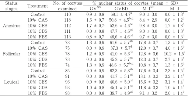
In experiment 1, a total 1,400 oocytes were selected from bitches ovaries at different stages of estrus. After 72 hr cultured, in anoestrus group was only oocytes from canine estrus serum treatment have developed to MII stages(1.7±1.3%) but still no significant different(p>0.05) in rate of meiotic resumption among treatment group. In follicular ovaries group, oocytes from 10% CES treatment has highest maturation rate to MII stages (10.2±1.5%) and there were significant differences(p<0.05) with another treatment groups. From luteal ovaries group, there were no significant differences(p>0.05) between serum treatment and rate of maturation to MII stages of oocytes were 2.3±1.5%; 3.2±1.4%;
3.1±1.4%; 1.0±1.4%; 2.0±1.4% for control, CAS, CES, CDS and FBS treatment, respectively(Table 1).
In experiment 2, a total 814 oocytes were collected from bitches ovaries at different stages of estrus. In follicular ovaries group, oocytes from 10% CES treatment has significantly higher(p<0.05) maturation rate to MII stages(10.0±1.8%) than another treatment in follicular stages group(Table 2).
In experiment 3, a total 1,406 oocytes from different stages of estrous cycle were selected.
In follicular stages of ovary were 7.8±1.3%;6.0
± 1.3%; 11.5 ± 1.2%; 8.8 ± 1.1%; 2.8 ± 1.2%
and 8.3 ± 1.5% for control, CAS, CES, CDS,
Table 1. Meiotic status of different reproductive stages recovered oocytes cultured in TCM 199 supplemented with canine serum and fetal bovine serum
Status
stages Treatment No. of oocytes examined
% nuclear status of oocytes (mean ± SD)
GV
NSGVBD M I
NSM II
Anestrus
Control 110 0.9 ± 0.8 68.1 ± 4.7
a9.0 ± 3.0 0.0 ± 1.3
b10% CAS 118 1.6 ± 0.7 50.8 ± 4.5
bcd8.4 ± 2.9 0.0 ± 1.2
b10% CES 112 1.7 ± 0.7 52.6 ± 4.6
bc9.8 ± 3.0 1.7 ± 1.3
b10% CDS 111 0.0 ± 0.8 47.7 ± 4.6
cd9.0 ± 3.0 0.0 ± 1.3
b10% FBS 113 0.8 ± 0.7 48.6 ± 4.6
bd9.7 ± 3.0 0.0 ± 1.3
bFollicular
Control 73 1.3 ± 0.9 61.6 ± 5.7
ab17.8 ± 3.7 1.3 ± 1.6
b10% CAS 75 0.0 ± 0.9 37.3 ± 5.7
d12.0 ± 3.7 4.0 ± 1.6
b10% CES 78 1.2 ± 0.9 41.0 ± 5.6
cd12.8 ± 3.6 10.2 ± 1.5
a10% CDS 73 0.0 ± 0.9 45.2 ± 5.7
cd12.3 ± 3.7 2.7 ± 1.6
b10% FBS 74 1.3 ± 0.9 48.6 ± 5.7
bcd10.8 ± 3.7 1.3 ± 1.6
bLeuteal
Control 85 0.0 ± 0.9 62.3 ± 5.3
ab17.6 ± 3.4 2.3 ± 1.5
b10% CAS 91 0.0 ± 0.8 41.7 ± 5.1
cd13.1 ± 3.3 3.2 ± 1.4
b10% CES 96 0.0 ± 0.8 40.6 ± 5.0
cd15.6 ± 3.2 3.1 ± 1.4
b10% CDS 93 1.0 ± 0.8 45.1 ± 5.1
cd11.8 ± 3.3 1.0 ± 1.4
b10% FBS 98 0.0 ± 0.8 39.7 ± 4.9
cd9.1 ± 3.2 2.0 ± 1.4
ba-d) Values with different superscripts are significantly different (p<0.05) NS means that there is not significant difference between treatments
Control, TCM 199 alone, CAS, canine anestrus serum, CES, canine estrus serum and CDS, canine diestrus serum
Table 2. Meiotic status of different ovarian stages recovered oocytes cultured in TCM 199 supplemented with canine estrus serum(CES) and fetal bovine serum (FBS)
Status
stages Treatment No. of oocytes examined
% nuclear status of oocytes(mean ± SD)
GV GVBD M I
NSM II
Anestrus
TCM 199 87 16.0 ± 3.0
b55.1 ± 5.2
c12.6 ± 4.0 0.0 ± 1.5
a10% CES 82 10.9 ± 11.7
b40.2 ± 5.3
ab14.6 ± 4.1 2.4 ± 1.5
ab20% CES 85 11.7 ± 3.1
b42.3 ± 5.2
abc12.9 ± 4.0 1.1 ± 1.5
ab10% FBS 85 15.2 ± 3.1
ab32.9 ± 5.2
a12.9 ± 4.0 1.1 ± 1.5
abFollicular
TCM 199 59 8.4 ± 3.7
ab52.5 ± 6.3
bc15.2 ± 4.8 0.0 ± 1.8
a10% CES 60 5.0 ± 3.7
a33.3 ± 6.2
a30.0 ± 4.8 10.0 ± 1.8
c20% CES 57 15.7 ± 3.8
b29.8 ± 6.4
a26.3 ± 4.9 3.5 ± 1.8
ab10% FBS 60 8.3 ± 3.7
ab46.6 ± 6.2
abc18.3 ± 4.8 0.0 ± 1.8
aLeuteal
TCM 199 63 4.7 ± 3.6
a60.3 ± 6.1
c17.4 ± 4.7 1.5 ± 1.7
ab10% CES 62 4.8 ± 3.6
a32.2 ± 6.1
a17.7 ± 4.7 4.8 ± 1.7
b20% CES 56 0.0 ± 3.8
a39.2 ± 6.4
ab14.2 ± 5.0 1.7 ± 1.8
ab10% FBS 58 3.4 ± 3.7
a29.3 ± 6.3
a18.9 ± 4.9 0.0 ± 1.8
aa-c) Values with different superscripts are significantly different (p<0.05) NS means that there is not significant difference between treatments
CAS, canine anestrus serum, CES, canine estrus serum and CDS, canine diestrus serum
FBS and E2 treatment respectively, and there were significant differences (p<0.05) between 10% CES and another treatment in this group.
In luteal groups were significant differences (p<0.05) between 10% CAS with E2 and TCM-199 with E2 treatment group. Within estrus cycle, there were significant differences (p<0.05) between follicular stages and another stages (Table 3).
Within estrus cycle stage, there were significant differences (p<0.05) between follicular stages and another stages in this experiment.
Table 3. Meiotic status of different ovarian stages recovered oocytes cultured in TCM 199 supplemented with canine serum and fetal bovine serum with 2ug/ml estradiol-17β
Status stages
Treatment with 2 ug/ml estradiol -17β
No. of oocytes examined
% nuclear status of oocytes(mean ± SD)
GV GVBD M I M II
Anestrus
TCM 199 110 11.4 ± 2.4
b67.0 ± 5.2
c6.8 ± 3.7
ab0.0 ± 2.0
a10% CAS 118 18.3 ± 2.4
c56.3 ± 5.3
bc11.4 ± 3.7
b1.1 ± 2.0
a10% CAS 118 18.3 ± 2.4
c56.3 ± 5.3
bc11.4 ± 3.7
b1.1 ± 2.0
a10% CDS 111 25.5 ± 2.4
d38.3 ± 5.3
a11.6 ± 3.8
b0.0 ± 2.0
a10% FBS 113 13.1 ±2.4
bc59.3 ± 5.1
bc0.0 ± 3.7
a0.0 ± 1.9
aFollicular
TCM 199 73 0.0 ± 2.8
a43.7 ± 6.1
a28.1± 4.4
cd7.8 ± 1.3
b10% CAS 75 1.5 ± 2.8
a53.0 ± 6.0
abc31.8 ± 4.3
d6.0 ± 1.3
b10% CAS 78 0.0 ± 2.7
a55.0 ± 5.9
bc21.7 ± 4.2
cd11.5 ± 1.2
c10% CDS 73 0.0 ± 2.5
a41.7 ± 5.5
a29.1 ± 3.9
cd8.8 ± 1.1
b10% FBS 74 1.4 ± 2.7
a47.1 ± 5.9
ab20.0 ± 4.2
cd2.8 ± 1.2
aLuteal
TCM 199 85 0.9 ± 2.2
a61.7 ± 4.8
c19.6 ± 3.5
bc3.9 ± 1.8
a10% CAS 91 1.9 ± 2.2
a44.5 ± 4.9
a10.8 ± 3.5
b6.9 ± 1.8
b10% CAS 96 0.9 ± 2.1
a38.4 ± 4.8
a15.3 ± 3.4
b4.8 ± 1.8
a10% CDS 93 0.1 ± 2.2
a48.1 ± 4.7
ab13.8 ± 3.4
b3.7 ± 1.8
a10% FBS 98 1.8 ± 2.3
a44.8 ± 4.7
a17.7 ± 3.4
b1.8 ± 1.8
aa-d) Values with different superscripts are significantly different (p<0.05)
CAS, canine anestrus serum, CES, canine estrus serum and CDS, canine diestrus serum