Introduction
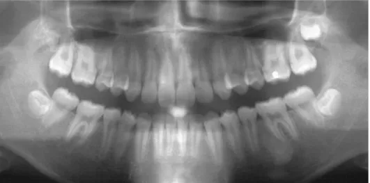
Idiopathic osteosclerosis (IO) in the jaws is defined as a local- ized radiopacity of unknown origin.1Most of these lesions are asymptomatic and are detected on radiographs taken for some other purposes (Fig. 1).1On ordinary radiographic examinations, osteosclerotic areas frequently present as isolated radiopacities located at various positions in the jaws.2
Radiographs are routinely acquired in the diagnostic record for orthodontic treatment planning. In conventional orthodontic examination, lateral cephalometric and panoramic radiographs are taken. However, very few studies have analyzed the preval- ence of IO on radiographs taken for orthodontic diagnosis and the Angle’s classification of malocclusion. Therefore, the purpose of this study was to evaluate the prevalence of the idiopathic osteosclerosis (IO) in Korean malocclusion patients according to age, sex, and the Angle’s classification of maloc- clusion.
Materials and Methods
The subjects of this study were 2,001 randomly selected patients, comprising 1,117 females and 884 males, with a mean age of 17.28 years (Table 1). The patients were selected from the Department of Orthodontics at Gangneung-Wonju National University Dental Hospital. No patients had known systemic disease, syndrome, or developmental defect. All pano- ramic radiographs were taken between 1998 and 2007 at the Department of Oral and Maxillofacial Radiology, Gangneung- Wonju National University Dental Hospital. Panoramic radio- graphs were taken with Cranex++Ceph (Soredex Orion Corp., Tuusula, Finland), and processed with FCR 5000R (Fuji Medi- cal System, Tokyo, Japan). The images were printed out with Fuji medical laser imager (Fuji Medical System, Tokyo, Japan).
The radiographs were read in table mounted, suitably masked, and investigated under constant ambient lighting condition using a magnifier with a 23 magnification (5′′ regular glass lens, Osung Corp, Seoul, Korea). Radiographs were evaluated from not more than 50 patients at a time to minimize the risk of false assessments caused by fatigue. Two orthodontists investigated the radiographs independently and recorded the observed IO.
A study on the prevalence of the idiopathic osteosclerosis in Korean malocclusion patients
Seung-Youp Lee, In-Woo Park*, Insan Jang, Dong-Soon Choi, Bong-Kuen Cha
Department of Orthodontics, College of Dentistry, Gangneung-Wonju National University
*Department of Oral and Maxillofacial Radiology, College of Dentistry, Gangneung-Wonju National University ABSTRACT
Purpose : This retrospective study was performed to investigate the prevalence of the idiopathic osteosclerosis (IO) in Korean malocclusion patients according to age, sex, and the Angle’s classification of malocclusion.
Materials and Methods : This study consisted of 2,001 randomly selected patients from the Department of Orthodon- tics at the Gangneung-Wonju National University Dental Hospital, Korea. The prevalence of IO in Korean malocclu- sion patients was recorded using their panoramic radiographs, and the following parameters were surveyed; age, sex, and the Angle’s classification of malocclusion. The chi-square test was analyzed to determine the statistical signifi- cance of differences in the prevalence of IO between age, sex, and the Angle’s classification of malocclusion.
Results : The prevalence of IO in the jaws was 6.7% in a total of 2,001 examined orthodontic patients. The majority of IO was found in the mandible (96.58%). The 30-39 age group showed the highest prevalence of IO (9.60%). There was a higher prevalence in females (6.89%) than in males (6.45%). The prevalence of IO in Angle Class I group (7.07%) was the most frequent, followed by Angle Class II group (6.72%), and Angle Class III group (6.40%). How- ever, there was no statistical significance in sex and Angle’s classification of malocclusion.
Conclusion : The prevalence of IO in malocclusion patients showed the differences between various age groups and most of them were found in the mandibular posterior area. However, sex and the type of malocclusion are not to be considered as a contributing factor of IO. (Korean J Oral Maxillofac Radiol 2010; 40 : 159-63)
KEY WORDS : Osteosclerosis; Radiography, Panoramic; Malocclusion
Received July 6, 2010; Revised August 4, 2010; Accepted September 27, 2010 Correspondence to : Prof. Bong-Kuen Cha
Department of Orthodontics, Gangneung-Wonju National University Dental Hospital, 120 Gangneung Daehangno, Gangneung, Gangwon, Korea
Tel) 82-33-640-3192, Fax) 82-33-640-3057, E-mail) [email protected]
From a set of 200 radiographs, agreement percentages and kappa indices for inter- and intraexaminer variations were calculated.
The intraexaminer agreement for the two observers for detect- ing IO was 98% (kappa 0.86) and 96.7% (kappa 0.82), respec- tively. The overall interexaminer agreement was 96%, corre- sponding to a kappa index of 0.83.
All radiographs were assessed for the presence of IO on the basis of the following criteria.1
1. A well-cleared radiopacity in the jaws that is located adja-
cent to sound teeth, adjacent to teeth with small restorations, or at a distance from the teeth.
2. Round or oval in shape and more than 3 mm in size.
3. No surrounding radiolucent boundary.
The following criteria3were excluded from this investigation:
1. Isolated radiopacities in edentulous regions because these could be residual condensing osteitis.
2. Radiopacities around teeth with deep caries, large restora- tions, or canal fillings (Fig. 2).
3. Radiopacities in patients with Gardner’s syndrome, famil- ial adenomatosis of the colon, and other systemic disease of bone.
The prevalence of IO was recorded and the following para- meters were surveyed; age, sex, location of IO in the jaws, and the Angle’s classification of malocclusion. The location of IO was classified as the following 4 areas; maxillary anterior and Fig. 1.Idiopathic osteosclerosis, a round, localized radiopacity, is de- tected between lower left second premolar and first molar.
Table 1.The distribution of 2,001 patients according to age and sex
Age Male Female Total
Under 9 207 243 450
10-19 431 499 930
20-29 174 238 412
30-39 46 79 125
Over 40 26 58 84
Fig. 2.Condensing osteitis, radiopa- city around tooth with deep caries, is detected on right lower first molar.
This lesion was excluded from this investigation, due to severe dental caries on right lower first molar.
posterior teeth area, and mandibular anterior and posterior teeth area.
The Angle’s classification of malocclusion for the patients was confirmed using the diagnostic dental models by 2 obser- vers. From a set of 200 pretreatment diagnostic dental models, the agreement percentages and kappa indices for inter- and intraexaminer variations were also calculated. The intraexaminer agreement for the two observers to confirm the Angle’s classi- fication of malocclusion was 100% (kappa 1.0) and 100% (kappa 1.0), respectively. The overall interexaminer agreement was 97%, corresponding to a kappa index of 0.86. Out of 2,001 patients, 509 patients showed Angle’s Class I malocclusion (25.3%), and 804 patients were Class II (39.9%), and 688 pati- ents were Class III malocclusion (34.8%).
The chi-square test was analyzed to determine the statistical significance of differences in the prevalence of IO between age, sex, location of IO in the jaws, and the Angle’s classifica- tion of malocclusion.
Results
Total of 146 IO were found in 134 patients (6.7%). Out of 134 patients, 10 patients had two IO, and one patient had three IO. There were no patients with 4 or more IO. Table 2 shows the prevalence of IO according to age. The 30-39 age group showed the highest prevalence of IO (9.60%), and children under 9 years showed the lowest prevalence of IO (3.11%).
There was significant difference in the prevalence of IO among age groups (p⁄0.01). Table 3 shows the prevalence of IO according to sex. The prevalence in females (6.89%) was higher than in males (6.45%), however there was no statistically signi- ficant difference (p==0.692).
Of the 146 lesions, 141 (96.6%) lesions were present in the mandible with only 5 (3.4%) lesions in the maxilla (Table 4).
And 112 (76.6%) lesions of the mandible were present at the posterior teeth area and 29 (20.0%) at the anterior area (Table 4). There was significant difference in the prevalence of IO
between the maxilla and mandible (p==0.000). Also, there was higher prevalence at the mandibular posterior area than the maxillary anterior, maxillary posterior, and mandibular anterior area.
The prevalence of IO in Angle Class I group (7.07%) was the most frequent, followed by Angle Class II group (6.72%), and Angle Class III group (6.40%) (Table 5). However, there was no statistical significance among the Angle’s classification of malocclusion (p==0.898).
Discussion
The prevalence of IO has been reported from 2.4% to 16.0%
in previous studies (Table 6).1-9In this study the prevalence of IO was 6.7%. The reason for these various results was pro- bably due to the difference in the ethnic group, type of radio- graphs, and definition of IO. Ahn et al.2used periapical radio- graphs reporting lower prevalence than our result. The preval- ence in a Caucasians by Geist and Katz1and Farman et al.10 was also lower than ours.
Table 2.The distribution and prevalence of 134 cases of idiopathic osteosclerosis according to age
Age No. of case No. of Prevalence
examined IO* (%)
Under 9 450 14 3.11
10-19 930 72 7.74
20-29 412 32 7.77
30-39 125 12 9.60
Over 40 84 4 4.76
*IO: idiopathic osteosclerosis, χ2==13.771, df==4, p==0.008
Table 3.The distribution and prevalence of 134 cases of idiopathic osteosclerosis according to sex
Sex No. of case No. of Prevalence
examined IO* (%)
Male 884 57 6.45
Female 1117 77 6.89
*IO: idiopathic osteosclerosis, χ2==0.157, df==1, p==0.692
Table 4.The distribution and prevalence of 146 lesions of idiopa- thic osteosclerosis according to quartet site
Distribution of IO* No. of Prevalence
IO* (%)
Maxilla Anterior teeth area 4 2.74
Posterior teeth area 1 0.68
Mandible Anterior teeth area 29 19.86
Posterior teeth area 112 76.71
*IO: idiopathic osteosclerosis, χ2==133.0, df==1, p==0.000
Table 5.The distribution and prevalence of 134 cases of idiopathic osteosclerosis according to the Angle’s classification of malocclu- sion
Classification of No. of case No. of Prevalence
malocclusion examined IO* (%)
Class I 509 36 7.07
Class II 804 54 6.72
Class III 688 44 6.40
*IO: idiopathic osteosclerosis, χ2==0.216, df==2, p==0.898
The present study found no difference of the incidence accord- ing to sex. This finding agrees with Park,5Garau et al.7, Kawai et al.9and Yonetsu et al.3whereas Geist and Katz1and McDon- nell11reported a female to male ratio of 1.5 : 1 and 2 : 1, respectively. Ahn et al.2showed that there was a higher preval- ence in women (3.12%) than in men (1.68%).
This study was also focused on the relation between the pre- valence of IO and age. Our result showed that there was a sta- tistical difference in the prevalence of IO among the age groups.
Some studies2-10reported that there was no statistical difference among age groups. On the other hand, Park5reported that relatively high occurrence of IO with aging. He reported that the prevalence of IO was 1.4% (10’s), 15.7% (20’s), 21.2%
(30’s), 22.7% (40’s), and 20.2% (50’s), respectively. Eversole et al.12also reported that there were higher prevalence in older age group. It was suggested that once IO was established in the early stage of life, it does not tend to regress.1,11,13In our study, the prevalence of IO was higher in older age group than in younger age group. However, in the age group over 40 years, the prevalence tended to decrease, and this might be originated from the insufficient samples.
Many previous studies reported higher prevalence of IO in the mandible than in the maxilla (Table 6). Our results showed
that 96.6% of IO was found in mandible, whereas only 3.4%
was found in the maxilla. And 76.6% of IO in the mandible was found in posterior area. Our result was in accordance with the previous reports.1-9,12,14The reason for high prevalence in the mandible has not been fully explained, however the dif- ference in the density between the mandible and the maxilla, and bending phenomenon of the mandible during mastication might be related with the occurrence of IO.
There were many previous studies on IO using radiographs, however no study about the Angle’s classification of malocclu- sion with findings of IO is cited in the literature. In the etiology of IO, Eversole et al.12reported that excessive occlusal forces could be a cause. Because the occlusal forces could be different between the types of malocclusion, we investigated the preva- lence of IO in three types of malocclusion patients. However, there was no significant difference in prevalence of IO among the Angle’s classification of malocclusion groups.
In conclusion, the prevalence of IO in the jaws was 6.7% in a total of 2,001 examined orthodontic patients. The majority of IO was found in the mandible (96.58%). The 30-39 age group showed the highest prevalence of IO (9.60%). And, there was a higher prevalence in females (6.89%) than in males (6.45%). The prevalence of IO in Angle Class I group (7.07%) Table 6.Prevalence of IOain the previous studies
Prevalence Number of Type of Excluding the
Age Most frequently of IOa subjects radiographs condensing osteitis detected location Sex
Geist and Katz1(1990) 5% 1921 Full mouth periapical
No - Mandibular
radiographs premolar region *
Ahn et al.2(1994) 2.43% 6220 Full mouth periapical
No NS Mandibular
radiographs premolar region *
Yonetsu et al.3(1997) 6.1% 1047 Panorama Yes - Mandibular first
molar region NS
Choi4(1995) 9.76% 7837 Panorama No NS Mandibular
molar region NS
Park5(1984) 16.0% 2160 Panorama No * Mandibular
premolar region NS MacDonald et al.6
Hong Kong in 1981 6.7%
Hong Kong in 1990 5.5%
Panorama Yes - Mandibular
London in 1990 2.7% premolar region *
Edinburgh in 1993 4.1%
Garau et al.7(2002) 8.3% 697 Panorama No NS Mandibular first
molar region NS
Williams et al.8(1998) 5.7% 1585 Full mouth periapical
No NS - -
radiographs
Kawai et al.9(1992) 9.7% 1203 Panorama No NS Mandibular first
molar region NS
aidiopathic osteosclerosis
*significant; NS, not significant
was the most frequent, followed by Angle Class II group (6.72%), and Angle Class III group (6.40%). However, there was no statistical significance in sex and Angle’s classification of mal- occlusion. The prevalence of IO in malocclusion patients show- ed the difference between the age groups, and most of them were found at the mandibular posterior area. However, sex and the type of malocclusion are not to be considered as a contribut- ing factor of IO.
References
1. Geist JR, Katz JO. The frequency and distribution of idiopathic osteosclerosis. Oral Surg Oral Med Oral Pathol 1990; 69 : 388-93.
2. Ahn SH, Choi M, Choi KS. A radiological study on the idiopathic osteosclerosis in the jaws. Korean J Oral Maxillofac Radiol 1994; 24 : 107-13.
3. Yonetsu K, Yuasa K, Kanda S. Idiopathic osteosclerosis of the jaws:
panoramic radiographic and computed tomographic findings. Oral Surg Oral Med Oral Pathol Oral Radiol Endod 1997; 83 : 517-21.
4. Choi KS. A study of idiopathic osteosclerosis in the panoramic radiographs. Korean J Oral Maxillofac Radiol 1995; 25 : 287-95.
5. Pak TW. A radiographic analysis of osteosclerosis of the jaws. Ko- rean J Oral Maxillofac Radiol 1984; 14 : 153-61.
6. MacDonald-Jankowski DS. Idiopathic osteosclerosis in the jaws of
Britons and of the Hong Kong Chinese: radiology and systematic review. Dentomaxillofac Radiol 1999; 28 : 357-63.
7. Garau V, Deschino A, Piras D, Cotti E. Idiopathic osteosclerosis in jaw bones. Clinical statistic study on a population of Sardinian origin.
Minerva Stomatol 2002; 51 : 377-83.
8. Williams TP, Brooks SL. A longitudinal study of idiopathic osteosclero- sis and condensing osteitis. Dentomaxillofac Radiol 1998; 27 : 275-8.
9. Kawai T, Hirakuma H, Murakami S, Fuchihata H. Radiographic investigation of idiopathic osteosclerosis of the jaws in Japanese dental outpatients. Oral Surg Oral Med Oral Pathol 1992; 74 : 237-42.
10. Alattar MM, Baughman RA, Collett WK. A survey of panoramic radio- graphs for evaluation of normal and pathologic findings. Oral Surg Oral Med Oral Pathol 1980; 50 : 472-8.
11. Austin BW, Moule AJ. A comparative study of the prevalence of mandibular osteosclerosis in patients of Asiatic and Caucasian origin.
Aust Dent J 1984; 29 : 36-43.
12. Farman AG, de V Joubert JJ, Nortje CJ. Focal osteosclerosis and apical periodontal pathoses in “European” and Cape coloured dental outpa- tients. Int J Oral Surg 1978; 7 : 549-57.
13. McDonnell D. Dense bone island. A review of 107 patients. Oral Surg Oral Med Oral Pathol 1993; 76 : 124-8.
14. Monahan R. Periapical and localized radiopacities. Dent Clin North Am 1994; 38 : 113-36.
15. Eversole LR, Stone CE, Strub D. Focal sclerosing osteomyelitis/focal periapical osteopetrosis: radiographic patterns. Oral Surg Oral Med Oral Pathol 1984; 58 : 456-60.