In vitro Activities of Polycalcium, a Mixture of Polycan and Calcium Lactate- Gluconate, on Osteoclasts and Osteoblasts
Jae-Suk Choi
1, Joo-Wan Kim
2, Ki-Young Kim
2, Hyung-Rae Cho
2, Yu-Mi Ha
1, Sae Kwang Ku
3, Kwang Keun Cho
4and In Soon Choi
1,5*
1
RIS Center, Industry-Academic Cooperation Foundation, Silla University, Busan 617-736, Korea
2
Glucan Corp. Research Institute, Marine Bio-industry Development Center 27, Busan 619-912, Korea
3
Department of Anatomy and Histology, College of Oriental Medicine, Daegu Haany University, 712-715, Korea
4
Department of Animal Resources Technology, Gyeongnam National University of Science and Technology, Chinju 660-758, Korea
5
Department of biological science, Silla University, Busan 617-736, Korea
Received May 17, 2011 /Revised July 27, 2011 /Accepted July 27, 2011The present study evaluated the beneficial effects of polycalcium (a mixture of Polycan and calcium lactate-gluconate 1:9 [g/g]) on osteoporosis using in vitro assays. Cell proliferation and alkaline phos- phatase activities of osteoblasts (human primary osteoblasts) and osteoclast differentiation of RAW264.7 cells (murine osteoclast progenitor cells) treated with different concentrations of poly- calcium for various periods were assessed. Osteoblast proliferation was stimulated and prevented RANKL-induced osteoclast differentiation of RAW264.7 cells. These results support the development of ideal anti-osteoporotic agents, such as polycalcium, that exhibit properties that accelerate bone for- mation and inhibit bone resorption.
Key words : Polycalcium, polycan, osteoclasts, osteoblasts
*Corresponding author
*Tel:+82-51-999-5348, Fax:+82-51-999-5344
*E-mail : [email protected]
Introduction
Normal bone metabolism relies on a balance between bone formation and resorption regulated by osteoblasts and osteoclasts, respectively. When bone resorption is favored over formation, pathological bone destruction leads to osteo- porosis [4,5]. The frequency of fractures significantly in- creases in patients with osteoporosis, and hip fractures pose a very serious problem because they often limit the patient’s quality of life [25].
Numerous attempts have been made to develop new agents that treat or prevent bone diseases [15]. The current anti-resorptive agents are extensively used, but there still re- mains a demand for a highly efficacious resorptive inhibitor with an excellent safety and efficacy profile. Anabolic agents that stimulate bone formation are less known than anti-re- sorptive agents [6]. Continuous trials to develop these agents have been conducted with an advanced understanding of osteoblast differentiation and bone formation. Various ap- proaches have searched for natural extracts that exert a ther- apeutic effect against bone loss.
Calcium (Ca) salts show anti-inflammatory activities
[8,18] and have potentially favorable preventive and ther- apeutic effects on osteoporosis [7,20]. However, most people do not consume sufficient amounts of Ca, even though it is an essential element of living organisms and is highly rec- ommended by nutrition experts [23,24].
Polycan (Glucan Corp., Korea), a commercial product of β -glucans that originated from Aureobasidium pullulans SM-2001, consists mostly of β-1,3/1,6-glucans and other or- ganic materials such as amino acids, mono- or di-un- saturated fatty acids (linoleic and linolenic acids), and fi- brous polysaccharides [16]. Recently, it has been reported that Polycan promotes fracture healing [14], exhibits anti-in- flammatory activity [11,12], and has anti-osteoporotic prop- erties, inhibiting bone loss and accelerating bone formation [17,19]. Several studies have shown that calcium phospha- tase and vitamin D affect Ca resorption, but do not directly stimulate bone metabolism, especially bone formation.
The aim of this study was to confirm the beneficial effects of polycalcium (a mixture of Polycan and calcium lac- tate-gluconate 1:9 [g/g]) on osteoporosis, specifically cell proliferation and the alkaline phosphatase (ALP) activity of osteoblasts, using in vitro assays. Osteoclast differentiation of RAW264.7 cells was also evaluated.
- Note -
Materials and Methods Reagents and cell culture
Dulbecco’s Modified Eagle Medium (DMEM), minimal es- sential medium alpha (α-MEM), fetal bovine serum (FBS), penicillin/streptomycin, and trypsin-EDTA, which were used for cell cultures, were purchased from Gibco-BRL (Grand Island, NY, USA). Human primary osteoblasts (hOBs) and murine osteoclast progenitor RAW264.7 cells were a kind gift from Professor Daewon Jeong of Yeungnam University. For osteoblast analysis, ascorbic acid and β -glycerophosphate were purchased from Sigma-Aldrich (St.
Louis, MO, USA) and an ALP assay kit (AnaSpec, Inc., Fremont, CA, USA) was used. For osteoclast analysis, re- combinant murine receptor activator of NF-κB ligand (RANKL) was purchased from BioVision (Mountain View, CA, USA).
Cell proliferation and ALP activity in human osteoblasts
The hOBs were grown in DMEM supplemented with 10%
FBS, 100 units/ml penicillin, and 100 μg/ml streptomycin in a humidified atmosphere of 5% CO
2. The cells were seed- ed at a density of 1×10
3per well in 96-well plates. After 24 hr, cells were transferred to fresh differentiation medium containing 0.05 mM ascorbic acid and 10 mM β -glycerophosphate every 3 days and were treated with dif- ferent concentrations of polycalcium. Cell proliferation was determined using a commercially available kit (Cell Counting Kit-8, Dojindo, Kumamoto, Japan) according to the manufacturer’s instructions. Briefly, hOBs (1×10
4cells/well) were incubated, and 10 μl 2-(2-methoxy-4-nitrophenyl)- 3-(4-nitrophenyl)-5-(2,4-disulfo-phenyl)-2H-tetrazolium monosodium salt (WST-8) was added to each well. After in- cubation for 1 hr, absorbance was measured at 450 nm using a microplate reader.
To measure ALP activity, hOBs were cultured in 24-well plates at a density of 1×10
4cells/well with a differentiation medium and were treated with different concentrations of polycalcium. Cells were extracted with 0.05% Triton X-100 in the assay buffer and centrifuged at 2,500× g for 10 min at 4°C. Using the supernatant, ALP activity was determined by the colorimetric method using p-nitrophenyl phosphate (PNP) as a substrate at a wavelength of 405 nm and the protein content was determined by a bicinchoninic acid (BCA) protein assay kit. ALP activity was expressed as pg
PNP/min/μg protein.
RANKL-induced osteoclast differentiation of RAW264.7 cells
RAW264.7 cells were grown in α-MEM supplemented with 10% FBS, 100 units/ml penicillin, and 100 μg/ml strep- tomycin in a humidified atmosphere of 5% CO
2, seeded on a 96-well plate at a density of 2×10
3cells/well, and treated with 100 ng/ml RANKL and with different concentrations of polycalcium. The medium was changed every 3 days and the number of undifferentiated RAW264.7 cells was eval- uated using a commercially available kit (Cell Counting Kit-8, Dojindo, Kumamoto, Japan).
Statistical analyses
Multiple comparison tests for different dose groups were conducted. Variance homogeneity was examined using the Levene test. If no significant deviations from homogeneity were detected, the data were analyzed by one-way ANOVA followed by the least-significant differences (LSD) mul- ti-comparison test to determine which pairs were sig- nificantly different. When there were significant deviations from homogeneity, a non-parametric comparison test (i.e., Kruskal-Wallis H test) was conducted. If significant differ- ences were observed, the Mann-Whitney U test was per- formed to determine which specific pairs were significantly different. Statistical analyses were conducted using SPSS for Windows (Release 14K, SPSS Inc., USA).
Results and Discussion
Effects of polycalcium on cell proliferation and ALP activity in hOBs
To investigate the effect of polycalcium on cell growth at concentrations of 0, 10
-5, 10
-3, 10
-1, and 10 mg/ml for a treatment period of 3, 7, or 10 days, a cell proliferation assay was performed. The growth of hOBs gradually increased at 7 and 10 days, whereas 10 mg/ml polycalcium significantly (p<0.05) increased growth for a treatment period of 3, 7, and 10 days (Fig. 1).
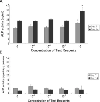
We also examined the effect of polycalcium on the ALP
activity of hOBs grown in differentiation medium at a con-
centration of 0, 10
-5, 10
-3, 10
-1, or 10 mg/ml for a treatment
period of 7 or 14 days. Polycalcium increased ALP activity
in a dose-dependent pattern. Significantly (p<0.05) increased
ALP activity was detected for a treatment period of 7 or
0 10-5 10-3 10-1 10 4.0
3.5 3.0 2.5 2.0 1.5 1.0 0.5 0.0
Fig. 1. Effects on osteoblast proliferation. A dose-dependent in- crease in the proliferation of human osteoblasts (hOB) after treatment with different concentrations of poly- calcium (mg/ml) for 10 days is shown. All data are rep- resented as the mean±S.D. of five independent experiments. *
p
<0.05 as compared to the control using the Mann-Whitney U test.14 days with 10 mg/ml polycalcium. When ALP activity was corrected with protein concentrations, there were no sig- nificant changes (Fig. 2). These results suggest that poly- calcium stimulates hOB proliferation dose-dependently, but does not promote hOB differentiation.
A
0 10-5 10-3 10-1 10 4.0
35 30 25 20 15 10 5 0
B
0 10-5 10-3 10-1 10 4.0
35 30 25 20 15 10 5 0
Fig. 2. Effects on alkaline phosphatase (ALP) activity. Polycalcium (mg/ml) increased ALP activity in a dose-dependent manner with significantly (
p
<0.05) high levels of activity after treatment of 10 mg/ml for 7 or 14 days (A).However, no significant changes in ALP activity were de- tected after correction for protein concentrations (B). All data are represented as the mean±S.D. of five in- dependent experiments. *
p
<0.05 as compared to the con- trol using the Mann-Whitney U test.RANKL-induced osteoclast differentiation of RAW264.7 cells
Different concentrations of polycalcium (0, 10
-5, 10
-3, and 10
-1mg/ml) were added to RAW264.7 cells after stimulation with RANKL. As shown in Figure 3, no osteoclast differ- entiation of RAW264.7 cells in the control (untreated) group was observed. However, the number of undifferentiated RAW264.7 cells decreased after RANKL treatment, suggest- ing that RANKL stimulated osteoclast differentiation. When polycalcium was administered at 0, 10
-5, 10
-3, 10
-1, or 10 mg/ml for a treatment period of 1 or 4 days, osteoclast dif- ferentiation of RAW264.7 cells was inhibited dose-depend- ently (Fig. 3). In general, these results indicate that poly- calcium directly inhibits RANKL-induced osteoclastogenesis.
Osteoblasts derived from mesenchymal cells synthesize and deposit bone matrix and increase bone mass [1]. The ontogenesis of bone-forming osteoblasts is a complex proc- ess and can essentially be divided into three steps: (1) com- mitment into osteoprogenitor cells and proliferation, (2) ex- tracellular matrix development and maturation, and (3) ma- trix mineralization [1,2]. Therefore, the proliferation and ac- tivity of osteoclasts have important roles in bone formation.
The efficacy of osteoblasts has been previously tested based on cell proliferation and activity [9].
Osteoclasts that originate from hematopoietic cells are key participants in bone remodeling because they are predom- inantly involved in bone resorption and can lead to an im- balance in bone remodeling. Osteoclastic bone resorption consists of multiple steps, including the differentiation of os- teoclast precursors, fusion into multinuclear osteoclasts, and the activation of mature osteoclasts to resorb bone [10].
Mature, multinucleated osteoclast-like cells (OCLs) maintain the proper actin cytoskeletal organization, ruffled border, and acidic condition during bone resorption [22]. The effi- cacy of anti-osteoporotic agents, therefore, has been con- tinually evaluated for the inhibition of osteoclastogenesis and its activity has been analyzed using the pit formation assay [9,21].
In the present study, we found that polycalcium stimu- lated the proliferation of hOB, and prevented RANKL-in- duced osteoclast differentiation of RAW264.7 cells in a dose-dependent manner. These results support the develop- ment of ideal anti-osteoporotic agents with properties that accelerate bone formation as well as inhibit bone resorption.
However, polycalcium did not affect hOB ALP activity,
which is an indication of osteoblast differentiation.
A
Centrol 10-5 mg/ml 10-3 mg/ml 10-1 mg/ml
B
0 10-5 10-3 10-1 1.5
1.0
0.5
0.0
Fig. 3. Effects on osteoclast differentiation. The number of un- differentiated RAW264.7 cells decreased after RANKL treatment (A). When polycalcium was administered at con- centrations of 0, 10-5, 10-3, or 10-1mg/ml for 4 days, osteo- clast differentiation of RAW264.7 cells was significantly in- hibited (A, B). All data are represented as the mean±S.D.
of five independent experiments. *
p
<0.05 as compared to the control using the Mann-Whitney U test.Therefore, the data suggest that polycalcium affects only hOB proliferation, not hOB differentiation. Specific genes, such as ALP, collagen I, and osteocalcin, are expressed dur- ing osteogenic differentiation [3,13]. The expression of osteo- genic genes during osteoblast differentiation should be fur- ther analyzed and the association of polycalcium and osteo- blast proliferation should be confirmed.
In summary, we conclude that polycalcium is a potential candidate for anti-osteoporosis therapy. Further studies us- ing in vitro and in vivo models are needed to elucidate the exact mechanism that confers the anti-osteoporotic effects of polycalcium.
Acknowledgment
This work was supported by a grant (No. 70007205) from the Ministry of Knowledge Economy, Republic of Korea.
References
1. Aubin, J. E. 2001. Regulation of osteoblast formation and function.
Reviews in Endocrine & Metabolic Disorders
2, 81-94.2. Benayahu, D. 2000. The hematopoietic microenvironment:
The osteogenic compartment of bone marrow: Cell biology and clinical application.
Hematology
4, 427-435.3. Chen D., M. Zhao, and G. R. Mundy. 2004. Bone morphoge- netic proteins.
Growth Factors
22, 233-241.4. Feldmann, M., F. M. Brennan, and R. N. Maini. 1996.
Rheumatoid arthritis.
Cell
85, 307-310.5. Goldring, S. R. and E. M. Gravallese. 2002. Pathogenesis of bone lesions in rheumatoid arthritis.
Curr
.Rheumatol
.Rep
. 4, 226-231.6. Gowen, M., J. G. Emery, and S. Kumar. 2000. Emerging therapies for osteoporosis.
Emerging Drugs
5, 1-43.7. Heaney, R. P., R. R. Recker, P. Watson, and J. M. Lappe.
2010. Phosphate and carbonate salts of calcium support ro- bust bone building in osteoporosis.
Am
.J
.Clin
.Nutr
. 92, 101-105.8. Hendry, J. A., B. G. Jeansonne, C. O. Dummett Jr, and W.
Burrell. 1982. Comparison of calcium hydroxide and zinc oxide and eugenol pulpectomies in primary teeth of dogs.
Oral
.Surg
.Oral
.Med
.Oral
.Pathol
. 54, 445-551.9. Hwang, Y. H., J. W. Lee, E. R. Hahm, K. C. Jung, J. H. Lee, C. H. Park, H. S. Rhee, J. M. Ryu, H. K. Kim, C. H. Yang, and I. Momordin. 2005. an inhibitor of AP-1, suppressed osteoclastogenesis through inhibition of NF-kappaB and AP-1 and also reduced osteoclast activity and survival.
Biochem
.Biophys
.Res
.Commun
. 337, 815-823.10. Jimi, E., S. Akiyama, T. Tsurukai, N. Okahashi, K.
Kobayashi, N. Udagawa, T. Nishihara, N. Takakashi, and T. Suda. 1999. Osteoclast differentiation factor acts as a mul- tifunctional regulator in murine osteoclast differentiation and function.
J
.Immun
. 163, 434-442.11. Kim, H. D., H. R. Cho, S. B. Moon, H. D. Shin, K. J. Yang, B. R. Park, H. J. Jang, L. S. Kim, H. S. Lee, and S. K. Ku.
2006. Effect of Exopolymers from
Aureobasidum pullulans
on formalin-induced chronic paw inflammation in mice.J
.Microbiol
.Biotechnol
. 16, 1954-1960.초록: In vitro 에서 polycalcium 복합조성물이 파골세포와 조골세포에 미치는 영향 최재석
1․김주완
2․김기영
2․문승배
2․하유미
1․구세광
3․조광근
4․최인순
1,5*
(
1신라대학교 RIS사업단,
2㈜글루칸,
3대구한의대학교 한의학과,
4경남과학기술대학교 동물소재공학과,
5신라 대학교 생물과학과)
본 실험에서는 폴리칸(베타-글루칸)과 칼슘 락테이트 글루코네이트 1:9 (g/g) 복합 조성물인 Polycalcium의 시 험관 내(in vitro) 골다공증에 대한 효과를 사람 유래 조골세포(human primary osteoblast)와 설치류 유래 파골 전구세포(raw264.7 cell)를 이용하여 평가하였다. Polycalcium이 조골세포에 미치는 영향을 확인한 결과, 10 mg/ml 농도의 polycalcium 처리군에서 무처리 대조군에 비해 유의성 있는 조골세포의 수적 증가가 각각 배양 3, 7 및 10일 후에 확인되었으며, 또한 10 mg/ml 농도의 polycalcium 처리군에서 무처리 대조군에 비해 유의성 있는 ALP함량의 증가가 확인되었다. Polycalcium이 파골세포에 미치는 영향을 확인한 결과, 각각 10
-5, 10
-3및 10
-1mg/ml polycalcium 처리군에서 무처리 대조군에 비해 유의성 있는 파골세포의 수적 감소가 배양 4일 후에 확인되었다. 이 같은 결과를 바탕으로, polycalcium이 조골세포의 증식 촉진 효과와 함께 파골세포 형성 억제 효 과가 있는 것으로 확인되었다.
12. Kim, H. D., H. R. Cho, S. B. Moon, H. D. Shin, K. J. Yang, B. R. Park, H. J. Jang, L. S. Kim, H. S. Lee, and S. K. Ku.
2007. Effects of β-glucan from
Aureobasidum pullulans
on acute inflammation in mice.Arch
.Pharm
.Res
. 30, 323-328.13. Komori, T. 2003. Requisite roles of Runx2 and Cbfb in skel- etal development.
J
.Bone Miner
.Metab
. 21, 193-197.14. Lee, H. S., H. R. Cho, S. B. Moon, H. D. Shin, K. J. Yang, B. R. Park, H. J. Jang, L. S. Kim, and S. K. Ku. 2008. Effect of β-glucan from
Aureobsidium pullulan
s on rat rib fracture healing.Lab
.Anim
.Res
. 24, 39-44.15. Rodan, G. A. and T. J. Martin. 2000. Therapeutic approaches to bone diseases.
Science
289, 1508-1514.16. Seo, H. P., J. M. Kim, H. D. Shin, T. K. Kim, H. J. Chang, B. R. Park, and J. W. Lee. 2002. Production of β -1,3/1,6-glucan by
Aureobasidium pullulans
SM-2001.Korean J
.Bitechnol
.Bioeng
. 17, 376-380.17. Shin, H. D., K. J. Yang, B. R. Park, C. W. Son, H. J. Jang, and S. K. Ku. 2007. Antiosteoporotic effect of Polycan, be- ta-glucan from
Aureobasidium
, in ovariectomized osteopor- otic mice.Nutrition
23, 853-860.18. Smith, M. M., P. Ghosh, Y. Numata, and M. K. Bansal. 1994.
The effects of orally administered calcium pentosan poly- sulfate on inflammation and cartilage degradation produced in rabbit joints by intraarticular injection of a hyaluro- nate-polylysine complex.
Arthritis
.Rheum
. 37, 125-136.19. Song, H. B., D. C. Park, G. M. Do, S. L. Hwang, W. K. Lee, H. S. Kang, B. R. Park, H. J. Jang, C. W. Son, E. K. Park, S. Y. Kim, and T. L. Huh. 2006. Effect of exopolymers of
Aureobasidium pullulans
on improving osteoporosis induced in ovariectomized mice.J
.Microbiol
.Biotechnol
. 16, 37-45.20. Sosa, M. and C. Bregni. 2003. Metabolism of the calcium and bioavailability of the salts of most frequent use.
Boll
.Chim
.Farm
. 142, 28-33.21. Suda, T., I. Nakamura, E. Jimi, and N. Takahashi. 1997.
Regulation of osteoclast function.
J
.Bone Miner
.Res
.12, 869-879.22. Suda, T., N. Udagawa, and N. Takahashi. 1996. Osteoclast generation, p. 87, In Raisz L. G., G. A. Rodan and J. P.
Bilezikian (eds.),
Principles of Bone Biology
. Academic Press, San Diego, CA. USA.23. The Korean Nutrition Society. 2010. Dietary reference in- takes for Koreans. 341-356.
24. The Ministry of Health and Welfare, Korea Centers for Disease Control and Prevention. 2009. Korea Health Statistics 2009: Korea national health and nutrition examina- tion survey (KNHANES IV-3). 28-31.
25. Yamaguchi, K., M. Yada, T. Tsuji, M. Kuramoto, and D.
Uemura. 1999. Suppressive effect of norzoanthamine hydro- chloride on experimental osteoporosis in ovariectomized mice.