Anisomycin, an Inhibitor of Protein Synthesis, Overcomes TRAIL Resistance in Human Hepatocarcinoma Cells via Caspases Activation and Bid Downregulation
Cheng-Yun Jin
1, Cheol Park
2, Su Hyun Hong
3and Yung Hyun Choi
3,4*
1
School of Pharmaceutical Science, Zhengzhou University, 100 Kexue Avenue, Zhengzhou, Henan 450001, China
2
Department of Molecular Biology, College of Natural Sciences, Dongeui University, Busan 614-714, Korea
3
Department of Biochemistry, Dongeui University College of Korean Medicine, Busan 614-052, Korea
4
Anti-Aging Research Center & Blue-Bio Industry Regional Innovation Center, Dongeui University, Busan 614-714, Korea
Received April 24, 2014 /Revised May 31, 2014 /Accepted June 23, 2014Anisomycin, also known as flagecidin, is an antibiotic produced by Streptomyces griseolus that inhibits protein synthesis by binding to the ribosomal 28S subunit. The tumor necrosis factor-related apopto- sis-inducing ligand (TRAIL) is a protein that induces apoptotic cell death. TRAIL primarily causes apoptosis in tumor cells by binding to death receptors. Many human cancer cell lines are refractory to TRAIL-induced cell death. In this study, we investigated whether anisomycin could enhance TRAIL-mediated apoptosis in TRAIL-resistant human hepatocarcinoma Hep3B cells. Treatment with anisomycin and TRAIL alone did not reduce cell viability in Hep3B cells. However, in the presence of TRAIL, the anisomycin concentration dependently reduced the cell viability. Our results indicate that anisomycin sensitizes Hep3B cells to TRAIL-mediated apoptosis and that this occurs, at least part- ly, via caspase activation. Interestingly, Bid knockdown by small interfering RNA significantly re- duced the induction of apoptosis in combination with anisomycin and TRAIL, indicating that aniso- mycin effectively acts to lower the threshold at which TRAIL-mediated truncated Bid triggers the mi- tochondrial-mediated apoptosis program in Hep3B cells. Therefore, the use of TRAIL in combination with anisomycin might provide an effective therapeutic strategy for the safe treatment of some TRAIL-resistant cancer cells.
Key words : Anisomycin, apoptosis, Bid, caspase, TRAIL
*Corresponding author
*Tel : +82-51-850-7413, Fax : +82-51-853-4036
*E-mail : [email protected]
This is an Open-Access article distributed under the terms of the Creative Commons Attribution Non-Commercial License (http://creativecommons.org/licenses/by-nc/3.0) which permits unrestricted non-commercial use, distribution, and reproduction in any medium, provided the original work is properly cited.
Journal of Life Science 2014 Vol. 24. No. 7. 769~776 DOI : http://dx.doi.org/10.5352/JLS.2014.24.7.769
서 론
최근 발견된 tumor necrosis factor (TNF)-related apopto- sis-inducing ligand (TRAIL)는 정상세포에서는 세포독성을 나타내지 않는 반면 , 암세포나 형질전환세포 또는 바이러스에 감염된 세포 등에서 선택적 apoptosis 유도한다는 점에서 재 조합 수용성 TRAIL의 의학적 유용성에 주목 받고 있다[2, 26].
아울러 몇몇 TRAIL 수용체의 역할에 관한 작용기전도 비교적 상세하게 알려져 있는데 , death receptor 4 또는 5(DR4/5)가 TRAIL과 세포막에서 결합하게 되면 death receptor-mediated extrinsic apoptosis 경로 개시 신호로 작용하여 caspase-8의 활성화를 유도한다 [2, 25]. Caspase-8의 활성화는 mitochon- dria-dependent 또는 independent한 경로에 의한 apoptosis를 유도하기 위한 실행자 caspase의 활성을 직·간접적으로 활성
화시킨다 . 특히 mitochondria-dependent apoptosis 경로의 경 유를 위해서는 caspase-8이 Bcl-2 family 단백질의 BH3-only member 중의 하나인 Bid를 truncation시킴으로서 mitochon- drial amplification loop을 자극한다[13, 23]. 그러나 간암세포 를 포함한 많은 암세포는 이미 TRAIL 저항성을 획득한 경우 가 많기 때문 [11, 12, 18, 28]에 TRAIL을 활용한 암 치료법에서 TRAIL 감수성 증대를 위한 대체 약물의 발굴이 시급히 요구 되어지고 있다 .
본 연구에서는 TRAIL 감수성 발굴의 일환으로 Streptomyces griseolus에 의하여 생산되는 pyrrolidine 계 항생제의 일종인 anisomycin [6]에 의한 TRAIL 저항성 극복 가능성에 대한 추 가적인 연구를 시행하였다 . Anisomycin은 28S rRNA와 직접 결합하여 작용을 방해함으로서 ribotoxic stress를 유발시키는 물질로서 그 과정에 mitogen-activated protein kinases (MAPKs)를 포함한 다양한 신호전달계의 활성을 동반한다[3, 8, 9]. 몇몇 선행 연구에서 anisomycin은 MAPKs 중, c-Jun N-terminal kinase (JNK)와 p38 MAPK의 활성을 유도하는 ri- botoxic stress 반응을 촉진함으로서 암세포 증식을 apoptosis 유도를 통하여 억제하는 것으로 보고된 바 있다 [4, 21, 24, 27].
최근 보고에 따르면 anisomycin은 몇몇 종류의 암세포에서
death receptor-mediated apoptosis 경로를 활성화시키는 것
으로 보고된 바 있는데 , 예를 들어 전립선 암세포에서 aniso- mycin은 JNK를 활성화시킴으로서 TRAIL에 의한 apoptosis 를 향상시킬 수 있는 것으로 나타났다 [19]. 그리고 교아종세포 (glioblastoma)에서 anisomycin은 JNK의 활성과 Bim의 발현 증대를 통하여 TRAIL 매개 및 Fas 수용체 매개 apoptosis 유발 을 증가시키는 것으로 알려져 있다 [27]. 또한 anisomycin에 의한 Bim의 발현 증가는 악성 중피종(malignant meso- thelioma) 세포에서 TRAIL 매개 apoptosis 유발을 증가시켰는 데 , 이는 anisomycin이 JNK의 활성 증대를 통하여 TRAIL 저 항성을 극복할 수 있음을 보여주는 결과 [1, 17]이며, anisomy- cin이 TRAIL 저항성 극복을 위한 매우 유용한 항암증진제로 서의 가능성을 보여 주는 다수의 선행 결과들을 잘 일치된다 [7, 16, 20, 22, 30]. 최근 본 연구실에서도 TRAIL 저항성 간암세 포에서 anisomycin은 Bid와 연관된 mitochondrial dysfunc- tion과 c-Jun/AP-1 활성을 동반하면서 TRAIL 저항성을 극복 할 수 있음을 보고한 바 있다 [10]. 본 연구에서는 간암세포에서 anisomycin의 TRAIL 저항성 극복 및 감수성 증대 가능성에 관한 추가적인 자료를 제시하기 위하여 선행연구에서 미진했 던 MAPKs 경로의 p38 MAPK 부분을 보완하고자 한다.
재료 및 방법
시약 및 항체
Anisomycin과 TRAIL은 Calbiochem (San Diego, CA, USA)과 KOMA Biotech Inc. (Seoul, Korea)에서 각각 구입 후 , Phosphate Buffered Saline (PBS)와 dimethyl sulfoxide (DMSO, Sigma-Aldrich Chemical Co., St Louis, MO, USA)에 녹여 stock solution을 만든 후 적정 농도로 배지에 희석하여 처리하였다 . 4',6-diamidino-2-phenylindole (DAPI), annexin V fluorescein isothiocyanate (FITC) 및 propidium iodide (PI) 는 Sigma-Aldrich에서 구입하였으며, 5,5′, 6,6′-tetrachloro- 1,1′,3,3′-tetraethyl-imidacarbocyanine iodide (JC-1)과 cas- pase 억제제(caspase-3 inhibitor, z-Asp-Glu-Val-Asp (DEVD)- fmk 및 caspase-8 inhibitor, z-Asp-Glu-Val-Asp (DEVD)-fmk) 는 Calbiochem에서 구입하였다. RPMI 1640 배지와 fetal bo- vine serum (FBS)는 Invitrogen Corp. (Carlsbad, CA, USA)와 GIBCO-BRL (Gaithersburg, MD, USA)에서 각각 구입하였다.
본 연구에 사용된 일차 항체는 Santa Cruz Biotechnology (Santa Cruz, CA, USA)에서 구입하였으며, peroxidase-labeled donkey anti-rabbit, sheep anti-mouse immunoglobulin 및 enhanced chemiluminescence (ECL) kit는 Amersham (Arlington Heights, IL, USA)에서 구입하였으며, 나머지 언급 되지 않은 시약들은 Sigma-Aldrich에서 구입하였다.
세포배양 및 세포 생존율의 측정
인체 간암 Hep3B 세포는 American Type Culture Collection
(Rockville, MD, USA)에서 구입하였으며, 10% FBS, 2 mM glutamine, 100 U/ml penicillin 및 100 μg/ml streptomycin가 함유된 RPMI1640 배지를 이용하여 37°C, 5% CO
2조건하에서 배양하였다 . Aanisomycin 및 TRAIL의 처리에 따른 Hep3B 세포의 생존율의 측정을 위하여 세포 배양용 6 well plate에 Hep3B 세포를 2×10
5개 /ml 씩 분주한 다음 24시간 동안 안정 화시킨 후 각각의 시료를 적정농도로 처리하여 배양하였다 . 24시간 경과 후 상층액을 제거하고 0.05% trypsin-EDTA를 처리하여 세포를 부유시킨 다음 phosphate-buffered saline (PBS)과 0.5% trypan blue solution을 각 well 당 1 ml 씩을 동량으로 첨가하여 2분간 처리하였다. 처리된 세포를 hemo- cytometer에 적용하여 염색이 되지 않은 살아있는 세포의 수 를 도립 현미경 (inverted microscope, Carl Zeiss, Germany)을 이용하여 계수하였다 . 측정은 모두 세 번을 하였으며, 이에 대한 평균값과 표준 오차를 분석하였다 .
DAPI staining에 의한 핵의 형태 관찰
Apoptosis 유발 여부 확인을 위한 핵의 형태적 변화를 관찰 하기 위하여 준비된 세포를 37% formaldehyde 용액과 PBS를 1:9의 비율로 섞은 fixing solution으로 30분 동안 고정하였 다 . 이를 PBS로 세척하고 0.2%의 Triton X-100 (Amresco, Solon, OH, USA)을 첨가하여 상온에서 10분 동안 반응시킨 후 2.5 μg/ml 농도의 DAPI 용액을 처리하여 세포의 핵을 염색 하였다 . 염색이 끝난 후 증류수 및 PBS로 세척하고 mounting solution을 처리한 후 형광 현미경(Carl Zeiss, Germany)을 이용하여 400배의 배율로 각 농도에 따른 암세포의 핵의 형태 변화를 관찰하였다 .
DNA flow cytometry 분석
Apoptosis의 정량적 분석을 위한 DNA flow cytometry 분 석을 위해 준비된 세포를 고정하고 염색하기 위하여 Cycle- TEST PLUS DNA REAGENT Kit (Becton Dickinson, San Jose, CA, USA)를 이용하였으며, 염색 후 4
oC, 암실에서 30분 동안 반응을 시켰다 . 반응이 끝난 세포를 35 mm mesh를 이용 하여 단일세포로 분리한 후 FACSCalibur (Becton Dickinson) 를 적용시켜 형광반응에 따른 Cellular DNA content 및 histo- gram을 CellQuest software 및 ModiFit LT 프로그램을 이용 하여 분석하였다 .
Mitochondrial membrane potential (MMP, Δψ
m)의 분석
Anisomycin과 TRAIL 처리에 의한 apoptosis 유발에서 미
토콘드리아의 관련성 여부를 확인하기 위하여 준비된 세포를
모은 다음 500 μl의 PBS에 부유시키고 10 μM의 JC-1 용액을
첨가하여 37
oC에서 20분 동안 반응시켰다. 반응이 끝난 후 원
심분리를 이용하여 상층액을 제거한 다음 차가운 PBS를 첨가
하고 35 mm mesh를 이용하여 단일세포로 분리한 후
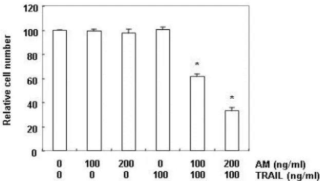
Fig. 1. Effect of anisomycin on the cell viability in TRAIL-re- sistant human hepatocellular carcinoma Hep3B cells.
Hep3B cells were treated with the indicated concen- trations of anisomycin (AM) in the presence or absence of 100 ng/ml TRAIL for 24 hr. After treatments, cell number was determined by Trypan blue exclusion assay. Each point represents the mean ± SD of three in- dependent experiments. The significance was de- termined using Student’s t-test (*,
p
<0.05 vs. untreated control).FACSCalibur에 적용시켜 MMP의 변화 정도를 분석하였다.
Western blot analysis에 의한 단백질 발현 분석 특정 단백질의 발현 변화를 확인하기 위하여 lysis buffer [25 mM Tris-Cl (pH 7.5), 250 mM NaCl, 5 mM EDTA, 1%
NP-40, 1 mM phenymethylsulfonyl fluoride (PMSF), 5 mM dithiothreitol (DTT)]를 이용하여 4
oC에서 1시간 동안 반응시 켜 준비된 세포를 용해시켰다 . Bio-Rad 단백질 정량 시약 (Bio-Rad, Hercules, CA, USA)을 이용하여 단백질 농도를 정 량 한 다음 동량의 Laemmli sample buffer (Bio-Rad)를 섞어 서 균일한 농도의 단백질 sample을 만들었다. 이렇게 만들어 진 단백질 sample은 sodium dodecyl sulphate (SDS)-poly- acrylamide gel을 이용한 전기영동을 실시하여 분자량에 따라 분리한 다음 nitrocellulose membrane (Schleicher and Schuell, Keene, NH, USA)으로 electroblotting에 의해 전이시 켰다 . 단백질이 전이된 nitrocellulose membrane은 5% skim milk 용액을 이용하여 비특이적인 단백질들에 대한 blocking 을 실시하고 1차 항체를 처리하고, 적정 2차 항체를 사용하여 반응시켰다 . 2차 항체에 대한 반응이 끝난 후 충분히 세척하고 암실에서 ECL 용액을 적용시킨 다음 X-ray film에 감광시켜 특정단백질의 발현 양을 분석하였다 .
In vitro caspase-8 및 -3의 활성 측정
다양한 조건에서 배양된 세포들의 caspase 활성을 측정하 기 위하여 150 μg의 단백질이 함유된 50 μl의 sample에 50 μ l의 reaction buffer [40 mM HEPES (pH 7.4), 20% glycerol (v/v), 1 mM EDTA, 0.2% NP-40 and 10 mM DL-DTT]를 혼합 한 다음 두 종류의 caspases 기질 5 μl를 첨가하여 37
oC, 암실에 서 3시간 동안 반응시켰다. 반응이 끝난 후 ELISA reader를 이용하여 405 nm 파장에서 caspases 활성 정도에 따른 변화를 측정하였다 . 실험에 사용된 기질은 caspase-3의 경우에는 DEVD- p-nitroaniline (pNA)이었고 caspase-8의 경우에는 IETD- pNA였다.
Small interference RNA (siRNA)의 처리
Bid의 발현을 억제하기 위하여 Bid siRNA 및 control RNA 는 Dharmacon (Chicago, IL, USA)에서 구입하였으며, LipofectAMINE 2000 (Invitrogen Corp.)을 이용하여 trans- fection시켰다. siRNA transfection 후 24시간 동안 배양된 세 포들의 Bid 발현을 Western blotting 방법으로 확인하고, 이들 세포에 anisomycin 및 TRAIL을 단독 혹은 동시 처리하여 apoptosis 유발의 정도를 비교하였다.
통계분석
모든 실험결과는 평균 ± 표준편차로 표시하였고 SigmaPlot (Systat Software Inc., San Jose, CA, USA)을 이용하여 Student
t-test를 이용하여 통계적 유의성을 얻었다.
결과 및 고찰
Anisomycin이 TRAIL 저항성 Hep3B 세포의 생존율 및 apoptosis에 미치는 영향
대표적인 TRAIL 저항성 간암세포의 일종인 Hep3B 세포
[12]에서 anisomycin이 TRAIL 저항성을 극복할 수 있는지의
가능성을 먼저 조사하기 위하여 세포독성을 나타내지 않는
범위에 해당되는 농도의 TRAIL과 anisomycin을 동시 처리하
였을 경우 생존율 억제 효과가 있는지의 유무를 비교하였다 .
Fig. 1의 결과에서 알 수 있듯이, anisomycin은 200 ng/ml 처
리군까지 Hep3B 세포의 생존에 큰 영향을 미치지 못하였지
만 , 세포독성을 나타내지 않는 100 ng/ml의 TRAIL과 동시에
처리하였을 경우 , anisomycin 농도 의존적으로 Hep3B 세포의
생존율 억제를 보여 주었다 . 이러한 anisomycin과 TRAIL의
동시 처리에 의한 생존율 저하가 apoptosis 유도와 연관되었
는지의 여부를 조사하기 위하여 세포 핵의 형태적 변화를 조
사한 결과 , anisomycin과 TRAIL이 단독으로 처리된 세포에
비하여 동시 처리군에서 전형적인 apoptosis가 유발된 세포에
서 관찰되는 염색질 응축에 의한 apoptotic body의 형성이 증
가되었으며 (Fig. 2A), apoptosis가 유발된 세포의 빈도에 해당
되는 sub-G1기에 속하는 세포의 빈도 역시 매우 증가되었다
(Fig. 2B). 또한 anisomycin과 TRAIL의 동시 처리에 따른
apoptosis 유도 증가가 미토콘드리아 기능 손상과 연관성이
있는지를 위하여 JC-1 염색법을 이용한 MMP 변화 여부로 조
사한 결과 , anisomycin 및 TRAIL 단독 처리군은 대조군의 경
우와 유사하였으나 , anisomycin과 TRAIL의 동시 처리군에
A
B
C
Fig. 2. Anisomycin sensitizes Hep3B cells to TRAIL-mediated apoptosis. Hep3B cells were treated for 24 hr with 200 ng/ml anisomycin in the absence or presence of 100 ng/ml TRAIL. (A) The nuclei stained with DAPI sol- ution were observed under fluorescent microscope using a blue filter (magnification ×400). (B and C) Apoptotic sub-G1 cell population and the loss of MMP were de- termined by a flow cytometer after DAPI (B) and JC-1 (C) staining, respectively. Results are expressed as per- centages of two independent experiments.
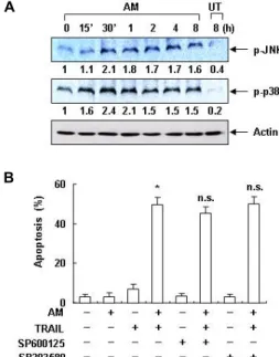
A
B
Fig. 3. Effects of anisomycin on the activation of JNK and p38 MAPK in Hep3B cells. (A) Hep3B cells were treated with 200 ng/ml anisomycin (AM) for the indicated times. The cell extracts were prepared for Western blot analysis of p-JNK and p-p38 MAPK. Actin was used as an internal control. The numbers represent the average densito- metric analyses as compared with actin in, at a mini- mum, 2 or 3 different experiments. (B) Hep3B cells were treated with 100 ng/ml anisomycin (AM) alone, 100 ng/ml TRAIL alone, or anisomycin plus TRAIL in the presence or absence of JNK and p38 MAPK inhibitors for 24 hr. The sub-G1 population was calculated to esti- mate the apoptotic cell population. Significance was de- termined using Student's t-test (∗,
p
<0.05 vs. untreated control; n.s., not significant).서는 MMP 소실의 증가가 매우 증가되었다(Fig. 2C). 이상의 결과는 TRAIL 저항성을 보이는 Hep3B 세포에서 anisomycin 은 TRAIL 매개 apoptosis 유도를 증가시켰음을 보여주는 것 이며 이러한 과정은 미토콘드리아의 기능 손상과 연관이 있음 을 알 수 있었다 .
Anisomycin에 의한 JNK 및 p38 MAPK의 활성에 미치 는 TRAIL의 영향
MAPKs 중, extracellular signal-regulated kinase (ERK)의 활성화가 세포의 생존과 증식 촉진에 더 큰 영향을 주는 반면 , JNK와 p38 MAPK의 활성화는 apoptosis 및 분화 유도와 연관 성이 깊은 것으로 알려져 있다 . 특히 JNK는 anisomycin의 세 포독성에 잠재적 기여 인자로 작용하는 것으로 알려져 왔으나 [5, 10, 28], 급성 림프구성 백혈병(acute lymphoblastic leuke- mia) [15], 악성 뇌교종(glioblastoma) [14] 뿐만 아니라 대장암 [29], 대식세포[4] 등에서 p38 MAPK의 활성 역시 anisomycin 에 의한 apoptosis 유도 활성 과정에 기여하는 것으로 보고되 어지고 있다 . 따라서 다음은 Hep3B 간암세포에서 anisomycin 에 의하여 JNK와 p38 MAPK이 활성화되는지의 여부와 TRAIL과의 동시 처리에 의한 apoptosis 유발에 미치는 영향 에 관하여 조사하였다 . Hep3B 세포에 anisomycin을 처리하였 을 경우 , JNK는 anisomycin 처리 30분 후부터, p38 MAPK은 15분 후부터 인산화가 증가되어 JNK 및 p38 MAPK이 aniso- mycin에 의하여 활성화되었음을 보여주었다(Fig. 3A). 그러나
JNK 선택적 억제제인 SP600125와 p38 MAPK 선택적 억제제 인 SB203580을 선처리한 후 anisomycin과 TRAIL을 처리하였 을 경우 , 두 억제제에 의한 apoptosis 증가 또는 감소에 유의적 인 변화가 관찰되지 않았다 (Fig. 3B). 이는 anisomycin에 의하 여 Hep3B 세포에서 비록 JNK와 p38 MAPK이 활성화되었으 나 TRAIL과의 동시 처리에 의한 apoptosis 유도에는 크게 관 여하지 않을 가능성을 보여주는 것이다 .
Anisomycin과 TRAIL 동시 처리에 의한 apoptosis 유 도에 미치는 caspase 활성 억제제의 영향
TRAIL과 연관된 death receptor-mediated extrinsic apop- tosis 활성과 직접 연관이 있는 caspase는 caspase-8로서 cas- pase-8이 활성화되면 하위 caspase에 해당되는 caspase-3의 활 성화됨으로서 다양한 기질단백질들의 분해가 개시된다 [2, 25].
따라서 anisomycin이 TRAIL의 존재 하에 extrinsic apoptosis
경로를 활성화시키는지의 여부를 조사하기 위하여 caspase-8
의 활성 정도를 조사하였다 . Fig. 4A의 결과는 caspase-8의 기
A B
C
Fig. 4. Caspase activation is involved in the sensitization to anisomycin/TRAIL-induced apoptosis in Hep3B cells. (A) Hep3B cells were treated for 24 with the indicated concentrations of anisomycin in the absence or presence of 100 ng/ml TRAIL for 24 hr. Relative activity of caspase-3 and -8 was determined using caspases activation kit was used according to the manu- facturer’s protocol. Each point represents the mean ± SD of three independent experiments. The significance was determined using Student’s t-test (∗,
p
<0.05 vs. untreated control). (B) Hep3B cells were treated with 100 ng/ml anisomycin (AM) alone, 100 ng/ml TRAIL alone, or anisomycin plus TRAIL in the presence or absence of caspase inhibitors for 24 hr. The cell extracts were prepared for Western blot analysis of caspase-8 and -3. Actin was used as an internal control. (C) To quantify the degree of apoptosis, cells grown under the same conditions as (B) were evaluated for sub-G1 DNA content using a flow cytometer. Significance was determined using Student’s t test (*,p
<0.05 vs. untreated control; (#,p
<0.05 vs. co-treatment with anisomycin and TRAIL).질과 caspas-3의 기질로서 IETD-pNA 및 DEVD-pNA를 이용 한 in vitro 활성 정량화 결과로서 anisomycin이나 TRAIL을 단독으로 처리할 경우 , caspase-8 뿐만 아니라 caspase-3의 활 성도 검출되지 않았다 . 그러나 TRAIL이 존재하는 조건에서 anisomycin을 처리할 경우 처리 시간 의존적으로 두 caspase 의 활성이 유의적으로 증가되었다 . 또한 anisomycin 및 TRAIL 단독 처리군에서는 pro-caspase-8의 발현엔 큰 변화가 없었으 나 , 동시 처리군에서 pro-caspase-8의 발현이 매우 감소되었 다 . Caspase-3의 경우에도 유사하게 anisomycin과 TRAIL의 동시 처리군에서 단독 처리군에 비하여 활성형 caspase-3의 발현이 증가되었다 . 그러나 caspase-8 및 -3의 선택적 억제제 (z-IETD-fmk 및 z-DEVD-fmk)를 처리할 경우, 완벽하지는 않 지만 , pro-caspase-8의 발현 감소가 다소 차단되거나 활성형 caspase-3의 발현이 상대적으로 감소되었다(Fig. 4B). 이는 상 대적 apoptosis 유발 빈도의 감소와 관련이 있었으며(Fig. 4C), 이상의 결과는 Hep3B 세포에서 TRAIL이 존재하는 조건에서 anisomycin은 extrinsic apoptosis 경로 활성을 유도하였음을
의미한다 .
Anisomycin과 TRAIL 동시 처리에 의한 apoptosis 유 도에서 Bid의 역할
Bcl-2 family에는 apoptosis 유도를 촉진하거나 억제하는 정 반대의 역할을 하는 다양한 단백질로 구성되어 있다 [13, 23].
이상의 결과에서 anisomycin이 TRAIL 존재 하에 caspase-8의
활성에 의한 caspase-3의 활성을 유도하였지만, caspase-3의
활성 유도가 caspase-8의 활성에 의한 직접적인 결과인지 mi-
tochondria-mediated intrinsic 경로를 증폭시킨 결과에 의한
것인지의 여부는 정확하지 않다 . 따라서 anisomycin이 TRAIL
존재하는 apoptosis를 유발하기 위하여 Bcl-2 family 단백질의
발현에 변화를 줄 수 있는지는 조사한 결과는 Fig. 5A에 나타
낸 바와 같다 . 제시된 결과에서 알 수 있듯이 anisomycin 처리
에 의하여 Bcl-2 family 단백질 중 대표적인 apoptosis 유도
단백질에 해당되는 Bax의 발현은 큰 변화가 없었으나, an-
ti-apoptotic Bcl-2의 경우에는 다소 감소하여 anisomycin이 처
A
C B
Fig. 5. Involvement of Bid on the apoptotic activity by combined treatment with anisomycin and TRAIL in Hep3B cells.
(A) Hep3B cells were treated for 24 with the indicated concentrations of anisomycin for 24 hr. Cell extracts were prepared for Western blot analysis of Bcl-2 family mem- ber proteins. (B) Cells were transfected with either con- trol or Bid-specific siRNA. Twenty-four hours post-trans- fection, cells were lysed, and then equal amounts of cell lysates were subjected to electrophoresis and analyzed by Western blot for Bid expression. (C) Hep3B cells trans- fected with control or Bid-specific siRNA were treated with or without 100 ng/ml anisomycin (AM) in the pres- ence or absence of 100 ng/ml TRAIL for 24 hr. The sub-G1 population was calculated to estimate the apop- totic cell population. Significance was determined using Student's t-test (∗,
p
<0.05 vs. untreated control). The numbers represent the average densitometric analyses as compared with actin in, at a minimum, 2 or 3 different experiments.리된 Hep3B 세포에서 Bcl-2에 대한 Bax의 상대적인 발현 증가 가 있었음을 알 수 있었다 . 따라서 Bax의 상대적 발현 증가는 최소한의 TRAIL 존재 하에 mitochondria-mediated intrinsic 경로 활성에 어느 정도 기여하였을 것으로 추정된다 . 아울러 anisomycin 처리에 의하여 truncation 된 Bid (tBid) 단백질은 비록 관찰할 수 없었지만 Bid 발현의 감소가 관찰되었기에 anisomycin과 TRAIL 동시 처리에 의한 apoptosis 유도에 Bid 의 역할에 관한 추가적인 실험을 실시하였다 . 이를 위하여 Bid siRNA를 처리하여 Bid의 발현을 차단(knockdown of Bid)시 킨 조건 (Fig. 5B)에서 anisomycin과 TRAIL 동시 처리에 의한 apoptosis 유도 정도를 control siRNA를 처리한 군과 비교하 였다 . Fig. 5C의 결과에서 알 수 있듯이 control siRNA가 처리
된 세포에서 anisomycin과 TRAIL의 동시에 처리에 의하여서 는 apoptosis 유가가 유의적으로 증가되었으나, Bid siRNA 처리군에서는 apoptosis가 거의 유도되지 않았다. 이는 TRAIL 저항성 Hep3B 세포에서 TRAIL 신호계 연관 apoptosis 상승 을 위한 mitochondria 신호계의 증폭은 Bid 의존적으로 일어 나고 있음을 보여주는 결과이다 .
이상의 결과에서 anisomycin은 TRAIL 저항성을 획득한 암 세포의 apoptosis 유도를 위한 촉진제로서 효율적으로 사용될 수 있음을 보여 주는 것이다 .
감사의 글
이 논문은 2014학년도 동의대학교 교내연구비에 의해 연구 되었음 (2014AA058).
References
1. Abayasiriwardana, K. S., Barbone, D., Kim, K. U., Vivo, C., Lee, K. K., Dansen, T. B., Hunt, A. E., Evan, G. I. and Broaddus, V. C. 2007. Malignant mesothelioma cells are rap- idly sensitized to TRAIL-induced apoptosis by low-dose anisomycin
via
Bim.Mol Cancer Ther
6, 2766-2776.2. Ashkenazi, A., Pai, R. C., Fong, S., Leung, S., Lawrence, D.
A., Marsters, S. A., Blackie, C., Chang, L., McMurtrey, A.
E., Hebert, A., DeForge, L., Koumenis, I. L., Lewis, D., Harris, L., Bussiere, J., Koeppen, H., Shahrokh, Z. and Schwall, R. H. 1999. Safety and antitumor activity of re- combinant soluble Apo2 ligand.
J Clin Invest
104, 155-162.3. Campos, C. B., Bédard, P. A. and Linden, R. 2003. Selective involvement of the PI3K/PKB/bad pathway in retinal cell death.
J Neurobiol
56, 171-177.4. Croons, V., Martinet, W., Herman, A. G., Timmermans, J.
P. and De Meyer, G. R. 2009. The protein synthesis inhibitor anisomycin induces macrophage apoptosis in rabbit athero- sclerotic plaques through p38 mitogen-activated protein kinase.
J Pharmacol Exp Ther
329, 856-864.5. Dampier, K., Hudson, E. A., Howells, L. M., Manson, M.
M., Walker, R. A. and Gescher, A. 2001. Differences between human breast cell lines in susceptibility towards growth in- hibition by genistein.
Br J Cancer
85, 618-624.6. Grollman, A. P. 1967. Inhibitors of protein biosynthesis. II.
Mode of action of anisomycin.
J Biol Chem
242, 3226-3233.7. Hadji, A., Clybouw, C., Auffredou, M. T., Alexia, C., Poalas, K., Burlion, A., Feraud, O., Leca, G. and Vazquez, A. 2010.
Caspase-3 triggers a TPCK-sensitive protease pathway lead- ing to degradation of the BH3-only protein puma.
Apoptosis
15, 1529-1539.8. Hemi, R., Paz, K., Wertheim, N., Karasik, A., Zick, Y. and Kanety, H. 2002. Transactivation of ErbB2 and ErbB3 by tu- mor necrosis factor-alpha and anisomycin leads to impaired insulin signaling through serine/threonine phosphorylation of IRS proteins.
J Biol Chem
277, 8961-8969.9. Iordanov, M. S., Pribnow, D., Magun, J. L., Dinh, T. H.,
Pearson, J. A., Chen, S. L. and Magun, B. E. 1997. Ribotoxic stress response: activation of the stress-activated protein kinase JNK1 by inhibitors of the peptidyl transferase re- action and by sequence-specific RNA damage to the al- pha-sarcin/ricin loop in the 28S rRNA.
Mol Cell Biol
17, 3373-3381.10. Jin, C. Y., Park, C., Hong, S. H., Han, M. H., Jeong, J. W., Xu, H., Liu, H., Kim, G. Y., Kim, W. J., Yoo, Y. H. and Choi, Y. H. 2013. Synergistic induction of TRAIL-mediated apop- tosis by anisomycin in human hepatoma cells via the BH3-only protein Bid and c-Jun/AP-1 signaling pathway.
Biomed Pharmacother
67, 321-328.11. Jin, C. Y., Park, C., Kim, G. Y., Lee, S. J., Kim, W. J. and Choi, Y. H. 2009. Genistein enhances TRAIL-induced apop- tosis through inhibition of p38 MAPK signaling in human hepatocellular carcinoma Hep3B cells.
Chem Biol Interact
180, 143-150.12. Kim, Y. S., Schwabe, R. F., Qian, T., Lemasters, J. J. and Brenner, D. A. 2002. TRAIL-mediated apoptosis requires NF-kappaB inhibition and the mitochondrial permeability transition in human hepatoma cells.
Hepatology
36, 1498- 1508.13. Kischkel, F. C., Lawrence, D. A., Chuntharapai, A., Schow, P., Kim, K. J. and Ashkenazi, A. 2000. Apo2L/TRAIL-de- pendent recruitment of endogenous FADD and caspase-8 to death receptors 4 and 5.
Immunity
12, 611-620.14. Li, J. Y., Huang, J. Y., Li, M., Zhang, H., Xing, B., Chen, G., Wei, D., Gu, P. Y. and Hu, W. X. 2012. Anisomycin in- duces glioma cell death via down-regulation of PP2A cata- lytic subunit
in vitro
.Acta Pharmacol Sin
33, 935-940.15. Liu, Y., Ge, J., Li, Q., Gu, L., Guo, X., Ma, Z. G. and Zhu, Y. P. 2013. Anisomycin induces apoptosis of glucocorticoid resistant acute lymphoblastic leukemia CEM-C1 cells via ac- tivation of mitogen-activated protein kinases p38 and JNK.
Neoplasma
60, 101-110.16. Maag, R. S., Mancini, M., Rosen, A. and Machamer, C. E.
2005. Caspase-resistant Golgin-160 disrupts apoptosis in- duced by secretory pathway stress and ligation of death receptors.
Mol Biol Cell
16, 3019-3027.17. Nakshatri, H., Rice, S. E. and Bhat-Nakshatri, P. 2004.
Antitumor agent parthenolide reverses resistance of breast cancer cells to tumor necrosis factor-related apoptosis-in- ducing ligand through sustained activation of c-Jun N-ter- minal kinase.
Oncogene
23, 7330-7344.18. Pei, Z., Chu, L., Zou, W., Zhang, Z., Qiu, S., Qi, R., Gu, J., Qian, C. and Liu, X. 2004. An oncolytic adenoviral vector of Smac increases antitumor activity of TRAIL against HCC in human cells and in mice.
Hepatology
39, 1371-1381.19. Sah, N. K., Munshi, A., Kurland, J. F., McDonnell, T. J., Su, B. and Meyn, R. E. 2003. Translation inhibitors sensitize prostate cancer cells to apoptosis induced by tumor necrosis
factor-related apoptosis-inducing ligand (TRAIL) by activat- ing c-Jun N-terminal kinase.
J Biol Chem
278, 20593-20602.20. Seo, B. R., Min, K. J., Kim, S., Park, J. W., Park, W. K., Lee, T. J. and Kwon, T. K. 2013. Anisomycin treatment enhances TRAIL-mediated apoptosis in renal carcinoma cells through the down-regulation of Bcl-2, c-FLIP (L) and Mcl-1.
Biochimie
95, 858-865.21. Shifrin, V. I. and Anderson, P. 1999. Trichothecene mycotox- ins trigger a ribotoxic stress response that activates c-Jun N-terminal kinase and p38 mitogen-activated protein kinase and induces apoptosis.
J Biol Chem
274, 13985-13992.22. Slipicevic, A., Øy, G. F., Rosnes, A. K., Stakkestad, Ø., Emilsen, E., Engesæter, B., Mælandsmo, G. M. and Flørenes, V. A. 2013. Low-dose anisomycin sensitizes melanoma cells to TRAIL induced apoptosis.
Cancer Biol Ther
14, 146-154.23. Sprick, M. R., Weigand, M. A., Rieser, E., Rauch, C. T., Juo, P., Blenis, J., Krammer, P. H. and Walczak, H. 2000.
FADD/MORT1 and caspase-8 are recruited to TRAIL re- ceptors 1 and 2 and are essential for apoptosis mediated by TRAIL receptor 2.
Immunity
12, 599-609.24. Stadheim, T. A. and Kucera, G. L. 2002. c-Jun N-terminal kinase/stress-activated protein kinase (JNK/SAPK) is re- quired for mitoxantrone- and anisomycin-induced apoptosis in HL-60 cells.
Leuk Res
26, 55-65.25. Wajant, H., Gerspach, J. and Pfizenmaier, K. 2005. Tumor therapeutics by design: targeting and activation of death receptors.
Cytokine Growth Factor Rev
16, 55-76.26. Wiley, S. R., Schooley, K., Smolak, P. J., Din, W. S., Huang, C. P., Nicholl, J. K., Sutherland, G. R., Smith, T. D., Rauch, C., Smith, C. A. and Goodwin, R. G. 1995. Identification and characterization of a new member of the TNF family that induces apoptosis.
Immunity
3, 673-682.27. Xia, S., Li, Y., Rosen, E. M. and Laterra, J. 2007. Ribotoxic stress sensitizes glioblastoma cells to death receptor induced apoptosis: requirements for c-Jun NH2-terminal kinase and Bim.
Mol Cancer Res
5, 783-792.28. Yamanaka, T., Shiraki, K., Sugimoto, K., Ito, T., Fujikawa, K., Ito, M., Takase, K., Moriyama, M., Nakano, T. and Suzuki, A. 2000. Chemotherapeutic agents augment TRAIL- induced apoptosis in human hepatocellular carcinoma cell lines.
Hepatology
32, 482-490.29. Yang, H., Choi, H. J., Park, S. H., Kim, J. S. and Moon, Y.
2009. Macrophage inhibitory cytokine-1 (MIC-1) and sub- sequent urokinase-type plasminogen activator mediate cell death responses by ribotoxic anisomycin in HCT-116 colon cancer cells.
Biochem Pharmacol
78, 1205-1213.30. Zhang, L., Zhu, H., Davis, J. J., Jacob, D., Wu, S., Teraishi, F., Gutierrez, A., Wang, Y. and Fang, B. 2004. Lack of p38 MAP kinase activation in TRAIL-resistant cells is not related to the resistance to TRAIL-mediated cell death.