• 검색 결과가 없습니다.

Anti-allergic Effect of Seungmagalgeun-tang through Suppression of NF-${\kappa}B$ and p38 Mitogen-Activated Protein Kinase Activation in the RBL-2H3 Cells

N/A
N/A
Protected

Academic year: 2021

Share "Anti-allergic Effect of Seungmagalgeun-tang through Suppression of NF-${\kappa}B$ and p38 Mitogen-Activated Protein Kinase Activation in the RBL-2H3 Cells"

Copied!
7
0
0

로드 중.... (전체 텍스트 보기)

전체 글

(1)

Anti-allergic Effect of Seungmagalgeun-tang through Suppression of NF-κB and p38 Mitogen-Activated Protein Kinase Activation in the RBL-2H3 Cells

Ji Hyo Lyu, Sun Ae Lyu

1

, Hwa Jung Yoon

1

, Woo Shin Ko

1

*

Clinical Research Center of Oriental Medicine, 1 : Department of Oriental Medicine, College of Oriental Medicine, Dongeui University

In previous report, Seungmagalgeun-tang (SGT) could exert its anti-inflammatory actions in the BV-2 microglial cells. However, study on the anti-inflammatory effect of SGT in mast cells has not been identified. Therefore, we examined on the anti-inflammatory effect of SGT on the phorbol 12-myristate 13-acetate (PMA) and calcium ionophore A23187-induced rat basophilic leukemia (RBL-2H3) cells. SGT inhibited the release of β-hexosaminidase and secretion and expression of pro-inflammatory cytokines such as tumor necrosis factor (TNF)-α and interleukin (IL)-4 on RBL-2H3 cells, without affecting cell viability. The protein expression level of nuclear factor (NF)-κB (p65) was decreased in the nucleus by SGT. In addition, SGT suppressed the degradation of inhibitory protein IκB-α protein, the activation of p38 mitogen-activated protein kinase (MAPK), and the expressions of cyclooxygenase (COX)-2 mRNA and protein level in RBL-2H3 cells. These results suggest that SGT could be involved anti-allergic effect by control of NF-κB (p65) translocation into the nucleus through inhibition of IκB-α degradation and suppression of COX-2 expression.

󰠏󰠏󰠏󰠏󰠏󰠏󰠏󰠏󰠏󰠏󰠏󰠏󰠏󰠏󰠏󰠏󰠏󰠏󰠏󰠏󰠏󰠏󰠏󰠏󰠏󰠏󰠏󰠏󰠏󰠏󰠏󰠏󰠏󰠏󰠏󰠏󰠏󰠏󰠏󰠏󰠏󰠏󰠏󰠏󰠏󰠏󰠏󰠏󰠏󰠏󰠏󰠏󰠏󰠏󰠏󰠏󰠏󰠏󰠏󰠏󰠏󰠏󰠏󰠏󰠏󰠏󰠏󰠏󰠏󰠏󰠏󰠏󰠏󰠏󰠏󰠏󰠏󰠏󰠏󰠏󰠏󰠏󰠏󰠏󰠏󰠏󰠏󰠏󰠏󰠏󰠏󰠏󰠏󰠏󰠏󰠏󰠏󰠏󰠏󰠏󰠏󰠏󰠏󰠏󰠏󰠏󰠏󰠏

Key words : Seungmagalgeun-tang, cytokines, nuclear factor kappa B, Inhibitor kappa B, cyclooxygenase-2, mitogen-activaed protein kinases

* To whom correspondence should be addressed at : Woo Shin Ko, Clinical Research Center of Oriental Medicine, Dongeui University, Busan, 614-054, Korea

․E-mail : wsko@deu.ac.kr, ․Tel : 051-850-7444

․Received : 2008/08/06 ․Revised : 2008/09/18 ․Accepted : 2008/10/27

Introduction

Seungmagalgeun-tang (SGT) is a traditional orient herbal medicine formula consisting of four ingredients. This formula has traditionally been used to treat shang han epidemicdiseases, smallpox and measles, urticaria, etc. by dispelling the superficial muscles, promoting eruption, tonifying the blood and clearing away heat and detoxification in the inflammatory diseases

1,2)

.

The mast cell is a tissue-based inflammatory cell of bone marrow origin that responds to the danger signals of innate and acquired immunity with both the immediate and delayed release of inflammatory mediators

3)

. Activated mast cells can produce histamine as well as a wide variety of other inflammatory mediators such as eicosanoids, proteoglycan, proteases, and several pro-inflammatory and chemotactic cytokines

4-6)

. β-hexosaminidase is also stored in secretory granules of mast cells, and is also released concomitantly with histamine when mast cells are immunologically activated

7)

.

Tumor necrosis factor (TNF)-α has been suggested to induce tissue damage and it also is considered a major initiator of inflammation

8,9)

. Interleukin (IL)-4 is a multi-functional cytokine that regulates both innate and adaptive immunity.

Inappropriate expression is associated with allergic disease,

autoimmunity, and failure to clear certain infections

10,11)

.

The expression of inflammatory cytokines depends on the

activation of a transcription factor, namely nuclear factor-kappa

B (NF-κB). The activation involves the phosphorylation,

ubiqutination, and degradation of Inhibitor kappa B (IκB),

leading to the nuclear migration of NF-κB

12)

. Cyclooxygenase

(COX) are two isoforms, COX-1 is expressed constitutively in

many types of cells, where it is believed to perform

housekeeping activities for normal cellular function, and the

other is (COX-2) induced in certain types of cells by a variety

of inflammatory stimulants. In general, the inflammatory

reactions are associated with an induction of COX-2

13)

. Some of

the mitogen-activated protein kinases (MAPKs) important to

mammalian cells include extracellular signal-regulated kinase

(ERK), c-jun N-terminal kinase (JNK), p38. p38 and NF-κB are

thought to play an important role of in the regulation of

pro-inflammatory molecules on cellular responses

14,15)

. Recent

studies in a variety of cultured cells indicate that induction of

COX-2 is regulated by the MAPK

16,13)

.

(2)

Rat basophilic leukemia (RBL-2H3) cells display properties of mucosal-type mast cells. Therefore, we used RBL-2H3 cells as a model cell line for allergic reactions. In the present study, we examined the anti-allergic effects of the Seungmagalgeun-tang (SGT) on RBL-2H3 cells. We found that SGT inhibited degranulation, TNF-α, IL-4, COX-2 secretion, NF-κB (p65), and MAPKs activations from the PMA plus A23187-induced RBL-2H3 cells.

Materials and Methods

1. Preparation of Seungmagalgeun-tang (SGT)

SGT was purchased from a local oriental herb store, Kwang Myoung Dang (Busan, Korea). Each of the SGT was identified and authenticated by Professor W.S. Ko, College of Oriental Medicine, Dongeui University (Busan, Korea) (Table 1). SGT, a one day dose for human adults were boiled with distilled water at 100℃, and the whole mixture is decocted until the volume is reduced by half. The extract water (400 ml) was filtered through 0.22 μm filter and the filtrate was freeze-dried (yield, 22.20 g) and kept at 4℃. The dried filtrate was dissolved in phosphate buffered saline (PBS) and filtered through 0.22 μm filter before use.

Table 1. Prescription of Seungmagalgeun-tang (SGT)

Herbs Dose

Puerariae radix 8 g

Paeoniae radix 4 g

Cimicifugae rhizoma 4 g

Glycyrrhizae radix 4 g

Zingiberis rhizoma 4 g

Allium 4 g

2. Reagents

Phorbol 12-myristate 13-acetate (PMA), calcium ionophore A23187, 3-(4,5-dimethylthiazol-2-yl)-2,5-diphenyltetrazolium bromide (MTT) and ρ-nitro-phenyl-N-β-D-glucosaminide were purchased from Sigma Chemical Co. (St. Louis, MO).

Dulbecco's Modified Eagle's Medium (DMEM) containing L-glutamine (200 mg/L) and FBS were purchased from Hyclone (Logan, UT). TNF ELISA kit (BD OptEIATM Rat TNF ELISA Set), IL-4 ELISA kit (BD OptEIATM Rat IL-4 ELISA Set) and anti-COX-2 monoclonal antibodies were purchased from BD Biosciences (Franklin Lakes, NJ). Anti-NF-κB (p65), IκB-α, β -actin, p38, ERK, JNK, and phosphorylated-p38, -ERK, -JNK polyclonal antibodies were purchased from Cell Signaling Technology (Beverly, MA). Anti-COX-1 monoclonal antibodies were purchased from Santa Cruz Biotechnology (Santa Cruz, CA). Phosphatase labeled affinity purified antibody to rabbit and mouse IgG and BCIP/NBT phosphatase substrate were

purchased from KPL (Gaithersburg, MD).

3. Cells culture

RBL-2H3 cells were cultured in Dulbecco's Modified Eagle's Medium (DMEM) with 10 % (v/v) heat-inactivated fetal bovine serum (FBS), 100 U/ml penicillin and 100 ㎍/ml streptomycin in a humidified incubator with 5 % CO

2

. In all experiments, RBL-2H3 cells were treated for 1 h with the presence of the indicated concentrations of SGT prior to stimulation with 50 nM PMA plus 1 μM A23187 in serum-free DMEM.

4. MTT assay

The cell viability of SGT was assessed using the MTT assay

17)

in the remaining cells after Griess reaction. The MTT solution (0.5 mg/ml) was added to each well. After incubation for 4 h at 37℃ and 5 % CO

2

, the supernatant were removed and formed fornazan crystals in viable cells were measured at 540 nm with a microplate reader. The percentage of cell viability was calculated against untreated cells. All experiments were performed in triplicate well.

5. β-hexosaminidase assay

β-hexosaminidase was measured in both supernatant and pellet fractions using a previously reported method

18)

. Briefly, RBL-2H3 cells (3 × 10

5

cells) were treated for 1 h with the presence of the indicated concentrations of SGT prior to stimulation with 50 nM PMA plus 1 μM A23187 and incubated at 37℃ for 50 min. After stimulation, 50 ㎕ of each sample was incubated with 50 ㎕ of 1 mM ρ-nitro-phenyl-N-β -D-glucosaminide dissolved in 0.1 M citrate buffer, pH 5, in 96 well microtiter plate at 37℃ for 1 h. The reaction was terminated with 200 ㎕/well of 0.1 M carbonate buffer, pH 10.5. The plate was read at 405 nm in an ELISA reader. The inhibition percentage of β-hexosaminidase release was calculated using the following equation:

β -hexosaminidase release(%)= A

405

of sup.

A

405

of sup. + A

405

of pellet ×100

where is A

405

is absorption of measured at 405 nm and sup. is supernatant.

6. Enzyme-linked immunosorbent assay for pro-inflammatory cytokines (TNF-α, IL-4)

Each cytokines concentration in RBL-2H3 cells were

measured with commercially available Rat TNF, IL-4 ELISA kit

(BD Biosciences), according to the manufacture's protocol.

(3)

Color development was measured at 450 nm using an automated microplate ELISA reader.

7. Isolation of total RNA from cells and Reverse Transcriptase-Polymerase Chain Reaction (RT-PCR)

Total RNA was isolated as per the manufacture's instructions. Briefly, cells were lysed additional Trizol reagent (Invitrogen, Carlsbad, CA) and the cell lysate was passed through the pipette several times. 0.2 ml of chloroform was added per 1 ml of Trizol reagent. The tubes were shaken vigorously and incubated at room temperature for 2-3 min.

The samples were centrifuged at 14,000 g for 20 min. The aqueous phase was transferred to a fresh tube and RNA was precipitated by the addition of 0.5 ml isopropanol. The RNA pellet was air-dried and resuspended in nuclease-free water.

The concentration of RNA was estimated spectrophoto- metrically. Three microgram RNAs were reverse-transcribed using M-MLV reverse transcriptase (Promega, Madison, WI).

Single stranded cDNA was amplified by PCR with primers (Table 2).

PCR amplifications were done in a 20 ㎕ PCR PreMix (Bioneer Co., Korea) containing 10 mM Tris-HCl, 40 mM KCl, 1.5 mM MgCl

2

, 250 μM dNTP, 1 unit of Taq polymerase.

Amplifications were carried out in a PCR machine (ASTEC PC802) using an initial denaturation at 95℃ for 5 min followed by 28 cycles of denaturation for 40 sec at 95℃, annealing for 50 sec at 55℃ and extension for 40 sec at 72℃. This was concluded with a final extension for 7 min at 72℃. Amplicons were separated in 1 % agarose gels in 0.5× TBE buffer at 100 V for 30 min, stained with ethidium bromide and visualised under UV light. GAPDH (Glyceraldehyde- 3-phosphate dehydrogenase) was used as an internal control to evaluate relative expressions of TNF-α, IL-4 and COX-2.

Table 2. Oligonucleotide primers used for PCR in this study.

Target gene

Oligonucleotide sequences (5’ to 3’ direction)

Expected size

Accession number TNF-α CCCTTTATCGTCTACTCCTC

GGTCACCAAATCAGCATTAT 664 bp D00475 IL-4 AACACTTTGAACCAGGTCAC

AGTGCAGGACTGCAAGTATT 330 bp X16058 COX-1 ACTGGTCTGCCTCAACACCA

CAAGGGTGAGACCCCAAGTT 223 bp S67721 COX-2 TGACCAGAGCAGAGAGATGA

CATAAGGCCTTTCAAGGAGA 250 bp S67722 GAPDH AACATGCCAAGTATGATGAC

AGGAGTAAGAAACCCTGCAC 269 bp XR008885

8. Western blot analysis

Cell extracts were prepared by detergent lysis procedure.

Cells (5 × 10

6

cells) were scraped, washed once with PBS and resuspended in lysis buffer. Equal amounts of protein were

separated electrophoretically using 10 % SDS-PAGE, and then the gel was transferred to nitrocellulose membranes. Blots were blocked for at least 2 h with 5 % non-fat dry milk. The blot was incubated with NF-κB (p65), IκB-α, β-actin, p38, ERK, JNK, and phosphorylated-p38, -ERK, -JNK polyclonal antibodies and COX-1, COX-2 monoclonal antibodies at 4℃

and secondary antibodies at room temperature were detected by the AP (BCIP/NBT phosphatase substrate) system according to the recommended procedure.

9. Preparation of nuclear extract

The treated cells were washed and centrifuged and then resuspended in hypotonic buffer and incubated on ice for 30 min. After centrifugation at 14,000 rpm, 4℃ for 20 min, the supernatant cytoplasmic protein was collected. The remnants were resuspended in extraction buffer, incubated on ice for 30 min, and then centrifuged (14,000 rpm, 4℃, 20 min), after which the supernatant nuclear extract was collected.

10. Statistical analysis

Data is presented as the mean ± SEM (standard error of mean) of at least three separate experiments. Comparisons between two groups were analyzed using Student’s t-test. P values less than 0.05 considered be statistically significant.

Results

1. SGT effect on the cell viability

The cell viability effect of SGT on RBL-2H3 cells was evaluated by MTT assay. As shown in Fig. 1, SGT concentrations from 0.5 mg/ml to 2.0 mg/ml had no effect on cell survival. These results suggest SGT inhibits PMA plus A23187-induced TNF-α, IL-4 and COX-2 production in RBL-2H3 cells without effect on the cell viability in each condition.

Fig. 1. Effect of SGT on the cell viability in RBL-2H3 cells. Cell viability

was evaluated by MTT assay. Data represent the mean ± SEM of three

independent experiments.

(4)

2. SGT inhibited degranulation in RBL-2H3 cells

Inhibitory effects of SGT on the release of β -hexosaminidase from RBL-2H3 cells were evaluated by the methods, as described in Materials and Methods. The release of β-hexosaminiase decreased significantly with all concentrations of SGT (Fig. 2).

Fig. 2. Effect of SGT on degranulation in RBL-2H3 cells . Cells were treated with the indicated concentration of SGT. Degranulation was assessed by β-hexosaminidase release into the supernatant. β-hexosaminidase released into the medium is presented as mean ± SEM (n=3). * P < 0.05, *** P < 0.005;

significantly different from the stimulated group.

3. SGT reduced pro-inflammatory cytokines (TNF-α and IL-4) levels

We examined whether SGT could regulate pro-inflammatory cytokines such as TNF-α and IL-4 in RBL-2H3 cells. Cells were pre-treated with various concentration of SGT and then PMA plus A23187 challenge for 8 h. Treatment with SGT dose-dependently blocked TNF-α and IL-4 secretion induced by PMA plus A23187 in RBL-2H3 cells (Fig. 3A, 3B). Also these cytokines expression decreased significantly by SGT in dose-dependent manner (Fig. 3C). In contrast to TNF-α and IL-4, the level of GAPDH mRNA expression remained the same under these conditions.

4. SGT inhibited the activation of NF-κB (p65) and the degradation of IκB-α

We examined the effects of SGT on NF-κB (p65) expression and IκB-α degradation using Western blot analysis.

RBL-2H3 cells were treated with SGT and PMA plus A23187 for 2 h. In the stimulated cells, the expression level of NF-κB (p65) was not changed in the cytoplasm (C-p65) and increased in the nucleus (N-p65). SGT inhibited the PMA plus A23187-induced the nuclear translocation of NF-κB (p65) (Fig.

4A). RBL-2H3 cells were treated with SGT and PMA plus A23187 for 30 min. As shown in Fig. 4B, PMA plus A23187 treatment caused degradation of IκB-α (lane 2). However, SGT inhibited degradation of IκB-α (lane 3).

Fig. 3. Effects of SGT on pro-inflammatory cytokines in RBL-2H3 cells. TNF-α (A) and IL-4 (B) concentration was measured from cell supernatants using ELISA method. Vertical bars represent as the mean ± SEM from 4 wells. * P < 0.05, ** P < 0.01, *** P < 0.005; significantly different from the stimulated group.

Total RNA was isolated, TNF-α and IL-4 mRNA expression was detected by RT-PCR analysis (C). Lane 1. negative control group; lane 2. positive control group (only treated stimulus); lane 3. SGT 0.5 mg/ml + stimulus; lane 4. SGT 1.0 mg/ml + stimulus; lane 5. SGT 2.0 mg/ml + stimulus.

Fig. 4. Effects of SGT on the expression of NF- κ B (p65) and the degradation of I κ B- α in RBL-2H3 cells. (A) The cell extract were assayed Western blot analysis for NF-κB (p65) in cytoplasmic (C-p65) and nuclear extracts (N-p65). (B) The cell extract were assayed Western blot analysis for IκB-α in cytoplamic extract. Lane 1. negativecontrol group; lane 2. positive control group (only treated stimulus); lane 3. SGT 2.0 mg/ml + stimulus.

5. SGT suppressed the expression of COX-2

RT-PCR analyses were performed to assess the effect of

SGT on PMA plus A23187-induced COX-1 and COX-2 mRNA

(5)

expressions. RBL-2H3 cells were treated with SGT and PMA plus A23187 for 4 h. The accumulation of COX-2 mRNA levels was inhibited significantly by SGT, whereas COX-1 mRNA levels showed no change after such treatment (Fig. 5A). In addition, the expression of protein level of COX-2 was reduced by SGT (Fig. 5B).

Fig. 5. Effects of SGT on the expression of COX-2 on RBL-2H3 cells. (A) Total RNA was isolated, COX-1, COX-2 mRNA was analyzed by RT-PCR analysis. Lane 1. negative control group; lane 2. positive control group (only treated stimulus); lane 3. SGT 0.5 mg/ml + stimulus; lane 4. SGT 1.0 mg/ml + stimulus; lane 5. SGT 2.0 mg/ml + stimulus. (B) The cellular proteins were collected and the COX-1, COX-2 was assayed by Western blot analysis. Lane 1.

negative control group; lane 2. positive control group (only treated stimulus); lane 3. SGT 2.0 mg/ml + stimulus.

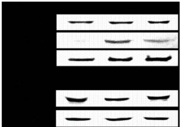
6. SGT inhibited the activation of p38 MAPK activation To determine the mechanisms of effect of SGT on the inflammatory cytokines expressions, we examined the effect of SGT on the MAPKs activation. RBL-2H3 cells were treated with SGT and PMA plus A23187 for 2 h. As shown in Fig. 6, SGT suppressed PMA plus A23187-induced p38 MAPK activation but did not affect the phosphorylation of ERK and JNK.

Fig. 6. Effects of SGT on the activation of p38 MAPK on RBL-2H3 cells. The cell extract were assayed Western blot analysis for the phosphorylation of p38, ERK, and JNK. Lane 1. negative control group; lane 2. positive control group (only treated stimulus); lane 3. SGT 2.0 mg/ml + stimulus.

Discussion

Mast cells play a central role in immediate hypersensitivity reactions and anaphylaxis, by producing and secreting a large spectrum of inflammatory mediators

19,20)

. Activation of mast cells leads to phosphorylation of tyrosine kinase and mobilization of internal Ca

2+

. This is followed by activation of protein kinase C, MAPKs, NF-κB, and releasing of inflammatory cytokines

15)

. And activated mast cells can production histamine, as well as a wide variety of other inflammatory mediators and several pro-inflammatory cytokines

8)

. Histamine, which is released from mast cells and basophils stimulated by an allergen, is usually determined as a degranulation marker in immediate allergic reactions in vitro experiments. When granulates in mast cells or basophils degranulate, an enzyme β-hexosaminidase is also released along with histamine. Thus, this enzyme activity is used as a marker of mast cell degranulation

21)

. The present study showed that SGT pre-treatment profoundly affected PMA plus A23187-induced degranulation of RBL-2H3 cells. These results may suggest SGT have anti-allergic action.

Mast cells concomitantly synthesize and release a variety of cytokines, including IL-3, IL-4, IL-5, IL-13, granulocyte macrophage colony-stimulating factor (GM-CSF) and TNF-α during the late-phase reaction. Several flavones (e.g., luteolin, apigenin, diosmetin and quercetin) obtained from nature have been reported to inhibit the release of TNF-α and IL-4

22)

. Similarly, we found that the SGT dose-dependently inhibited TNF-α and IL-4 secretions in activated RBL-2H3 cells. Also mRNA expression of these cytokines significantly suppressed by SGT.

Most of the inflammatory genes over-expressed in asthma, such as those encoding pro-inflammatory cytokines, chemokines, adhesion molecules, and inflammatory enzymes, contains κB sites for NF-κB within their promoter, suggesting that these genes are controlled predominantly by NF-κB.

Pro-inflammatory molecules are regulated at the level of transcription and are involved in the inflammatory cascade

23,24)

. In most cells, NF-κB is sequestered in the cytoplasm in an inactive form because of tight association with its inhibitors, the IκBs. Activation of NF-κB is achieved through signal-induced phosphorylation of IκB at specific amino-terminal serine residues by the IκB kinase (IKK) complex. This phosphorylation triggers IκB degradation via the ubiquitin-proteasome pathway, resulting in NF-κB translocation into the nucleus

25)

. SGT suppressed translocation into the nucleus of NF-κB and inhibited degradation of IκB-α.

COX-2 is rapidly expressed in several cell types in

(6)

response to growth factors, cytokines, and pro-inflammatory molecules andhas emerged as a major player in inflammatory reactions in peripheral tissues

26)

. Pro-inflammatory cytokines are dependent upon NF-κB activation and NF-κB can promote COX-2 gene transcription, COX-2 inhibitors may exert an anti-inflammatory effect by inhibition of NF-κB

27)

. In this study, the expression of COX-2 mRNA and protein was decreased by treatment of SGT. Therefore, SGT may be used as anti-allergic agent.

The MAPK signaling cascade plays an essential role in the initiation of inflammatory responses

28)

. The exact signaling pathways among three types of MAPKs such as p38, ERK, JNK, are unclear. However, p38 MAPK is thought to play an important role in regulation of inflammatory responses.

Activation of p38 MAPK is essential for the expression of the pro-inflammatory cytokines

15)

. We determined the effect of SGT on the PMA plus A23187-stimulated activation of MAPKs in RBL-2H3 cells. In previous study, the effect of Gallic acid on PMA plus A23187 stimultaneously activated p38, ERK and JNK MAPKs in HMC-1 cells. Gallic acid specially inhibited the activation of p38 MAPK

15)

. We also confirmed that among all three MAPKs, SGT suppressed the activation of p38 MAPK but not ERK or JNK.

In conclusion, SGT inhibited translocation of NF-κB (p65) into the nucleus through regulation of IκB-α degradation.

Therefore, SGT suppressed the TNF-α, IL-4, and COX-2 secretions, and inhibited inflammatory cytokines production via p38 MAPK pathway inhibition. Overall SGT may affect anti-allergic action.

Acknowledgement

This work was supported by Dongeui University Grant (2007AA128).

References

1. Heo, J. DongEuiBoGam. Bub, Seoul. in press, p 731, 937, 1999.

2. Lyu, S.A., Lee, S.Y., Lee, S.J., Son, S.W., Kim, M.O., Kim, G.Y., Kim, Y.H., Yoon, H.J., Kim, H., Park, D.I., Ko, W.S.

Seungma-galgeun-tang attenuates proinflammatory activities through the inhibition of NF-κB signal pathway in the BV-2 microglial cells. J Ethnopharm. 107: 59-66, 2006.

3. Kim, H.M. Antiallergy drugs form oriental medicines. Int J Orient Med. 1: 1-7, 2000.

4. Burd, P.R., Rogers, H.W., Gordon, J.R., Martin, C.A., Jayaraman, S., Wilson, S.D., Dvorak, A.M., Galli, S.J., Dorf,

M.E. Interleukin 3-dependent and –independent mast cells stimulated with IgE and antigen express multiple cytokines. J Exp Med. 170: 245-257, 1989.

5. Plaut, M., Pierce, J.H., Watson, C.J., Hanley-Hyde, J., Nordan, R.P., Paul, W.E. Mast cell lines produce lymphokines in response to cross-linkage of Fc epsilon RI or to calcium ionophores. Nature. 339: 64-67, 1989.

6. Bradding, P., Feather, I.H., Wilson, S., Bardin, P.G., Heusser, C.H., Holgate, S.T., Howarth, P.H.

Immunolocalization of cytokines in the nasal mucosa of normal and perennial rhinitic subjects. The mast cells as a source of IL-4, IL-5, and IL-6 in human allergic mucosal inflammation. J Immunol. 151: 3853-3865, 1993.

7. Schwartz, L.B., Lewis, R.A., Seldin, D., Austen, K.F. Acid hydrolases and tryptase from secretory granules of dispersed human lung mast cells. J Immunol. 126:

1290-1294, 1981.

8. Kim, H.M., Lee, Y.M. Role of TGF-beta 1 on the IgE-dependent anaphylaxis reaction. J Immunol. 162:

4960-4965, 1999.

9. Jeong, H.J., Koo, H.N., Na, H.J., Kim, M.S., Hong, S.H., Eom, J.W., Kim, K.S., Shin, T.Y., Kim, H.M. Inhibition of TNF-α and IL-6 production by aucubin through blockade of NF-κB activation in RBL-2H3 mast cells. Cytokines 18(5):252-259, 2002.

10. Weiss, D.L. and Brown, M.A. Regulation of IL-4 production in mast cell: a paradigm for cell-type-specific gene expression. Immunol Rev. 179: 35-47, 2001.

11. Gregory D. Gregory, Shveta S. Raju, Susan Winandy, Melissa A. Brown. Mast cell IL-4 expression is regulated by lkaros and influences encephalitogenic Th1 responces in EAE. J Clin Invest. 116(5):1327-1336, 2006.

12. Moon, P.D., Lee, B.H., Jeong, H.J., An, H.J., Park, S.J., Kim, H.R., Ko, S.G.., Um, J.Y., Hong, S.H., Kim, H.M. Use of scopoletin to inhibit the production of inflammatory cytokines through inhibition of the IκB/NF-κB signal cascade in the human mast cell line HMC-1. European Journal of Pharmacology 555: 218-225, 2007.

13. Hundley Thomas R., Anjana R. Prasad, Michael A. Beaven.

Elevated levels of cyclooxygenase-2 in antigen-stimulated mast cells is associated with minimal activation of p38 mitogen-activated protein kinase. J Immunol. 167:

1629-1636, 2001.

14. Azzolian, A., Bongiovanni, A., Lampiasi, N. Substance P induces TNF-alpha and IL-6 production through NF kappa B in peritoneal mast cells. Biochim Biophys Acta. 1643:

75-83, 2003.

15. Kim, S.H., Jun, C.D., Suk, K.H., Choi, B.J., Lim, H.J., Park,

(7)

S.J., Lee, S.H., Shin, H.Y., Kim, D.K., Shin, T.Y. Gallic acid inhibits histamine release and pro-inflammatory cytokine production in mast cells. Toxicological Sciences 91(1):123-131, 2006.

16. Guan, Z., Buckman, S.Y., Pentland, A.P., Templeton, D.J., Morrison, A.R. Induction of cyclooxygenase-2 by the activated MEKK1→SEK1/MKK4→p38 mitogen-activated protein kinase pathway. J Biol Chem. 274: 12901-12908, 1998.

17. Skehan, P. Assays of cell growth and cytotoxicity. In:

Studzinski, G.P. (Ed.), Cell Growth and Apoptosis. Oxford University press, New York, p 180, 1998.

18. Schwartz, L.B., Austen, K.F., Wasserman, S.I. Immunologic release of β-hexosaminidase from purified rat serosal mast cells. J Immunol. 123: 1445-1450, 1979.

19. Steven, R.L. and Austen, K.F. Recent advance in the cellular and molecular biology of mast cells. Immunol Today. 10: 381-385, 1989.

20. Galli, S.J., Gordon, J.R. Wershil, B.K., 1991. Cytokine production by mast cells and basophils. Curr Opin Immunol. 3: 865-872, 1991.

21. Cheong, H., Choi, E.J., Yoo, G.S., Kim, K.M., Ryu, D.Y.

Desacetylmatricarin, an anti-allergic component from Taraxacum platycarpum. Planta Med. 64(6):577-588, 1998.

22. Kimata, M., Inagaki, N., Nagai, H. Effects of luteolin and other flavonoids on IgE-mediated allergic reactions. Planta Med. 66(1):25-29, 2000.

23. Christman, J.W., Sadikot, R.T., Blackwell, T.S. The role of nuclear factor-kappa B in pulmonary diseases. Chest. 117:

1482-1487, 2000.

24. Sarkar, A., Sreenivasan, Y., Manna, S.K. α-Melanocyte- stimulating hormone induces cell death in mast cells : involvement of NF-κB. FEBS Lett. 549: 87-93, 2003.

25. Karin, M. and Ben-Neriah, Y. Phosphorylation meets ubiquitnation: the control of NF-[kappa] B activity. Annu Rev Immunol. 18: 621-663, 2000.

26. Minghetti, L. Cyclooxygenase-2 (COX-2) in Inflammatory and Degenerative Brain Diseases. J Neuropathol Exp Neurol. 63(9):901-910, 2004.

27. Hou R.C., Chen, Y.S., Huang, J.R., Jeng, K.G. Cross-linked bromelain inhibits lipopolysaccharide-induced cytokine production involving cellular signaling suppression in rats.

J Agric Food Chem. 54(6):2193-2198, 2006.

28. Adwanikar, H., Karim, F., Gereau, R.W. Inflammation persistently enhances nocifensive behaviors mediated by spinal group Ⅰ mGluRs through sustained ERK activation.

Pain. 111: 125-135, 2004.

수치

Table  1.  Prescription  of  Seungmagalgeun-tang  (SGT)
Table  2.  Oligonucleotide  primers  used  for  PCR  in  this  study.
Fig.  4.  Effects  of  SGT  on  the  expression  of  NF- κ B  (p65)  and  the  degradation  of  I κ B- α  in  RBL-2H3  cells
Fig.  6.  Effects  of  SGT  on  the  activation  of  p38  MAPK  on  RBL-2H3  cells.   The  cell  extract  were  assayed  Western  blot  analysis  for  the  phosphorylation  of  p38,  ERK,  and  JNK

참조

관련 문서

Just as we achieved good control with a joint-based controller that was based on a linearizing and decoupling model of the arm, we can do the same for the Cartesian case.

In this study, we tested whether a potent AMP-activated protein kinase (AMPK) activator, nectandrin B inhibits VSMC proliferation and neointiima formation and tried to

In the region of Auerbach’s plexus (AP) of small intestine, the staining intensity of the ICC-IR cells was reduced in the HFD group compared to the control group.. The numbers

Conclusions: Thus, 25-HC induced odontoclast differentiation through the odontoclastogenesis factor-mediated activation of NF-κB and upregulated inflammatory

 Often found connected to other molecules on the outsides of cells --- cellular recognition, cell signaling, cell

Background : Janus tyrosine kinase 2 (JAK2) V617F mutation has been described in a high proportion of patients with Philadelphia

Activating mutation in the tyrosine kinase JAK2 in polycythemia vera, essential thrombocythemia and myeloid metaplasia with myelofibrosis.. A gain-of-function

Glutamate excitotoxicity induced by excessive activation of NMDA receptor causes various damage to cells, which leads to cell death.. In previous studies, increased ROS