206 Ann Dermatol
Received March 3, 2009, Revised June 1, 2009, Accepted for publication August 12, 2009
Corresponding author: Hyun Jeong Park, M.D., Department of Derma- tology, St. Mary’s Hospital, College of Medicine, The Catholic University of Korea, 62 Yeouido-dong, Yeongdeungpo-gu, Seoul 150-713, Korea. Tel: 82-2-3779-1391, Fax: 82-2-783-7604, E-mail:
Ann Dermatol Vol. 22, No. 2, 2010 DOI: 10.5021/ad.2010.22.2.206
CASE REPORT
Atypical Porokeratosis Developing Following Bone Marrow Transplantation in a Patient with
Myelodysplastic Syndrome
Sang Hee Cha, M.D., Hyun Jeong Park, M.D., Jun Young Lee, M.D., Baik Kee Cho, M.D.
Department of Dermatology, College of Medicine, The Catholic University of Korea, Seoul, Korea
Porokeratosis is an abnormal disease of keratinization of epidermis. It is clinically characterized by margins covered with keratin layer and it typically has an atrophied macule with a protruded, circular form. Histopathologically, it shows the findings of cornoid lamella. Risk factors for its development include organ transplantation, long-term use of corticosteroids, immunocompromised status, including AIDS, and exposure to ultraviolet light. We herein report a case of atypical porokeratosis in a 38-year-old man who developed porokeratosis involving multiple sites following bone marrow transplantation for myelodysplastic syndrome.
(Ann Dermatol 22(2) 206∼208, 2010) -Keywords-
Bone marrow transplantation, Myelodysplastic syndrome, Porokeratosis
INTRODUCTION
Porokeratosis is a disorder of epidermal keratinization that is inherited as an autosomal dominant trait1. Since it was first described by Mibelli in 1893, it has been classified into 6 types as follows, based on clinical characteristics:
porokeratosis of Mibelli, disseminated superficial poro- keratosis (DSP), disseminated superficial actinic porokera- tosis, porokeratosis palmaris et plantaris disseminata, linear porokeratosis, and punctate porokeratosis1.
In cases in which porokeratosis developed in immuno- compromised patients, it is typically seen as a dissemi- nated superficial form which occurs in such multiple sites as the arm, leg and trunk1. To date, many cases have been reported of porokeratosis developing following kidney transplantation. Only 10 cases of prokeratosis developing following bone marrow transplantation (BMT) have been described in the literature, most of which developed following BMT for leukemia. We experienced a case of atypical porokeratosis involving multiple sites, following BMT for myelodysplastic syndrome (MDS).
CASE REPORT
A 38-year-old man presented with an 8-year-history of asymptomatic reddish-brown papules and plaques. The patient was diagnosed with MDS in August 1996, and underwent allogenic BMT following conditioning with busulphan and total body irradiation in February 1997.
Two weeks following BMT, cutaneous graft-versus-host disease developed. After treatment with methylpredni- solone for 11 months, the lesions resolved completely.
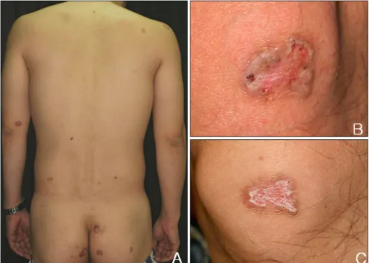
The initial dose of methylprednisolone was 125 mg and was tapered throughout the treatment course. Then, without further specific treatment for MDS, the skin lesions reappeared 3 years following BMT. The lesions were multiple, reddish-brown, well-defined papules and plaques 0.5∼2.5 cm in size, appearing on both extremities and the trunk (Fig. 1A). Their centers were atrophied and colored pink, and they had raised, brown, keratinized borders (Fig. 1B). The number of lesions increased by 1∼
2 per year, and the sizes of the lesions also increased. The patient denied a past history of excessive exposure to sunlight. No notable findings were identified in the family history. Physical examination and laboratory tests also
Atypical Porokeratosis Developing Following BMT
Vol. 22, No. 2, 2010 207 Fig. 1. (A) Asymptomatic, multiple, slowly spreading, 0.5∼2.5 cm, brow- nish to erythematous papules and plaques widely distributed on the body for 8 years. (B) Erythematous plaque with central atrophy and peripheral ridge. (C) Lesion was follo- wed clinically without specific the- rapy for 6 months.
Fig. 2. Typical cornoid lamella characteristic of porokeratosis (H&E, x100).
revealed no significant findings. Histopathology showed characteristic cornoid lamellae, which appeared as columns of parakeratosis extending at about a 45o angle from a focus of dyskeratotic keratinocytes. There was no granular layer beneath the parakeratotic column (Fig. 2). A biopsy confirmed the diagnosis of porokeratosis. After topical application of 20% urea for several months, the skin lesions showed slight improvement (Fig. 1C).
DISCUSSION
Immunosuppressive therapy has been reported to exa- cerbate or initiate the development of porokeratosis. Recently,
with the widespread use of organ transplantation and immunosuppressive treatments, the incidence of porokera- tosis has increased to 0.34∼10.68% of patients who have undergone organ transplantation2. The latency period between organ transplantation and the appearance of porokeratosis ranges from 4 months to 14 years2. Clini- cally, immunosuppression-associated porokeratosis is more often characterized by multiple rather than single lesions3. Most lesions are found on the legs, arms, and trunk1. Although the exact type of porokeratosis has not always been specified, 50% of reported cases have been identified as DSP3.
Recently, there have been many cases reported with atypical clinical characteristics of porokeratosis. In 1997, Herranz et al.4 reported that most of the cases were the mixed type of porokeratosis of Mibelli, and that DSP developed in 11 patients following kidney transplantation.
In our case, the distribution and number of lesions were similar to those of DSP. However, based on the findings of well-defined keratinized lesions that were several centi- meters in size and asymmetrical, a diagnosis of porokera- tosis of Mibelli could not be ruled out. Therefore, our case was diagnosed as an atypical mixed type of porokeratosis.
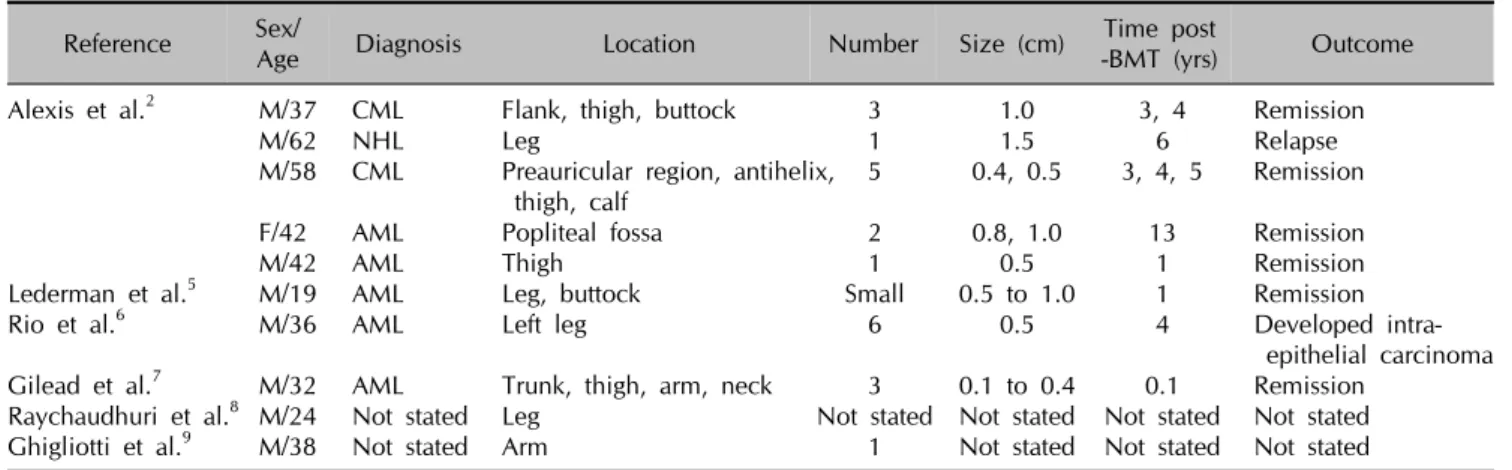
Only 10 cases of porokeratosis after BMT have been reported in the English literature, and they are summarized in Table 12,5-9. Most cases of porokeratosis following BMT were associated with leukemia. To date, immunodeficiency disorders associated with porokeratosis have been reported to include AIDS and precancerous blood disorders, as
SH Cha, et al
208 Ann Dermatol
Table 1. Cases of porokeratosis following bone marrow transplantation reported in the English literature Reference Sex/
Age Diagnosis Location Number Size (cm) Time post
-BMT (yrs) Outcome
Alexis et al.2 M/37 CML Flank, thigh, buttock 3 1.0 3, 4 Remission
M/62 NHL Leg 1 1.5 6 Relapse
M/58 CML Preauricular region, antihelix, 5 0.4, 0.5 3, 4, 5 Remission thigh, calf
F/42 AML Popliteal fossa 2 0.8, 1.0 13 Remission
M/42 AML Thigh 1 0.5 1 Remission
Lederman et al.5 M/19 AML Leg, buttock Small 0.5 to 1.0 1 Remission
Rio et al.6 M/36 AML Left leg 6 0.5 4 Developed intra-
epithelial carcinoma Gilead et al.7 M/32 AML Trunk, thigh, arm, neck 3 0.1 to 0.4 0.1 Remission
Raychaudhuri et al.8 M/24 Not stated Leg Not stated Not stated Not stated Not stated
Ghigliotti et al.9 M/38 Not stated Arm 1 Not stated Not stated Not stated
CML: chronic myeloid leukemia, NHL: non-Hodgkin’s lymphoma, AML: acute myeloid leukemia.
seen in our case3. MDS is a disorder associated with abnormal B cell, NK cell, and CD4 (+) T cell function;
therefore this bone marrow stem cell disorder can result in immunodeficiency.
It remains unclear whether the development of porokera- tosis in our patient was affected by the immunodeficiencies associated with MDS or by the immunosuppressive treat- ment associated with the BMT. It is assumed, however, that the pathophysiology of porokeratosis is similar in either case3. To explain this, several hypotheses have been proposed. First, loss of immunosurveillance caused by immunosuppression allows for the proliferation of abnormal keratinocyte clones, which leads to porokeratosis1,3. To support this, Manganoni et al.10 provided evidence that the number and function of Langerhans cells were decreased and the expression of the HLA-DR antigen was reduced in the lesions. It has also been hypothesized that poro- keratosis develops because of the higher rates of mitotic division in abnormal clones than in normal keratinocytes.
In most porokeratosis cases, treatment is not necessary. It has also been reported that lesions have spontaneously healed in immunocompromised patients after the primary malignancy was treated. In approximately 7∼11.6% of patients, however, porokeratosis is associated with malig- nancies such as squamous cell carcinoma, basal cell carcinoma, and Bowen’s disease. In particular, linear porokeratosis and porokeratosis of Mibelli are highly associated with these malignancies11. Therefore, in immunocompromised patients who develop porokera- tosis, the possibility of malignant transformation must be considered, and such cases must be followed by close observation. Histopathology is required to confirm malig- nant transformation in suspicious cases.
REFERENCES
1. Schamroth JM, Zlotogorski A, Gilead L. Porokeratosis of Mibelli. Overview and review of the literature. Acta Derm Venereol 1997;77:207-213.
2. Alexis AF, Busam K, Myskowski PL. Porokeratosis of Mibelli following bone marrow transplantation. Int J Dermatol 2006;45:361-365.
3. Levin RM, Heymann WR. Superficial disseminate porokera- tosis in a patient with myelodysplastic syndrome. Int J Dermatol 1999;38:138-139.
4. Herranz P, Pizarro A, De Lucas R, Robayna MG, Rubio FA, Sanz A, et al. High incidence of porokeratosis in renal transplant recipients. Br J Dermatol 1997;136:176-179.
5. Lederman JS, Sober AJ, Lederman GS. Immunosuppression:
a cause of porokeratosis? J Am Acad Dermatol 1985;13:
75-79.
6. Rio B, Magana C, Le Tourneau A, Bachmeyer C, Levy V, Hamont N, et al. Disseminated superficial porokeratosis after autologous bone marrow transplantation. Bone Marrow Transplant 1997;19:77-79.
7. Gilead L, Guberman D, Zlotogorski A, Vardy DA, Klaus SN.
Immunosuppression-induced porokeratosis of Mibelli:
complete regression of lesions upon cessation of immuno- suppressive therapy. J Eur Acad Dermatol Venereol 1995;5:
170-172.
8. Raychaudhuri SP, Smoller BR. Porokeratosis in immuno- suppressed and nonimmunosuppressed patients. Int J Dermatol 1992;31:781-782.
9. Ghigliotti G, Nigro A, Gambini C, Farris A, Burroni A, de Marchi R. Mibelli's porokeratosis after bone marrow trans- plantation. Ann Dermatol Venereol 1992;119:968-970.
10. Manganoni AM, Facchetti F, Gavazzoni R. Involvement of epidermal Langerhans cells in porokeratosis of immuno- suppressed renal transplant recipients. J Am Acad Dermatol 1989;21:799-801.
11. Anzai S, Takeo N, Yamaguchi T, Sato T, Takasaki S, Terashi H, et al. Squamous cell carcinoma in a renal transplant recipient with linear porokeratosis. J Dermatol 1999;26:
244-247.