Received January 27, 2015, Revised October 21, 2015, Accepted for publication November 16, 2015
Corresponding author: Byung-Soo Kim, Department of Dermatology, Pusan National University School of Medicine, 179 Gudeok-ro, Seo-gu, Busan 49241, Korea. Tel: 82-51-240-7338, Fax: 82-51-245-9467, E-mail: dockbs@
pusan.ac.kr
This is an Open Access article distributed under the terms of the Creative Commons Attribution Non-Commercial License (http://creativecommons.
org/licenses/by-nc/4.0) which permits unrestricted non-commercial use, distribution, and reproduction in any medium, provided the original work is properly cited.
Copyright © The Korean Dermatological Association and The Korean Society for Investigative Dermatology
Ann Dermatol Vol. 28, No. 4, 2016 http://dx.doi.org/10.5021/ad.2016.28.4.470
ORIGINAL ARTICLE
Change in Antimicrobial Susceptibility of Skin-Colonizing Staphylococcus aureus in Korean Patients with Atopic
Dermatitis during Ten-Year Period
Jung-Min Park1, Ju-Hyun Jo2, Hyunju Jin1, Hyun-Chang Ko1,3, Moon-Bum Kim1,3, Jung-Min Kim4, Do-Won Kim5, Ho-Sun Jang6, Byung-Soo Kim1,3
1Department of Dermatology, Pusan National University School of Medicine, 2Bon Dermatologic Clinic, 3Biomedical Research Institute, Pusan National University Hospital, Busan, Departments of 4Microbiology and 5Dermatology, Kyungpook National University School of Medicine, Deagu, 6Jang Ho Sun Dermatologic Clinic, Busan, Korea
Background: A small subset of adolescents atopic dermatitis (AD) tends to persist. This also leads to get more antibiotics exposure with advancing years. Antibiotic resistance has been regarded as a serious problem during Staphylococcus aureus treatment, especially methicillin-resistant S. aureus (MRSA). Objective: It was investigated the S. aureus colo- nization frequency in the skin lesions and anterior nares of adolescent AD patients and evaluated the changes in S. aur- eus antimicrobial susceptibility for years. Methods: Patients who visited our clinic from September 2003 to August 2005 were classified into group A, and patients who visited from August 2010 to March 2012 were classified into group B. To investigate the differences with regard to patients’ age and disease duration, the patients were subdivided into groups according to age. Lesional and nasal specimens were examined. Results: Among the 295 AD patients, the total S.
aureus colonization rate in skin lesions was 66.9% (95/142) for group A and 78.4% (120/153) for group B. No significant changes in the systemic antimicrobial susceptibilities of S.
aureus strains isolated from adolescent AD patients were ob-
served during about 10-year period. The increased trend of MRSA isolation in recent adolescent AD outpatients suggest that the community including school could be the source of S. aureus antibiotic resistance and higher fusidic acid resist- ance rates provides evidence of imprudent topical use.
Conclusion: Relatively high MRSA isolation and fusidic acid resistance rates in recent AD patients suggest that the com- munity harbors antibiotic-resistant S. aureus. (Ann Dermatol 28(4) 470∼478, 2016)
-Keywords-
Anti-bacterial agent, Antimicrobial resistance, Antimicro- bial susceptibility, Atopic dermatitis, Staphylococcus aur- eus
INTRODUCTION
Atopic dermatitis (AD) is a genetically determined, chroni- cally relapsing inflammatory skin disease with multiple pathogenic factors. While AD occurs most commonly dur- ing infancy and children, a smaller subset of adolescents has persistent or new-onset AD. The association between Staphylococcus aureus infection and AD is well demon- strated by many investigators1,2. S. aureus can be found from dermatitic lesions of more than 90% of patients with AD, but also from approximately 70% taken from un- affected areas1-3. S. aureus plays an important role as a triggering factor3,4. The relationship between AD and ex- acerbation mechanism by S. aureus is mainly due to the superantigens (sAgs) and sAgs-specific immunoglobulin E that stimulate various numbers of different T-cell clones
and cytokine secretion. It has been often proposed that bacterial skin infections are uncommon in AD, while AD patients are commonly colonized with S. aureus. Recently lesional S. aureus colonization correlates positively with AD clinical severity, and anti-staphylococcal antibiotic therapy can reduce the severity of AD characteristic in- flammation5,6 .
The anterior nares are an important S. aureus colonization reservoir. High rate (75%∼90%) of nasal carriage of S. aureus has been reported in adults and children with AD.
In contrast, nasal colonization has ranged from 10% to 50% in normal controls. Thus S. aureus carriage in the nose should be targeted for decolonization7,8.
Antibiotic resistance is increasing around the world to date and has been regarded as an important issue during S. aureus treatment since a long time ago, especially me- thicillin-resistant S. aureus (MRSA). MRSA is more difficult to treat because it is resistant to a number of widely used antibiotics. The increasing incidence of community-ac- quired MRSA (CA-MRSA) in skin infections presents major challenges in the treatment direction. It also raises con- cerns that the eczematous skin lesions of AD patients might be favorable CA-MRSA reservoirs9,10. Topical fusidic acid and mupirocin have been commonly prescribed to eradicate different skin infections via over-the-counter pharmacy in Korea. Although many Asian countries have high MRSA infection rates, there have been no pub- lications about changes in the prevalence of anti- biotic-resistant S. aureus, including CA-MRSA, in AD patients. Moreover, few studies have dealt with adolescent AD about S. aureus colonization and its susceptibilities to various antibiotics.
In present study, it was investigated the S. aureus colo- nization frequency in the skin lesions and anterior nares of AD patients and evaluated the changes in S. aureus anti- microbial susceptibility for years. Differences were also analyzed with regard to patient age and disease duration.
Also, we investigated the prevalence of topical fusidic acid- and mupirocin-resistant S. aureus in adolescent AD.
MATERIALS AND METHODS
Patients
Adolescent AD patients with no evidence of skin in- fection, who visited the outpatient clinic of the Department of Dermatology, Pusan National University Hospital (Busan, Korea), were enrolled in the study. AD was diagnosed according to the Hanifin and Rajka diag- nostic criteria11. Total 295 patients who initially visited our clinic from September 2003 to August 2005 were clas- sified into group A, and patients who initially visited from
August 2010 to March 2012 were classified into group B.
To investigate the differences with regard to patient age and disease duration, the patients were subdivided into groups according to age (younger than 18 years and older than 18 years) and disease duration (less than 1 year, 1∼5 years, and more than 5 years). At the first visit, the patient age and disease duration were estimated and AD severity was assessed according to the SCORing Atopic Dermatitis (SCORAD) index12. The exclusion criteria were the pres- ence of other skin or allergic diseases; a recent (within 4 weeks) history of inpatient hospital admission; recent (within 4 weeks) treatment with antibiotics, systemic corti- costeroids, or immunosuppressants; and treatment with topical antibiotics in the previous 2 weeks.
Methods
The study protocol was approved by the Pusan National University Hospital Institutional Review Board (IRB no.
1409-012-035). Lesional skin specimens were obtained by rolling sterile cotton-tipped swab sticks (transport medium swab; Micromedia Co., Seoul, Korea) over the most af- fected skin areas twice for at least 5 seconds each. Nasal swabs were obtained by reaching upward toward the top of both anterior nares with sterile cotton-tipped swab stick, followed by a 360o twist to sweep the entire vestibule. The swab specimens were immediately placed in Amie’s medium (Micromedia Co.) and were streaked on sheep blood agar plates (Asan Medical Co., Seoul, Korea), incubated at 35oC, and examined at 24 and 48 hours. Colonies were identified in a blind manner by oth- er investigator. In some selected samples of two groups, antibiotic susceptibility tests were performed with the same Vitek 2 system (BioMérieux, Durham, NC, USA) ac- cording to the manufacturer’s instructions.
For group A, a panel of 8 antibiotics (clindamycin, eryth- romycin, habekacin, oxacillin, gentamicin, penicillin, bac- trim, and vancomycin) was used to test for gram-positive bacteria. For group B, a panel of 11 antibiotics or combi- nations (ciprofloxacin, fusidic acid, rifampin, teicoplanin, tetracycline, nitrofurantoin, quinupristin/dalfopristin, line- zolid, telithromycin, mupirocin, and tigecycline) was add- ed to the previous panel.
Statistical analysis
The Shapiro-Wilk normality test was performed to eval- uate differences between the groups with regard to patient age, disease duration, and severity, using the Predictive Analytics Software package (PASW for Windows; IBM Co., Armonk, NY, USA). The chi-square and Fisher’s exact tests were performed to estimate differences in the colo- nization rates between groups that were subdivided ac-
Table 2. Total colonization rates (%) of Staphylococcus aureus and methicillin-resistant S. aureus (MRSA) in lesional skin and anterior nares
Colonization Lesional skin Anterior nares
Group A Group B p-value Group A Group B p-value
S. aureus 66.9 (95/142) 78.4 (120/153) 0.03 64.1 (91/142) 63.9 (62/97) 0.05
MRSA 3.1 (2/64) 10.4 (11/106) >0.05 7.0 (4/57) 13.0 (3/23) >0.05
Values are presented as percentage (number/total number).
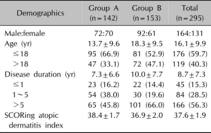
Table 1. Demographics of atopic dermatitis patients
Demographics Group A (n=142)
Group B (n=153)
Total (n=295)
Male:female 72:70 92:61 164:131
Age (yr) 13.7±9.6 18.3±9.5 16.1±9.9
≤18 95 (66.9) 81 (52.9) 176 (59.7) >18 47 (33.1) 72 (47.1) 119 (40.3) Disease duration (yr) 7.3±6.6 10.0±7.7 8.7±7.3 ≤1 23 (16.2) 22 (14.4) 45 (15.3) 1∼5 54 (38.0) 30 (19.6) 84 (28.5) >5 65 (45.8) 101 (66.0) 166 (56.3) SCORing atopic
dermatitis index
38.4±1.7 36.9±2.0 37.6±1.9
Values are presented as number only, mean±standard deviation, or number (%).
cording to time period, age, and disease duration.
Statistical significance was defined as a p-value of <0.05.
RESULTS
Clinical and demographic data
The clinical and demographic data for groups A and B are presented in Table 1. Overall, 142 and 153 patients were enrolled in groups A and B, respectively. The mean pa- tient age in group A was 13.7 years, and the mean disease duration was 7.3 years. The mean patient age in group B was 18.3 years, and the mean disease duration was 10.0 years. The difference in clinical severity according to the SCORAD index was not significant between the 2 groups.
Colonization of S. aureus
Among the 295 AD patients, the total S. aureus colo- nization rate in skin lesions was 66.9% (95/142) for group A and 78.4% (120/153) for group B (p=0.03). In group A, 142 samples were taken from the nares. In group B, 97 sampling were done in the nares. In the nasal swabs, S. aureus was found to colonize 64.1% (91/142) of the group A patients and 63.9% (62/97) of the group B pa- tients (p=0.05). To analyze antibiotics sensitivity, 64 sam- ples and 57 samples were used at the lesion and nares in group A. One hundred and six samples and 23 samples
were used in each of group B.
In group A, 2 of 64 (3.1%) and 4 of 57 (7.0%) patients car- ried MRSA in the lesional skin and the anterior nares, re- spectively, whereas in group B, 11 of 106 (10.4%) and 3 of 23 (13.0%) patients carried MRSA in the lesional skin and the anterior nares, respectively (p>0.05; Table 2).
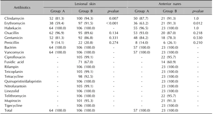
Changes in S. aureus antimicrobial susceptibility Changes in S. aureus antimicrobial susceptibility in ado- lescent AD patients over time are shown in Table 3. In le- sional skin, the rates of S. aureus susceptibility to clinda- mycin and erythromycin increased significantly in group B when compared with those in group A. The rates of sus- ceptibility of S. aureus in lesional skin to fusidic acid and mupirocin, which are the main topical agents used to treat AD skin infections, were 67.0% and 95.3%, respectively in group B. The susceptibility rates of S. aureus in the an- terior nares to fusidic acid and mupirocin were 60.9% and 91.3%, respectively. The rate of susceptibility of S. aureus in nasal swabs to erythromycin increased significantly dur- ing about 10-year period. For samples of both lesional skin and anterior nares, the penicillin susceptibility rate in- creased over time but remained much lower than those for other antibiotics.
The MRSA colonization rates did not significantly differ between groups A and B (p>0.05). All isolated MRSA strains were susceptible to habekacin, bactrim, and vanco- mycin (Table 4).
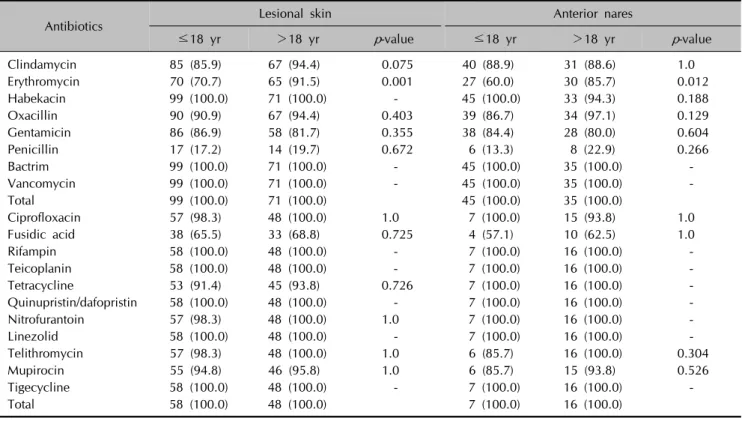
S. aureus antimicrobial susceptibility was also analyzed according to the patient age (younger than 18 years versus older than 18 years; Table 5). In patients older than 18 years, S. aureus in samples of both the lesional skin and anterior nares was significantly more susceptible to eryth- romycin, compared to the susceptibility in patients young- er than 18 years. Low susceptibility rates to penicillin and fusidic acid were observed regardless of the patient age.
Table 6 shows the antimicrobial susceptibility of S. aureus with regard to the disease duration. For lesional skin sam- ples, erythromycin susceptibility rate was significantly lower in patients with disease duration of less than 5 years than in those with disease duration of more than 5 years.
Table 3. Change in Staphylococcus aureus antimicrobial susceptibility
Antibiotics Lesional skin Anterior nares
Group A Group B p-value Group A Group B p-value
Clindamycin 52 (81.3) 100 (94.3) 0.007 50 (87.7) 21 (91.3) 1.0
Erythromycin 38 (59.4) 97 (91.5) <0.001 36 (63.2) 21 (91.3) 0.012
Habekacin 64 (100.0) 106 (100.0) - 55 (96.5) 23 (100.0) 1.0
Oxacillin 62 (96.9) 95 (89.6) 0.134 53 (93.0) 20 (87.0) 0.218
Gentamicin 52 (81.3) 92 (86.8) 0.331 48 (84.2) 18 (78.3) 0.530
Penicillin 9 (14.1) 22 (20.8) 0.274 8 (14.0) 6 (26.1) 0.210
Bactrim 64 (100.0) 106 (100.0) - 57 (100.0) 23 (100.0) -
Vancomycin 64 (100.0) 106 (100.0) - 57 (100.0) 23 (100.0) -
Ciprofloxacin - 105 (99.1) - - 22 (95.7) -
Fusidic acid - 71 (67.0) - - 14 (60.9) -
Rifampin - 106 (100.0) - - 23 (100.0) -
Teicoplanin - 105 (99.1) - - 23 (100.0) -
Tetracycline - 98 (92.5) - - 23 (100.0) -
Quinupristin/dafopristin - 106 (100.0) - - 23 (100.0) -
Nitrofurantoin - 105 (99.1) - - 23 (100.0) -
Linezolid - 106 (100.0) - - 23 (100.0) -
Telithromycin - 106 (100.0) - - 22 (95.7) -
Mupirocin - 101 (95.3) - - 21 (91.3) -
Tigecycline - 106 (100.0) - - 23 (100.0) -
Total 64 (100.0) 106 (100.0) 57 (100.0) 23 (100.0)
Values are presented as number (%).
Table 4. Change in methicillin-resistant Staphylococcus aureus antimicrobial susceptibility
Antibiotics Lesional skin Anterior nares Group A Group B Group A Group B Clindamycin 2 (100.0) 9 (81.8) 3 (75.0) 2 (66.7) Erythromycin 1 (50.0) 9 (81.8) 2 (50.0) 2 (66.7) Habekacin 2 (100.0) 11 (100.0) 4 (100.0) 3 (100.0) Gentamicin 2 (100.0) 9 (81.8) 3 (75.0) 2 (66.7)
Penicillin 0 0 0 0
Bactrim 2 (100.0) 11 (100.0) 4 (100.0) 3 (100.0) Vancomycin 2 (100.0) 11 (100.0) 4 (100.0) 3 (100.0)
Ciprofloxacin - 10 (90.9) - 3 (100.0)
Fusidic acid - 11 (100.0) - 3 (100.0)
Rifampin - 11 (100.0) - 3 (100.0)
Teicoplanin - 11 (100.0) - 3 (100.0)
Tetracycline - 9 (81.8) - 3 (100.0)
Quinupristin/
dafopristin
- 11 (100.0) - 3 (100.0)
Nitrofurantoin - 11 (100.0) - 3 (100.0)
Linezolid - 11 (100.0) - 3 (100.0)
Telithromycin - 10 (90.9) - 2 (66.7)
Mupirocin - 9 (81.8) - 2 (66.7)
Tigecycline - 11 (100.0) - 3 (100.0)
Total 2 (100.0) 11 (100.0) 4 (100.0) 3 (100.0) Values are presented as number (%).
Low susceptibility rates to penicillin and fusidic acid were observed regardless of the disease duration.
DISCUSSION
S. aureus may not be of primary importance in AD patho- genesis but is an important triggering and/or aggravating factor in cutaneous AD inflammation due to S. aureus sAgs13. S. aureus colonization rate is significantly higher in AD patients than in normal controls because the stratum corneum of AD patients is highly susceptible to colo- nization by various bacteria including S. aureus10,14. Also, several studies have showed that AD keratinocytes pro- duce lower amounts of antimicrobial peptides and this may increase the colonization and infection with S. aur- eus15. This study revealed the increasing prevalence of S. aureus colonization in adolescent AD skin (from 66.9% to 78.4% [p=0.03]) during about 10-year period. This is con- sistent with the increased S. aureus colonization rates over time that were previously described in cross-sectional studies10,16. The increased S. aureus colonization rate in- dicates the importance of determining the antibiotic sus- ceptibility of S. aureus and controlling AD inflammations effectively.
Prolonged or imprudent antibiotic use may induce the de- velopment of antibiotic-resistant S. aureus strains13,17.
Table 5. Staphylococcus aureus antimicrobial susceptibility with respect to age in patients with atopic dermatitis
Antibiotics Lesional skin Anterior nares
≤18 yr >18 yr p-value ≤18 yr >18 yr p-value
Clindamycin 85 (85.9) 67 (94.4) 0.075 40 (88.9) 31 (88.6) 1.0
Erythromycin 70 (70.7) 65 (91.5) 0.001 27 (60.0) 30 (85.7) 0.012
Habekacin 99 (100.0) 71 (100.0) - 45 (100.0) 33 (94.3) 0.188
Oxacillin 90 (90.9) 67 (94.4) 0.403 39 (86.7) 34 (97.1) 0.129
Gentamicin 86 (86.9) 58 (81.7) 0.355 38 (84.4) 28 (80.0) 0.604
Penicillin 17 (17.2) 14 (19.7) 0.672 6 (13.3) 8 (22.9) 0.266
Bactrim 99 (100.0) 71 (100.0) - 45 (100.0) 35 (100.0) -
Vancomycin 99 (100.0) 71 (100.0) - 45 (100.0) 35 (100.0) -
Total 99 (100.0) 71 (100.0) 45 (100.0) 35 (100.0)
Ciprofloxacin 57 (98.3) 48 (100.0) 1.0 7 (100.0) 15 (93.8) 1.0
Fusidic acid 38 (65.5) 33 (68.8) 0.725 4 (57.1) 10 (62.5) 1.0
Rifampin 58 (100.0) 48 (100.0) - 7 (100.0) 16 (100.0) -
Teicoplanin 58 (100.0) 48 (100.0) - 7 (100.0) 16 (100.0) -
Tetracycline 53 (91.4) 45 (93.8) 0.726 7 (100.0) 16 (100.0) -
Quinupristin/dafopristin 58 (100.0) 48 (100.0) - 7 (100.0) 16 (100.0) -
Nitrofurantoin 57 (98.3) 48 (100.0) 1.0 7 (100.0) 16 (100.0) -
Linezolid 58 (100.0) 48 (100.0) - 7 (100.0) 16 (100.0) -
Telithromycin 57 (98.3) 48 (100.0) 1.0 6 (85.7) 16 (100.0) 0.304
Mupirocin 55 (94.8) 46 (95.8) 1.0 6 (85.7) 15 (93.8) 0.526
Tigecycline 58 (100.0) 48 (100.0) - 7 (100.0) 16 (100.0) -
Total 58 (100.0) 48 (100.0) 7 (100.0) 16 (100.0)
Values are presented as number (%).
Table 6. Staphylococcus aureus antimicrobial susceptibility with respect to disease duration in patients with atopic dermatitis
Antibiotics Lesional skin Anterior nares
≤1 yr 1∼5 yr >5 yr p-value ≤1 yr 1∼5 yr >5 yr p-value
Clindamycin 18 (81.8) 41 (89.1) 93 (91.2) 0.567 10 (100.0) 23 (92.0) 38 (84.4) 0.407 Erythromycin 16 (72.7) 31 (67.4) 88 (86.3) 0.023 5 (50.0) 16 (64.0) 36 (80.0) 0.108 Habekacin 22 (100.0) 46 (100.0) 101 (99.0) 1.0 10 (100.0) 24 (96.0) 44 (97.8) 0.783 Oxacillin 19 (86.4) 40 (87.0) 98 (96.1) 0.053 8 (80.0) 23 (92.0) 42 (93.3) 0.409 Gentamicin 18 (81.8) 40 (87.0) 86 (84.3) 0.824 9 (90.0) 22 (88.0) 35 (77.8) 0.582
Penicillin 3 (13.6) 6 (13.0) 22 (21.6) 0.408 1 (10.0) 5 (20.0) 8 (17.8) 0.919
Bactrim 22 (100.0) 46 (100.0) 102 (100.0) - 10 (100.0) 25 (100.0) 45 (100.0) - Vancomycin 22 (100.0) 46 (100.0) 102 (100.0) - 10 (100.0) 25 (100.0) 45 (100.0) -
Total 22 (100.0) 46 (100.0) 102 (100.0) 10 (100.0) 25 (100.0) 45 (100.0)
Ciprofloxacin 12 (100.0) 23 (100.0) 70 (98.6) 1.0 1 (50.0) 4 (100.0) 17 (100.0) 0.087 Fusidic acid 7 (58.3) 15 (65.2) 49 (69.0) 0.700 1 (50.0) 4 (100.0) 9 (52.9) 0.825
Rifampin 12 (100.0) 23 (100.0) 71 (100.0) - 2 (100.0) 4 (100.0) 17 (100.0) -
Teicoplanin 12 (100.0) 23 (100.0) 71 (100.0) - 2 (100.0) 4 (100.0) 17 (100.0) - Tetracycline 10 (83.3) 20 (87.0) 68 (95.8) 0.104 2 (100.0) 4 (100.0) 17 (100.0) - Quinupristin
/dafopristin
12 (100.0) 23 (100.0) 71 (100.0) - 2 (100.0) 4 (100.0) 17 (100.0) -
Nitrofurantoin 12 (100.0) 23 (100.0) 70 (98.6) 1.0 2 (100.0) 4 (100.0) 17 (100.0) -
Linezolid 12 (100.0) 23 (100.0) 71 (100.0) - 2 (100.0) 4 (100.0) 17 (100.0) -
Telithromycin 12 (100.0) 23 (100.0) 70 (98.6) 1.0 2 (100.0) 3 (75.0) 17 (100.0) 0.261 Mupirocin 11 (91.7) 23 (100.0) 67 (94.4) 0.353 2 (100.0) 3 (75.0) 16 (94.1) 0.462 Tigecycline 12 (100.0) 23 (100.0) 71 (100.0) - 2 (100.0) 4 (100.0) 17 (100.0) -
Total 12 (100.0) 23 (100.0) 71 (100.0) 2 (100.0) 4 (100.0) 17 (100.0)
Values are presented as number (%).
Although careful antibiotic use has been often suggested, to our knowledge, no reports have investigated changes in antibiotics susceptibility only in adolescent AD patients.
Diamantis et al.18 reported a comparison of antibiotic re- sistance patterns in pediatric dermatology patients infected by S. aureus in 2005∼2007 (66% of children with AD) versus 2008∼2009 (72.4% with AD). They found an in- crease in S. aureus antibiotic resistance except to methi- cillin, which surprisingly decreased. Other pediatric der- matology clinic in North Carolina also conducted the anti- biotic susceptibility profiles in S. aureus cutaneous in- fections between 2005 and 200719. The subjects of study included 66% of AD patients, and they demonstrated the following resistance patterns: penicillin (86%), erythromy- cin (46%), methicillin (32%), clindamycin (22%), gentami- cin (3%), vancomycin (0%), and trimethoprim-sulfame- thoxazole (0%).
In the patients with CA-MRSA infections, traditional MRSA risk factors are absent and resistance is usually limited to β-lactam antibiotics16. Following the first report in 1961 in England, the incidence of MRSA has increased pro- gressively20. Recently, MRSA infections have been de- scribed in patients without established risk factors who are living in the community, especially AD patient. In a pre- vious report, 4.2% of those obtained from the general out- patient pediatric population showed methicillin resistance and in our study, the frequency of CA-MRSA-positive skin samples was 10.4% despite smaller samples21,22.
This agrees with earlier findings that the CA-MRSA preva- lence was 7.4% to 18.4% of skin cultures from AD pa- tients9,10. In our study, increased trend of MRSA isolation rates were observed in both skin lesions and anterior nares although these increases were statistically insignificant.
Moreover, MRSA colonization rates in healthy individuals were reported as 0%∼9% in previous studies, and these rates are comparable with those of AD patients21-23. Since S. aureus has a predilection for damaged skin and AD pa- tients are frequently exposed to antimicrobials, the rela- tively lower rate of MRSA colonization observed in our study might be meaningful. But, increased caution during MRSA infection management is required in AD patients, as they can be sources of CA-MRSA. All MRSA strains in this study were susceptible to vancomycin, the treatment of choice for MRSA infections. However, vancomycin use should be reserved for MRSA infections which is based by culture.
The present study verified the low rates of S. aureus pen- icillin susceptibility in adolescent AD outpatients (14.0%
∼26.1%), regardless of the time period, age, or disease duration. This finding is consistent with a previous study, which reported that 13% of AD patients were sensitive to
penicillin16. The penicillin susceptibility rate remains much lower than that of other antibiotics, even though penicillin usage is restricted in AD patients in Korea. This is because the rates of declining resistance appear to be slower than that of emerging resistance and appear to vary with different agent classes24.
Fusidic acid has been widely used as topical antimicrobial to treat bacterial superinfections in AD patients until now.
In our study, relatively low susceptibility rates (60.9%∼
67.0%) to fusidic acid were observed regardless of the pa- tient age and disease duration. In Korea, topical fusidic acid antimicrobial has been classified over-the counter drug and its low susceptibility provides evidence of im- prudent topical use. Our results suggest that another agent should be used for the treatment of adolescent AD pa- tients with suspected S. aureus infections. In a British study published in 2009, 41% of S. aureus isolates from dermatology patients were fusidic acid-resistant, com- pared with a 50% resistance rate in 2001, due to usage re- strictions and a significant decrease in the use of topical fusidic acid25,26. The authors supposed that a lag period might occur before fusidic acid resistance is absent from the community. Topical fusidic acid use should be re- stricted due to the current high level of resistance. A rela- tively high susceptibility (91.3%∼95.3%) to mupirocin was demonstrated in our study, regardless of the time peri- od, patient age, and disease duration. This finding was consistent with previous results, which suggested that 4%
of isolates from AD patients were mupirocin-resistant27. These results indicate that topical fusidic acid has been used more extensively than mupirocin. However, the po- tential for the development of bacterial resistance to mu- pirocin ointment should not be ignored, and thus caution regarding its use is needed to retain the high antimicrobial effects28,29.
Despite the concerns of many dermatologists, there were no significant changes in S. aureus antimicrobial suscepti- bility in AD, except for erythromycin and clindamycin, during the recent 10-year period. In the 1990s, eryth- romycin was the first-line treatment for bacterial infections in AD patients, but its use has decreased after reports of high erythromycin resistance rates in S. aureus and recom- mended usage restrictions30,31. According to our data, the erythromycin susceptibility rate increased significantly during the period from 59.4% to 91.5% for the lesional skin samples and from 63.2% to 91.3% for the anterior nare samples. Previous reports conducted in the USA, Europe, and Asia indicated that 51%∼76% of S. aureus strains were erythromycin-susceptible in 199932-34. Hoeger’s study35 of antimicrobial susceptibility, which was pub- lished in 2004, revealed that the rate of S. aureus eryth-
romycin resistance remained low in 82% of AD patients.
Thus, erythromycin should no longer be recommended as a therapeutic agent for S. aureus-infected AD patients.
In previous studies conducted in 1997 and 1999, a 91%∼
97% clindamycin susceptibility rate was demonstrated in Singapore and Europe36,37. In 2008, Niebuhr et al.30 re- ported that clindamycin has been recommended as a first-line therapy (alternative to cephalexin or cefuroxime) and for staphylococcal skin infections in Germany, and another study conducted in 2005 and 2006 revealed a clindamycin susceptibility rate of 79% for S. aureus35,36. Interestingly, in our study, the clindamycin susceptibility rate in lesional skin samples increased significantly from 81.3% to 94.3% during the period. In Korea, reports de- scribed relatively low S. aureus susceptibility rates to clin- damycin (48%∼82.6%) and recommended clindamycin usage restrictions; our data reflect those efforts37,38. Clindamycin acts against a variety of anaerobic bacteria, but broad antibiotic coverage is not required in AD, as S. aureus is the most frequent skin infection-inducing micro- organism30. Therefore, clindamycin should also no longer be suggested as a therapeutic agent for S. aureus in- fections in AD patients.
According to our age-based analysis, for both the lesional skin and nasal cultures, patients younger than 18 years have significantly lower susceptibility rates against eryth- romycin than do patients older than 18 years. Our results are comparable to those of Arkwright et al.39, who studied age-related changes in the S. aureus prevalence on af- fected AD skin. The authors found that children older than 5 years had a higher prevalence of erythromycin-resistant S. aureus (35%) than did younger children (26%). This dis- cordance of results between the 2 studies might be due to differences in the age groups and distributions. In our study, the group of patients who were younger than 18 years included more group A patients with significantly higher erythromycin resistance rates. According to our dis- ease duration-based analysis, different resistant patterns have shown in various antimicrobials, although we did not find a statistic difference. In our population, patients with disease duration of less than 5 years were less sus- ceptible to erythromycin than patients with disease dura- tion of more than 5 years. It was possible that eryth- romycin was no more used as a therapeutic agent for S. aureus-infected AD patients in different outpatient settings.
A previous study by Ewing et al.40 supported the idea that antibiotic therapy is not helpful in AD patients who do not present signs of bacterial infection. Moreover, the MRSA incidence rate increased after a 4-week systemic antibiotic therapy course. Continuous antibiotic use with the intent to clear S. aureus colonization in AD may ultimately result
in the failures of these antibiotics to treat severe infections, which are not uncommon in AD41. The therapeutic rec- ommendation for bacterial infections in AD patients in- cludes a combination therapy of topical anti-inflammatory drugs and topical/systemic antibiotics during the early stage when clinical signs of a secondary bacterial infection are present42. Recently, cephalexin, a first-generation cephalosporin, was found to be a good first-line antibiotic for the treatment of secondary S. aureus infections in AD due to its restricted antimicrobial spectrum, which com- prises gram-positive bacteria and a limited number of gram-negative strains30.
In conclusion, despite our concerns, no significant changes in the antimicrobial susceptibilities of S. aureus strains isolated from AD patients were observed during a 10-year period. These results indicate that in medical soci- ety, a high level of attention is focused on the misuse and abuse of antibiotics. However, the increased trend of MRSA isolation and fusidic acid resistance rates in recent AD outpatients suggest that the community including school could be the source of S. aureus antibiotic resist- ance and its imprudent prescription. To appropriately treat skin infections in adolescent AD, proper antibiotic use through periodic reviews and understandings of changes in microorganisms and antimicrobial sensitivities is neces- sary to avoid the excessive use of broad-spectrum empiric antibiotics.
REFERENCES
1. Aly R, Maibach HI, Shinefield HR. Microbial flora of atopic dermatitis. Arch Dermatol 1977;113:780-782.
2. David TJ, Cambridge GC. Bacterial infection and atopic eczema. Arch Dis Child 1986;61:20-23.
3. Roll A, Cozzio A, Fischer B, Schmid-Grendelmeier P. Mic- robial colonization and atopic dermatitis. Curr Opin Allergy Clin Immunol 2004;4:373-378.
4. Kim BS, Kim JY, Lim HJ, Lee WJ, Lee SJ, Kim JM, et al.
Colonizing features of Staphylococcus aureus in early childhood atopic dermatitis and in mothers: a cross- sectional comparative study done at four kindergartens in Daegu, South Korea. Ann Allergy Asthma Immunol 2011;
106:323-329.
5. Williams JV, Vowels BR, Honig PJ, Leyden JJ. S. aureus isolation from the lesions, the hands, and the anterior nares of patients with atopic dermatitis. Pediatr Dermatol 1998;15:194-198.
6. Kim BS, Park JY, Song CH, Kim JY, Lim HJ, Lee HS, et al.
Clarifying the transmission route of Staphylococcus aureus colonizing the skin in early childhood atopic dermatitis.
Ann Allergy Asthma Immunol 2012;109:448-453.
7. Herz U, Bunikowski R, Renz H. Role of T cells in atopic dermatitis. New aspects on the dynamics of cytokine
production and the contribution of bacterial superantigens.
Int Arch Allergy Immunol 1998;115:179-190.
8. Lever R, Hadley K, Downey D, Mackie R. Staphylococcal colonization in atopic dermatitis and the effect of topical mupirocin therapy. Br J Dermatol 1988;119:189-198.
9. Chung HJ, Jeon HS, Sung H, Kim MN, Hong SJ.
Epidemiological characteristics of methicillin-resistant Sta- phylococcus aureus isolates from children with eczematous atopic dermatitis lesions. J Clin Microbiol 2008;46:991-995.
10. Huang JT, Abrams M, Tlougan B, Rademaker A, Paller AS.
Treatment of Staphylococcus aureus colonization in atopic dermatitis decreases disease severity. Pediatrics 2009;
123:e808-e814.
11. Hanifin JM, Rajka G. Diagnostic features of atopic dermatitis.
Acta Derm Venereol Suppl (Stockh) 1980;92:44-47.
12. Kunz B, Oranje AP, Labrèze L, Stalder JF, Ring J, Taïeb A.
Clinical validation and guidelines for the SCORAD index:
consensus report of the European Task Force on Atopic Dermatitis. Dermatology 1997;195:10-19.
13. Taskapan MO, Kumar P. Role of staphylococcal superan- tigens in atopic dermatitis: from colonization to inflammation.
Ann Allergy Asthma Immunol 2000;84:3-10; quiz 11-12.
14. Sung HC, Jung HD, Park KD, Lee WJ, Lee SJ, Kim DW. A quantitative culture study of Staphylococcus aureus in adolescent and adult patients with atopic dermatitis using the contact-plate sampling technique. Korean J Dermatol 2007;45:673-679.
15. Donald YML, Lawrence FE, Mark B. Atopic dermatitis (Atopic eczema). In: Goldsmith LA, Katz SI, Gilchrest BA, Paller AS, Leffell DJ, Wolff K, editors. Fitzpatrick’s derma- tology in general medicine. New York: McGraw-Hill Medical, 2012:168.
16. Goh CL, Wong JS, Giam YC. Skin colonization of Staphylococcus aureus in atopic dermatitis patients seen at the National Skin Centre, Singapore. Int J Dermatol 1997;
36:653-657.
17. Ellis MW, Lewis JS 2nd. Treatment approaches for com- munity-acquired methicillin-resistant Staphylococcus aureus infections. Curr Opin Infect Dis 2005;18:496-501.
18. Diamantis ML, Ortega-Loayza AG, Morrell DS. Update on the characterization of Staphylococcus aureus skin infections in a pediatric dermatology tertiary health care outpatient facility: antibiotic susceptibility patterns and decreased methicillin resistance. J Am Acad Dermatol 2011;64:440- 441.
19. Ortega-Loayza AG, Diamantis SA, Gilligan P, Morrell DS.
Characterization of Staphylococcus aureus cutaneous infections in a pediatric dermatology tertiary health care outpatient facility. J Am Acad Dermatol 2010;62:804-811.
20. Eriksen KR. "Celbenin"-resistant staphylococci. Ugeskr Laeger 1961;123:384-386.
21. Fridkin SK, Hageman JC, Morrison M, Sanza LT, Como- Sabetti K, Jernigan JA, et al; Active Bacterial Core Sur- veillance Program of the Emerging Infections Program Network. Methicillin-resistant Staphylococcus aureus disease in three communities. N Engl J Med 2005;352:1436-1444.
22. Matiz C, Tom WL, Eichenfield LF, Pong A, Friedlander SF.
Children with atopic dermatitis appear less likely to be infected with community acquired methicillin-resistant Staphylococcus aureus: the San Diego experience. Pediatr Dermatol 2011;28:6-11.
23. Fritz SA, Garbutt J, Elward A, Shannon W, Storch GA.
Prevalence of and risk factors for community-acquired methicillin-resistant and methicillin-sensitive staphylococcus aureus colonization in children seen in a practice-based research network. Pediatrics 2008;121:1090-1098.
24. Hsueh PR. Decreasing rates of resistance to penicillin, but not erythromycin, in Streptococcus pneumoniae after introduction of a policy to restrict antibiotic usage in Taiwan. Clin Microbiol Infect 2005;11:925-927.
25. Mitra A, Mohanraj M, Shah M. High levels of fusidic acid-resistant Staphylococcus aureus despite restrictions on antibiotic use. Clin Exp Dermatol 2009;34:136-139.
26. Shah M, Mohanraj M. High levels of fusidic acid-resistant Staphylococcus aureus in dermatology patients. Br J Dermatol 2003;148:1018-1020.
27. Kedzierska A, Kapińska-Mrowiecka M, Czubak-Macugowska M, Wójcik K, Kedzierska J. Susceptibility testing and resistance phenotype detection in Staphylococcus aureus strains isolated from patients with atopic dermatitis, with apparent and recurrent skin colonization. Br J Dermatol 2008;159:1290-1299.
28. Jones JC, Rogers TJ, Brookmeyer P, Dunne WM Jr, Storch GA, Coopersmith CM, et al. Mupirocin resistance in patients colonized with methicillin-resistant Staphylococcus aureus in a surgical intensive care unit. Clin Infect Dis 2007;45:541-547.
29. Park SY, Kim SM, Park SD. The prevalence, genotype and antimicrobial susceptibility of high- and low-level mupirocin resistant methicillin-resistant Staphylococcus aureus. Ann Dermatol 2012;24:32-38.
30. Niebuhr M, Mai U, Kapp A, Werfel T. Antibiotic treatment of cutaneous infections with Staphylococcus aureus in patients with atopic dermatitis: current antimicrobial resis- tances and susceptibilities. Exp Dermatol 2008;17:953-957.
31. Park CW. Pharmacologic treatment of atopic dermatitis. J Korean Med Assoc 2006;49:1046-1053.
32. Sugeng MW, Ang P, Tan HH, Goh CL. Characteristics of bacterial skin infections in children compared to adults at a tertiary dermatologic center. Int J Dermatol 1999;38:582- 586.
33. Jones ME, Schmitz FJ, Fluit AC, Acar J, Gupta R, Verhoef J.
Frequency of occurrence and antimicrobial susceptibility of bacterial pathogens associated with skin and soft tissue infections during 1997 from an International Surveillance Programme. SENTRY Participants Group. Eur J Clin Microbiol Infect Dis 1999;18:403-408.
34. Doern GV, Jones RN, Pfaller MA, Kugler KC, Beach ML.
Bacterial pathogens isolated from patients with skin and soft tissue infections: frequency of occurrence and antimicrobial susceptibility patterns from the SENTRY Antimicrobial Surveillance Program (United States and Canada, 1997).
SENTRY Study Group (North America). Diagn Microbiol Infect Dis 1999;34:65-72.
35. Hoeger PH. Antimicrobial susceptibility of skin-colonizing S. aureus strains in children with atopic dermatitis. Pediatr Allergy Immunol 2004;15:474-477.
36. Schöfer H, Brockmeyer N, Dissemond J, Effendy I, Esser S, Geiss HK, et al. Staphylococcal infections of the skin and mucous membranes. Guideline of the German Dermatologic Society, Study Group of Dermatologic Infectiology. J Dtsch Dermatol Ges 2005;3:726-734.
37. Bae EY, Lee JD, Cho SH. Isolation of causative micro- organism and antimicrobial susceptibility in impetigo.
Korean J Dermatol 2003;41:1278-1285.
38. Hong HJ, Lee CH, Park CO, Jung IW, Lee SH, Ko KS, et al.
A clinical study on staphylococcus aureus bacteremia.
Korean J Med 1997;53:359-370.
39. Arkwright PD, Daniel TO, Sanyal D, David TJ, Patel L.
Age-related prevalence and antibiotic resistance of pathogenic staphylococci and streptococci in children with infected atopic dermatitis at a single-specialty center. Arch Dermatol 2002;138:939-941.
40. Ewing CI, Ashcroft C, Gibbs AC, Jones GA, Connor PJ, David TJ. Flucloxacillin in the treatment of atopic dermatitis. Br J Dermatol 1998;138:1022-1029.
41. Benenson S, Zimhony O, Dahan D, Solomon M, Raveh D, Schlesinger Y, et al. Atopic dermatitis--a risk factor for invasive Staphylococcus aureus infections: two cases and review.
Am J Med 2005;118:1048-1051.
42. Lin YT, Wang CT, Chiang BL. Role of bacterial pathogens in atopic dermatitis. Clin Rev Allergy Immunol 2007;33:167- 177.