INTRODUCTION
A brief period of ischemia followed by reperfusion (ischemic preconditioning, IPC) can attenuate lethal injury to the heart as a result of prolonged period of ischemia and/or reperfusion (1-3). IPC has been shown to limit infarct size in ischemic myocardium to a remarkable degree in a variety of laborato- ry animals (4-9). Recent evidences suggest that this power- ful endogenous cellular adaptation is conserved in a human heart (10). Activation of adenosine A1(11), 1adrenergic (12), bradykinin B2receptors (13), opening of KATPchannels (14), preservation of myocardial high-energy phosphate (15), and attenuation of ischemia-induced acidosis (16) have been pro- posed as responsible mechanisms of IPC, but the exact mech- anisms still remain inconclusive.
Ashraf et al. (17) have demonstrated that repetitive Ca2+
depletion and repletion for short duration (Ca2+precondition- ing, CPC) can protect the heart against ischemia/reperfusion injury via the activation of protein kinase C (PKC) in a rat heart. It has been well documented that the activation of PKC plays an important role in IPC-induced cardioprotection against the development of myocardial infarction and post-ische- mic contractile dysfunction (3, 18, 19). However, the rela- tionship between CPC and PKC and/or PKC isozymes in a rabbit heart remains unclear. In this respect, this study was performed to determine whether CPC is cardioprotective as IPC, and to assess whether PKCs are involved in Ca2+-mediat- ed preconditioning in rabbit hearts, if CPC is cardioprotective.
MATERIALS AND METHODS Preparation of the heart
This study followed guidelines for the use of laboratory animals from the American Physiological Society. Male New Zealand White rabbits weighing 1.5 to 2.0 kg were used.
They were fed ad libitum with balanced diet and water, and housed in an acclimated room throughout the experiments.
The animal was stunned by a blow to the neck 30 min after intraperitoneal heparinization (300 IU/kg). The heart was rapidly isolated, transferred to a Langendorff perfusion appa- ratus (size 5, Hugo Sachs Elektronik, March-Hugstetten, Ger- many), and perfused with a constant flow rate (30 mL/min) of oxygenated Tyrode solution (containing in mmol/L: NaCl 140, KCl 4.4, CaCl21.0, MgCl21.0, HEPES buffer 3.0, and glucose 10.0) at 37 . The pH of the perfusate was equili- brated a physiological range (7.3-7.4). A latex balloon connect- ed to a pressure transducer was inserted into the left ventri- cular cavity through the mitral opening. Eight to 10 mmHg of the initial left ventricular end-diastolic pressure (LVEDP) was loaded, and the heart was constantly paced at 150 beats/
min with an electrical stimulator (Harvard Apparatus, Eden- bridge, U.K.). Hemodynamic parameters including left ven- tricular developed pressure (LVDP) and differentiation of the left ventricular pressure were monitored throughout the experi- ment as previously described (5).
Department of Anatomy and Cell Biology, Sungkyunkwan University School of Medicine, Suwon, Korea
Ho-dirk Kim, M.D.
Department of Anatomy and Cell Biology, Sungkyunkwan University School of Medicine, Suwon 440-746, Korea
Tel : +82.31-299-6071, Fax : +82.31-299-6089 E-mail : [email protected]
337
Recent studies demonstrated that brief period of Ca2+depletion and repletion (Ca2+
preconditioning, CPC) has strong protective effects against ischemia in a rat heart.
CPC and classic preconditioning (IPC) were compared in relation with infarct size and protein kinase C (PKC) isozymes. Isolated Langendorff-perfused rabbit hearts were subjected to 45-min ischemia (Isc) followed by 120-min reperfusion (R) with or without IPC, induced by 5-min Isc and 10-min R. In the CPC hearts, 5-min Ca2+
depletion and 10-min repletion (CPC) were given before 45-min Isc, with or with- out concurrent PKC inhibition (calphostin C, 200 nmol/L). IPC enhanced recovery of LV function, while CPC did not. Infarct size was significantly reduced by both CPC and IPC (p<0.05 vs. ischemic control). Membrane PKC was significantly in- creased from 2.53 0.07 (baseline, nmol/g tissue) to 3.11 0.07, 3.34 0.11, 3.15 0.09, and 3.06 0.08 by IPC, IPC and 45-min Isc, CPC and 45-min Isc, respec- tively (p<0.01). Immunoblots of membrane PKC were increased by IPC, IPC and 45-min Isc, and CPC. These effects were abolished by PKC inhibition. Thus, activation of PKC may have trigger role in the mechanism of cardioprotective effect by CPC.
Key Words : Ischemic Preconditioning Myocardial; Myocardial Infarction; Reperfusion Injury; Protein Kinase C; Rabbits; Heart
Received : 9 January 2003 Accepted : 14 March 2003
Experimental protocol
Experimental protocols are shown in Fig. 1. After commenc- ing retrograde aortic perfusion, hearts were allowed to stabi- lize for 50 min, and baseline hemodynamics were measured.
Coronary flow was measured by timed collection of coronary venous effluent as previously described (5). The hearts were randomly assigned to the following experimental groups: 1) in the non-preconditioned ischemic control (n=7), hearts were subjected to 45-min global ischemia followed by 120-min reperfusion; 2) in the ischemic preconditioning (IPC) group (n=8), hearts were preconditioned with 5-min global ischemia followed by 10-min reperfusion, and then subjected to 45- min global ischemia and 120-min reperfusion; 3) in the Ca2+
preconditioning (CPC) group (n=10), hearts were precondi- tioned with 5-min Ca2+-free perfusion followed by 10-min perfusion with Tyrode solution (containing 1.0 mmol/L CaCl2), and then subjected to 45-min global ischemia and 120-min reperfusion; 4) in the CPC and PKC inhibitor-treated (CPC PKCi) group (n=8), the experimental method was as similar as in the CPC group, except calphostin C (200 nmol/L) was given for 10 min during CPC regimen. We did not determine the effect of calphostin C in the non-preconditioned heart in which 45-min ischemia was given as in this study, since 200 nmol/L of calphostin C did not affect post-ischemic functional and biochemical data in those hearts of our previous study (20).
To determine the changes of PKC activity, supplement ex- periments (n=5 each) were added: experiments stopped just after baseline stabilization (base), 45-min global ischemia (Isc), IPC regimen (IPC), IPC regimen and 45-min global ischemia (IPC Isc), CPC regimen (CPC), CPC regimen and 45-min global ischemia (CPC Isc), CPC regimen and PKCi treat- ment (CPC PKCi) and CPC regimen, PKCi treatment and 45-min global ischemia (CPC PKCi Isc). All heart speci- mens were frozen in liquid nitrogen and stored at -80 before biochemical and molecular assays.
Any heart without established baseline for prolonged per- fusion for more than 50 min with poor contractility and per- sistent arrhythmia (ventricular tachycardia or fibrillation per- sisting for more than 20 min) on initial perfusion or reperfu- sion were excluded from the further study.
Determination of infarct size and lactate dehydroge- nase (LDH) leakage
Infarct area was determined by the method previously de- scribed (5). In brief, after the experiment, the heart was per- fused with 1% triphenyltetrazolium chloride (in phosphate buffer) for 20 min at room temperature and immersed over- night in 10% neutral formalin. Heart slices were photographed and the infarct area was determined by computerized mor- phometry.
LDH leakage was determined by the measurement of its activity in the timely collected coronary effluent with a diag- nostic kit (Sigma).
PKC assay and Western blot analysis of PKC isozymes
The left ventricle including interventricular septum was homogenized in 3 vols of buffer solution (containing in mmol/
L: Tris-HCl 20.0, sucrose 250.0, iodoacetic acid 1.0, phenyl- methylsulfonyl fluoride 1.0, EDTA 1.0, EGTA 1.0, and - mercaptoethanol 10.0; pH 7.4 at 4 ) using Ultra-Turrax homogenizer (10,000 rpm, 3 times for 30 sec each). Crude homogenate was centrifuged (360 g at 4 for 10 min). The supernatant was centrifuged at 100,000 g at 4 for 1 hr and the resulting supernatant was used for the measurement of cytosol PKC activity. Pellet was resuspended in 2 vols of the same buffer containing 0.3 vol% of Triton X-100 and gen- tly stirred at 4 for 1 hr, centrifuged at 100,000 g for 1 hr, and the resulting supernatant was used for measurement of membrane PKC activity. PKC activity was determined by using a commercial kit (PKC Assay System, RPN 77A, Amer- sham) as previously described (22, 23), and the protein con- centration in each fraction was determined by using DC Pro- tein Assay (Bio-Rad).
Cytosol and membrane PKC samples (each sample contain- ing 50 g of protein) were processed by SDS-PAGE (7.5%
separating gel) at 25 mA constant current for 5 hrs. Bio-Rad Kaleidoscope prestained standards were used to determine molecular masses. Proteins were transferred from gels to PVDF membrane (Hoefer Scientific). The PVDF membrane was in- cubated in blocking buffer (pH 7.6) containing 5% skim milk, 25 mmol Tris-HCl, 100 mmol NaCl and 0.1% Tween 20 for 1 hr at room temperature, and then incubated for 1 hr at room temperature with primary monoclonal antibodies ( , , , , and , Transduction Laboratory) at indicated concentrations (1:1,000). After intensive washing with 5% skim milk-free blocking buffer, blots were incubated for 1 hr in buffer con-
Control
IPC
CPC
CPC+PKCi
B
B
B
B
45-min Ischemia 2 hr Reperfusion
TTC staining 5-min Ischemia-10-min Reperfusion
5-min Ca2+-depletion
10-min Ca2+-repletion
10-min CalC (200 nmol/L)
TTC staining
TTC staining
TTC staining 2 hr Reperfusion
2 hr Reperfusion
2 hr Reperfusion 45-min Ischemia
45-min Ischemia
45-min Ischemia
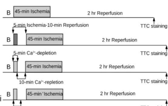
Fig. 1.Schematic illustration of the experimental protocol. Hearts were subjected to 5-min global ischemia and 10-min reperfusion in the ischemic preconditioned (IPC) group and to 5-min Ca2+-deple- tion and 10-min repletion in the Ca2+-preconditioned (CPC) group, before 45-min ischemia. In the CPC+PKCi (PKC inhibitor) group, calphostin C (CalC) was given for 10 min and washed out for 5 min during CPC. B, baseline. TTC: triphenyltetrazolium.
taining goat anti-mouse peroxidase conjugate (1:1,000, Bio- Rad). Finally, immunoreactive bands were visualized by en- hanced chemiluminiscence method (Amersham).
Statistics
Data were presented as mean SEM; they were analyzed by paired t-test within the same experimental group and by one-way ANOVA for repeated measures within different ex- perimental groups. When significant differences were obse- rved, the mean values were evaluated with Tukey s post-hoc test. A p value less than 0.05 was considered significant.
RESULTS Functional parameters
The baseline values of LVDP, dP/dt, LVEDP and coro- nary flow were not different between experimental groups.
Ischemia caused a rapid drop in LVDP, dP/dt and coronary flow; these parameters stayed at zero point during ischemia for 45-min. On reperfusion, IPC enhanced LVDP and dP/
dt recovery, but CPC did not. LVDP and dP/dt recovery on reperfusion were greater in the IPC group (>90% of pre-
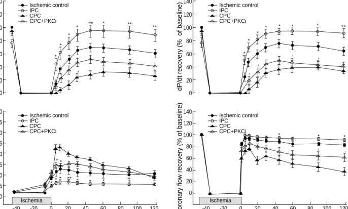
ischemic value) than in the ischemic control group (<70% of preischemic value), however, these parameters in the CPC and CPC PKCi groups were less than 50% of preischemic values (Fig. 2). Coronary flow recovery in the IPC group was greater than in the ischemic control group. It reached near to pre- ischemic value and maintained approximately at >90% of preischemic value in the IPC group, but difference between the IPC and ischemic control groups (80-90% of preischemic value) was not evident. In contrast, CPC and CPC+PKCi depressed the coronary flow recovery. Coronary flow recovery was smaller in the CPC group (<60% of preischemic value) than that in the ischemic control group (Fig. 2), but the dif- ference was not evident between the CPC and CPC PKCi groups. In all experimental groups, LVEDP was increased by 35 min after ischemia and further increased immediately after the onset of reperfusion. The increased levels of LVEDP last- ed throughout reperfusion both in the ischemic control and IPC group, however, the increase was less prominent in the IPC group (p<0.05). This result means that IPC attenuated myocardial contracture during reperfusion. In the CPC group, LVEDP reached a plateau (>100 mmHg, p<0.01 vs ischemic control group) in the earlier period of reperfusion and pro- gressively decreased afterwards (approximately 50 mmHg at the end of reperfusion).
LVDP recovery (% of baseline)
Ischemic control IPC
CPC CPC+PKCi 140
120 100 80 60 40 20 0
Fig. 2.Changes of the recovery rate of left ventricular developed pressure (LVDP), contractility (+dP/dt), left ventricular end-diastolic pres- sure (LVEDP), and coronary flow during ischemia and reperfusion. *p<0.05, **p<0.01, ischemic control vs. IPC; #p<0.05, CPC+PKCi vs.
CPC; p<0.05, ischemic control vs. CPC+PKCi.
LVEDP (mmHg)
Time (min) Ischemic control
IPC CPC CPC+PKCi 200
175 150 125 100 75 50 25 0
-40 -20 0 20 40 60 80 100 120
Coronary fiow recovery (% of baseline)
Time (min) Ischemic control
IPC CPC CPC+PKCi 140
120 100 80 60 40 20 0
-40 -20 0 20 40 60 80 100 120
dP/dt recovery (% of baseline)
Ischemic control IPC
CPC CPC+PKCi 140
120 100 80 60 40 20 0
*
*
*
*
*
* * * * *
*
** * * *
* *
*
* *
** * **
**
** **
Ischemia Ischemia
Infact size and LDH leakage
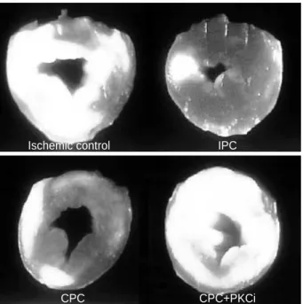
Infarct size (Fig. 3, 4) was expressed as a percentage of the left ventricle including the interventricular septum. We did not assume risk area since the entire myocardium might be at risk in global ischemia. Compared with the ischemic con- trol (45.2 2.4%), the infarct size was reduced by IPC (21.0 1.3%, p<0.05) and CPC (25.5 2.2%, p<0.05); however, infarct size was not reduced or limited by PKCi treatment (CPC PKCi, 37.9 2.5%).
LDH leakage at the baseline in the coronary effluent of the
ischemic control, IPC, CPC, and CPC PKCi groups were 6.00 0.62, 5.29 0.50, 5.50 0.54, 5.81 0.60 unit/mL, respectively. It was significantly increased in the ischemic control and CPC PKCi groups on reperfusion (p<0.05 vs CPC group, 5-20 min), however, no significant difference was found between the IPC and CPC groups (Fig. 5).
PKC activity and Western blot analysis of PKC isozymes
Total PKC activity in the baseline, after 45-min ischemia, IPC, IPC and 45-min ischemia, CPC, CPC and 45-min isch- emia, CPC PKCi, and CPC PKCi and 45-min ischemia were 8.57 0.06, 8.31 0.04, 8.75 0.12, 8.81 0.10,
Fig. 3.Heart slices stained with triphenyltetrazolium chloride (TTC).
Infarct (white area) was limited by IPC and CPC but infarct size-lim- iting effect is abolished by PKC inhibition with calphostin C (CPC +PKCi).
Ischemic control IPC
CPC CPC+PKCi
50
40
30
20
10
Infarct size (% of left ventricle) 0
Ischemiccontrol
IPC CPC
CPC+PKCi
*
*
Fig. 4.Infarct size as percentage of the left ventricle. Infarct size was reduced by IPC and CPC but reduction of infarct size is ab- olished by PKC inhibition with calphostin C (CPC+PKCi). *p<0.05, vs. ischemic control.
LDH (unit/L coronary effluent)
Reperfusion time (min)
Ischemic control IPC
CPC CPC+PKCi 100
80
60
40
20
0B 0 20 40 60 80 100 120
Fig. 5.Lactate dehydrogenase (LDH) leakage in the coronary efflu- ent. During early period on reperfusion, LDH leakage is signifi- cantly increased in the ischemic control and CPC+PKCi. B, base- line. *p<0.05, vs. CPC+PKCi.
8
6
4
2
Protein kinase C (nmol/g tissue) 0
Baseline 45-min Isc
IPC
IPC+45-min Isc CPC
CPC+45-min Isc
CPC+PKCiCPC+PKCi+45-min Isc Cytosol
Membrane
Fig. 6.Protein kinase C (PKC) activity. PKC activities of the cytosol fractions are not significantly different between the experimental groups; however, those of the membrane fractions increase by ischemic preconditioning (IPC), IPC and 45-min ischemia (IPC+
45-min Isc), Ca2+preconditioning (CPC), and CPC and 45-min ischemia (CPC+45-min Isc). *p<0.01, vs. baseline.
* * * *
8.68 0.14, 8.76 0.06, 8.61 0.05, 8.52 0.12 nmol/g tissue, respectively. It was slightly increased after the IPC and CPC regimen, however, there was no significant difference. As shown in the Fig. 6, cytosol PKC activity was not different between groups and membrane PKC activity was not changed after 45-min ischemia, CPC PKCi, and CPC PKCi and 45-min ischemia; however, membrane PKC activity was sig- nificantly increased after IPC, IPC and 45-min ischemia, CPC, and CPC and 45-min ischemia (p<0.01), from 2.53 0.07 nmol/g tissue (baseline) to 3.11 0.07 (IPC), 3.34 0.11 (IPC 45-min Isc), 3.15 0.09 (CPC), and 3.06 0.08 nmol/ g tissue (CPC 45-min Isc). These results indi- cate that PKC was activated and translocated from the cytosol to the cell membrane by IPC or CPC.
Five PKC isozymes ( , , , , and ) were analyzed by We- stern blot with corresponding monoclonal antibodies against each isozymes. Distribution and intensity of PKC isozymes except PKC were not different between experimental groups (data not shown). In comparison with the baseline, the mem- brane fraction of PKC increased after IPC, IPC and 45-min ischemia, and CPC (Fig. 7).
DISCUSSION
As already established, the myocardial cell uses Ca2+as the essential final step in excitation-contraction coupling, the pro- cess by which depolarization of the cell surface membrane ini- tiates the interactions between the contractile proteins that lead to tension development and shortening in the wall of the heart. Ca2+also mediates stimulus-secretion coupling in a vari- ety of non-motile cells. As indicated, Ca2+ions, by carrying signals generated at the cell surface to a variety of intracellu- lar organelles and proteins, can be viewed as the most impor- tant of the intracellular messengers (24). It is generally believed that a high Ca2+influx during ischemia/reperfusion (Ca2+over- load) causes pathological changes in the ischemia/reperfusion injury; however, in a rat heart, Ashraf et al. (17) have demon- strated that repetitive Ca2+depletion and repletion for a short duration (Ca2+preconditioning, CPC) had induced a signifi- cant functional recovery and remarkable preservation of cell
structure against ischemia/reperfusion injury. Our results are partly consistent with Ashraf et al. (17). However, functional recovery by CPC was not seen, although infarct size was sig- nificantly reduced by IPC and CPC; LVDP and dP/dt recov- ered only 25-30% of the baseline values, compared with those in the IPC group (80-90% of baseline values) or even in the ischemic control group (60-70% of baseline values). The rea- son of depressed functional recovery by CPC in this study is unclear. Differences in species and experimental methods could be causing factors, but they are not satisfactory explanations.
Since the early increase of LVEDP on reperfusion after 45-min ischemia in the CPC group was evident, it was doubtful whe- ther the CPC regimen in this study would cause Ca2+paradox injury, in which necrosis of the cell is a common feature, or not.
However, functional parameters after Ca2+depletion and reple- tion in the CPC group were not significantly different from the baseline values, and LDH leakage was not different between the IPC and CPC groups after 45-min ischemia. Moreover, infarct size was reduced or limited by CPC as seen in the IPC group. From these results, it could be concluded that 5-min Ca2+
depletion and 10-min Ca2+repletion did not cause Ca2+para- dox injury. Reduced response or sensitivity of contractile pro- teins to normal Ca2+concentration or reduced energy produc- tion on reperfusion due to myocardial stunning by CPC could be another possible factor, although intracellular Ca2+and ener- gy metabolism were not considered. They can cause a retarded functional recovery with diastolic abnormalities.
The physiological function of protein kinases is not yet fully understood, but PKC is activated upon external stimulation of the myocyte by various ligands including hormones, neuro- transmitters, and growth factors. Many lines of evidence in- dicate that PKC is involved in IPC of the heart. In this study, membrane PKC activity was increased by IPC and CPC, while PKC inhibition did not increase the activity. These results indicate that PKC was activated by IPC and CPC, and translo- cated from the cytosol to the cell membrane. It has been sug- gested that the elevation of Ca2+iduring IPC, in part, result- ed from Ca2+entry via voltage-dependent Ca2+channel (8, 25) triggers activation of PKC. However, cytosol PKC was not significantly decreased in both IPC and CPC in this study as in a rat heart (19), in contrast to other studies (8, 26). The reason for this is unclear, however, there is a possibility that a small change in the cytosol PKC by CPC or IPC did not affect the total amount of cytosol PKC, since the amount of cytosol PKC is much greater than that of membrane PKC.
In addition, in this study, immunoblots of the membrane PKC were evidently increased by IPC, IPC and 45-min is- chemia and by CPC, while concurrent PKC inhibition abol- ished the increase of membrane PKC . These results strong- ly suggest that the activation of membrane PKC is closely related with the infarct size-limiting effects of IPC and CPC.
There are some evidences that specific PKC isozymes were responsible for the mechanism of IPC or CPC. Ping et al. (27) described that IPC caused selective translocation of PKC and
Cyto Memb
Base 45-min Isc
IPC
IPC+45-min Isc CPC
CPC+45-min Isc
CPC+PKCiCPC+PKCi+45-min Isc
Fig. 7.Western blot analyses of PKC . Membrane (Memb) PKC is increased by IPC, IPC and 45-min ischemia (IPC+45-min Isc), and by CPC, while cytosol fraction between groups is not signifi- cantly different. Base, baseline; PKCi, PKC inhibition with cal- phostin C.
PKC without demonstrable changes in total myocardial PKC activity in the conscious rabbit heart, and our previous study (20) demonstrated that PKC has a trigger role for cardioprotective effect of IPC in the isolated rabbit heart. In contrast, Miyawaki et al. (19) demonstrated that immunolo- calization of PKC and PKC increased in the cell membrane of rat hearts. However, there is a controversy over the differ- ent PKC-coupled receptor systems, which might be involved in IPC. PKC activator did not reduce infarct size in canine hearts (22), and in porcine hearts, even the infarct size reduced by PKC inhibitor (28). These inconsistent results might have been derived from the following possibilities: 1) activation of PKC was not sufficient to phosphorylate certain key proteins;
2) expression of specific PKC isozymes was not significant to participate in the protection; or 3) mechanical factors of IPC or CPC protocol itself caused an aggravation of myocardial stunning. These experimental data provide that the activation of PKC may not be an universally applicable mechanism for the cardioprotective effect of IPC or CPC in common labora- tory animals, however, from this study, it is conceivable that a single dose of CPC could not enhance functional recovery but could protect the heart from infarction. Activation of mem- brane PKC may be responsible for trigger role in the car- dioprotection by both IPC and CPC.
ACKNOWLEDGMENT
This study was supported by the research grant from Sung- kyunkwan University School of Medicine (No. 2002-0145- 000), 2002-2003.
REFERENCES
1. Murry CE, Jennings RB, Reimer KA. Preconditioning with ischemia:
a delay of lethal cell injury in ischemic myocardium. Circulation 1986;
74: 1124-36.
2. Sumeray MS, Yellon DM. Ischaemic preconditioning reduces infarct size following global ischaemia in the murine myocardium. Basic Res Cardiol 1998; 93: 384-90.
3. Yabe K-I, Tanonaka K, Koshimizu M, Katsuno T, Takeo S. A role of PKC in the improvement of energy metabolism in preconditioned heart.
Basic Res Cardiol 2000; 95: 215-27.
4. Cave AC, Hearse DJ. Ischemic preconditioning and contractile func- tion: Studies with normothermic and hypothermic global ischemia. J Mol Cell Cardiol 1992; 24: 1113-23.
5. Kim H, Kim DJ, Chung HS, Shim SJ, Yoo UH, Rah BJ, Kim HD. Evi- dence of protein kinase C translocation by ischemic preconditioning in global ischemia model. J Korean Med Sci 1998; 13: 473-82.
6. Kitakaze M, Hori M, Takashima S, Sato H, Kamada T. Augmentation of adenosine production during ischemia as a possible mechanism of myocardial protection in ischemic preconditioning. Circulation 1991;
84 (Suppl I): I-306.
7. Li Y, Kloner RA. The cardioprotective effects of ischemic precon- ditioning are not mediated by adenosine receptors in rat hearts. Cir- culation 1993; 87: 1642-6.
8. Node K, Kitakaze M, Sato H, Minamino T, Komamura K, Shinozaki Y, Mori H, Hori M. Role of intracellular Ca2+in activation of protein kinase C during ischemic preconditioning. Circulation 1997; 96: 1257- 65.
9. Schott RJ, Rohmann S, Braun ER, Schaper W. Ischemic precondition- ing reduces infarct size in swine myocardium. Circ Res 1990; 66: 1133- 42.
10. Matsubara T, Minatoguchi S, Matsuo H, Hayakawa K, Segawa T, Ma- tsuno Y, Watanabe S, Arai M, Uno Y, Kawasaki M, Noda T, Take- mura G, Nishigaki K, Fujiwara H. Three minute, but not one minute, ischemia and nicorandil have a preconditioning effect in patients with coronary artery disease. J Am Coll Cardiol 2000; 35: 345-51.
11. Liu GS, Thornton JD, Van Winkle DM, Stanley AWH, Olsson RA, Downey JM. Protection against infarction afforded by precondition- ing is mediated by A1-adenosine receptors in rabbit heart. Circulation 1991; 84: 350-6.
12. Mitchell MB, Meng X, Ao L, Brown JM, Harken AH, Banerjee A.
Preconditioning of isolated rat heart is mediated by protein kinase C.
Circ Res 1995; 76: 73-81.
13. Goto M, Liu Y, Yang X, Ardell JL, Cohen MV, Downey JM. Role of bradykinin in protection of ischemic preconditioning in rabbit hearts.
Circ Res 1995; 77: 611-21.
14. Schultz JEJ, Hsu AK, Gross GJ. Morphine mimics the cardioprotec- tive effect of ischemic preconditioning via a glibenclamide-sensitive mechanism in the rat heart. Circ Res 1996; 78: 1100-4.
15. Vuorinen K, Ylitalo K, Peuhkurinen K, Raatikainen P, Ala-Rami A, Hassinen IE. Mechanism of ischemic preconditioning in rat myocardi- um: roles of adenosine, cellular energy state, and mitochondrial F1F0- ATPase. Circulation 1995; 91: 2810-8.
16. De Albuquerque CP, Gernstenblith G, Weiss RG. Importance of me- tabolic inhibition and cellular pH in mediating preconditioning con- tractile and metabolic effects in rat heart. Circ Res 1994; 74: 139-50.
17. Ashraf M, Suleiman J, Ahmad M. Ca2+preconditioning elicits a unique protection against the Ca2+paradox injury in rat heart. Role of adeno- sine. Circ Res 1994; 74: 360-7.
18. Armstrong SC, Hoover DB, Delacey MH, Ganote CE. Translocation of PKC, protein phosphatase inhibition and preconditioning of rab- bit cardiomyocytes. J Mol Cell Cardiol 1996; 28: 1479-92.
19. Miyawaki H, Zhou X, Ashraf M. Calcium preconditioning elicits st- rong protection against ischemic injury via protein kinase C signaling pathway. Circ Res 1996; 79: 137-46.
20. Kim H, Kim HC, Chung ST, Kim TH, Kim DJ, Rah BJ, Kim HD. Car- dioprotective effect of the ischemic preconditioning: its relation to acti- vation of protein kinase C. Korean Circ J 1999; 29: 602-12.
21. Belkin MB, Brown RD, Wright JG, LaMorte WW, Hobson RW. A new quantitative spectrophotometric assay of ischemia-reperfusion injury in skeletal muscle. Am J Surg 1988; 156: 83-6.
22. Przyklenk K, Sussman MA, Simkhovich BZ, Kloner RA. Does ische- mic preconditioning trigger translocation of protein kinase C in the canine model? Circulation 1995; 92: 1546-57.
23. Simkhovich BZ, Przyklenk K, Hale SL, Patterson M, Kloner RA. Dir- ect evidence that ischemic preconditioning does not cause protein ki- nase C translocation in rabbit heart. Cardiovasc Res 1996; 32: 1064- 70.
24. Katz A. Physiology of the Heart, 2nd ed, New York: Raven Press; 1992.
pp. 178-9.
25. Miyawaki H, Ashraf M. Ca2+as a mediator of ischemic precondition- ing. Circ Res 1997; 80: 790-9.
26. Ping P, Zhang J, Zheng Y-T, Li RCX, Dawn B, Tang X-L, Takano H, Balafanova Z, Bolli R. Demonstration of selective protein kinase
C-dependent activation of Src and Lck tyrosine kinases during ischemic preconditioning in conscious rabbits. Circ Res 1999; 85: 542-50.
27. Ping P, Zhang J, Qui Y, Tang X-L, Manchikalapudi S, Cao X, Bolli R. Ischemic preconditioning induces selective translocation of pro- tein kinase C isoforms and in the heart of conscious rabbits with- out subcellular redistribution of total protein kinase C activity. Circ Res 1997; 81: 404-14.
28. Vogt A, Barancik M, Weihrauch D, Arras M, Podzuweit T, Schaper W. Protein kinase C inhibitors reduce infarct size in pig hearts in vivo.
Circulation 1994; 90 (Suppl I): I-647.