INTRODUCTION
Apoptosis is an active mechanism that leads to cell death.
Tight regulation is essential to ensure a delicate balance bet- ween life and death, and the loss of apoptosis might proceed to a wide variety of diseases. Cancer also involves cellular de- fects that halt apoptosis in its development and progression of cancer (1). Hence, many studies have demonstrated the role of different apoptosis regulators in rendering tumor cells resistant to apoptosis both in vitro and in vivo. The upregu- lation of anti-apoptotic proteins would certainly assist the survival of tumor cells (2-5). Various anti-apoptotic proteins are expressed in different tumors, and their expression may be related with unfavorable prognostic features at diagnosis (6-14) and poor treatment responses (14-18).
Over the last decade, a complex network of pro- and anti-
apoptotic proteins that govern the tight regulation of the apoptosis pathways have been revealed (19-22). Among anti- apoptotic proteins, a group of proteins, known as the inhibi- tor of apoptosis protein (IAP), are the only cellular factors that act both on the initiator and effector caspases (23-26). To date, eight human IAPs have been identified: NAIP, cIAP1, cIAP2, XIAP, survivin, apollon, ILP-2, and livin (27). As their name implies, the IAP family proteins can inhibit the apoptosis induced by a variety of stimuli. Therefore, the overex- pression of various IAPs is regarded as an unfavorable factor in various malignancies. However, in breast cancer, the clin- ical relevance of IAP overexpression has not been evaluated with the exception of survivin. The overexpression of sur- vivin in breast cancer is associated with the presence of unfa- vorable prognostic factors at diagnosis and a poor clinical outcome.
Jaewon Choi, Yu Kyeong Hwang*, Young Jin Choi�, Ki Eun Yoo, Jeong Han Kim�, Seok Jin Nam�, Jung Hyun Yang�, Sang Jin Lee�, Keon Hee Yoo, Ki Woong Sung, Hong Hoe Koo, Young-Hyuck Im�
Department of Pediatrics, Samsung Medical Center, Sungkyunkwan University School of Medicine, Seoul;
Division of Immunotherapy*, Mogam Biotechnology Research Institute, Yongin; Division of Breast and Endocrine Surgery, Department of Surgery�, Samsung Medical Center, Sungkyunkwan University School of Medicine, Seoul; Genitourinary Cancer Branch�, National Cancer Center, Ilsan; Division of Hematology-Oncology, Department of Medicine�, Samsung Medical Center, Sungkyunkwan University School of Medicine, Seoul, Korea
Jaewon Choi and Yu Kyeong Hwang equally contributed to this work.
Address for correspondence Seok Jin Nam, M.D.
Department of Surgery, Samsung Medical Center, Sungkyunkwan University School of Medicine, 50 Irwon-dong, Gangnam-gu, Seoul 135-710, Korea Tel : +82.2-3410-3478, Fax : +82.2-3410-0040 E-mail : [email protected]
*This work was supported by the Samsung Biomedi- cal Research Institute, #C-A5-317-1.
S17
Neuronal Apoptosis Inhibitory Protein is Overexpressed in Patients with Unfavorable Prognostic Factors in Breast Cancer
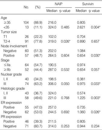
Neuronal apoptosis inhibitory protein (NAIP) is a recently identified inhibitor of apop- tosis protein. However, the clinical relevance of NAIP expression is not completely understood. In an attempt to determine the clinical relevance of NAIP expression in breast cancer, the levels of NAIP and survivin expression were measured in 117 breast cancer samples and 10 normal breast tissues using quantitative reverse- transcriptase-polymerase chain reaction. While there was no evidence of NAIP expression in the normal breast tissue, NAIP was expressed in all breast cancer samples. The level of NAIP expression in breast cancer was significantly higher (257 times) than in the universal tumor control. There was a strong correlation bet- ween the level of NAIP expression and the level of survivin expression (p=0.001).
The level of NAIP expression in patients with a large tumor (≥≥T2) and patients with an unfavorable histology (nuclear grade III) was significantly higher than in those patients with a small tumor (T1) and patients with a favorable histology (nucle- ar grade I, II) (p=0.026 and p=0.050, respectively). Although the level of NAIP ex- pression was higher in patients with other unfavorable prognostic factors, it was not significant. The three-year relapse-free survival rate was not significantly the patients showing high NAIP expression and patients showing low NAIP expres- sion (86.47±±4.79% vs. 78.74±±6.57%). Further studies should include the expres- sions of NAIP in a larger number of patients and for a longer period of follow-up to evaluate correlation with metastasis and treatment outcome. In conclusion, NAIP is overexpressed in breast cancer patients with unfavorable clinical features such as stage and tumor size, suggesting that NAIP would play a role in the disease manifestation.
Key Words : Breast Cancer; Neuronal Apoptosis Inhibitory rotein (NAIP); Apoptosis; Prognostic Factor;
Clinical Relevance
Received : 24 January 2007 Accepted : 25 June 2007
Neuronal apoptosis inhibitory protein (NAIP), which is a member of the IAPs, is expressed in mammalian cells and inhibits the apoptosis induced by a variety of signals. This gene is homologous to two baculovirus IAPs (28). NAIP has been linked to the inherited disease, spinal muscular atrophy (SMA), which occurs in children and manifests as a degen- eration of the motor neurons (29). NAIP may play a role in the mechanisms of resistance of tumor cells to various chemo- therapeutic agents. Moreover, the strong expression of IAPs, particularly Survivin and NAIP, is observed in the bone mar- row of AMLL (30, 31). However, little is known about the clinical relevance of NAIP expression in breast cancer.
In this context, this study examined the clinical relevance of NAIP expression in breast cancer using quantitative reverse transcriptase-polymerase chain reaction (RT-PCR). The result showed that overexpression of NAIP was associated with the unfavorable clinical features of breast cancer.
MATERIALS AND METHODS Patients and clinical evaluations
One hundred and seventeen patients, who were newly diag- nosed as breast cancer at Samsung Medical Center from Au- gust 2003 to December 2004, were enrolled in this study.
All the patients were diagnosed with a breast carcinoma pre- operatively by radiological findings and tissue biopsy, and they did not receive any form of treatment prior to surgery.
All the patients underwent a potentially curative resection, and the tumor specimens were sent to a pathologist for an evaluation of various prognostic factors including histologi- cal subtype, lymph node involvement, expression of estro- gen receptor (ER), and progesterone receptor (PR). A bone scan was performed routinely for an evaluation of the stage.
The Institutional Review Board in the Samsung Medical Center approved this study, and informed consent was ob- tained from the patients or guardians.
RNA isolation and cDNA synthesis
The tumor tissue specimens were taken from the periphery of the tumor mass resected in the operating room and stored at -70℃in a RNAlater reagent (Ambion, Austin, U.S.A.).
The tumor tissue specimens were homogenized using a rotor- stator homogenizer, DIAX 900 (Heidoph, Schwabach, Ger- many). The total RNA was extracted using a Qiamp kit (Qi- agen, Chatsworth, U.S.A.) according to the manufacturer’s protocol. After treatment with DNA-free� (Ambion) to remove the chromosomal DNA, the complementary DNA (cDNA) was synthesized using SuperScript III Reverse Tran- scriptase (Invitrogen, Carlsbad, U.S.A.) with oligo (dT) 15- mer primer and stored at -20℃until use.
Quantitative Real-time RT-PCR
The mRNA expression levels of the IAPs and glyceralde- hyde-3-phosphate dehydrogenase (GAPDH) were measured by quantitative RT-PCR using the ABI PRISM 7000 sequ- ence detector system (Applied Biosystems, Foster City, CA, U.S.A.). The real-time PCR amplification was carried out using the pre-developed assay-on-demand gene expression set for the NAIP gene (Hs00244967_m1, GeneBank acces- sion number NM_004536, Applied Biosystems), Survivin gene (Hs00153353_m1, GeneBank accession number NM_
001168, Applied Biosystems), and TaqMan�GAPDH Con- trol Reagents (Applied Biosystems) for the GAPDH gene in combination with the TaqMan� Universal PCR Master Mix (Applied Biosystems).
All the reactions were performed in triplicate using a 20 L sample containing 50 ng of cDNA. The reaction proto- col involved heating at 50℃for 2 min and then at 95℃for 10 min, followed by 40 cycles of amplification cycles (15 sec at 95℃and 1 min at 60℃). The analysis was performed using ABI PRISM 7000 Sequence Detection software (App- lied Biosystems). The expression level of the IAP genes in the unknown samples was calculated as the ratios of IAP ver- sus GAPDH. The IAP and GAPDH mRNA levels were quantified using a standard curves made from known serial dilution of Universal Human Reference RNA (Invitrogen, Carlsbad, U.S.A.). The standard curves were generated by assuming a linear relationship between the first cycle num- ber, at which the fluorescence signal increased significantly (Ct value), and the logarithm of the starting quantity. A ne- gative control without the template was included in each experiment.
Statistical analysis
The differences in the level of IAP expression with respect to the established clinicopathological prognostic factors and treatment outcome (occurrence of a relapse) were analyzed using a Mann-Whitney U test. The Spearman’s rank corre- lation test was used to assess the gene co-expression patterns of the NAIP and survivin in breast cancer tissues. The patients were categorized into two groups according to the NAIP expression levels (≥median or <median). The relapse-free survival rates (RFS) in each group were estimated using the Kaplan-Meier method and compared using the log-rank test. p values <0.05 were considered significant.
RESULTS Patient characteristics
One hundred and seventeen patients were enrolled in this study, and their clinical characteristics are listed in Table 1.
The median age was 59 yr (range 24-76). Thirteen patients (11.1%) were younger than 35 yr old. Ductal type was the most common histological subtype (77.8%). The tumor size was larger than T1 in 91 patients (77.8%). A lymph node metastasis was present in 57 patients (48.7%). The stage was higher than IIa in 52 patients (44.4%). The nuclear grade
was III in 74 patients (63.2%) and the histological grade was III in 58 patients (49.6%). There were 62 (53.0%) and 71 (60.7%) patients with ER- and PR-positive tumors, res- pectively.
Expression levels of NAIP were very high in breast cancer
While there was no evidence of NAIP expression in the normal breast tissue, NAIP was expressed in all the breast cancer samples. The level of NAIP expression in breast can- cer was significantly higher than in universal tumor control.
Fig. 1 shows the relative levels of NAIP and survivin expres- sion compared with the universal tumor cell control. While the median levels of survivin expression were 0.8 times that of the control, the median level of NAIP expression was very high (257 times that of the control) (Fig. 1A). In addition, the level of NAIP expression was strongly correlated with that of survivin (p<0.001, Fig. 1B).
NAIP expression was associated with the presence of unfavorable prognostic factors
Table 1 and Fig. 2 show the level of NAIP expression with respect to the prognostic factors. The level of NAIP expres- sion in patients with a large tumor (≥T2), and an unfavor- able histology (nuclear grade III) was significantly higher than in the patients with a small tumor (T1) and a favorable histology (nuclear grade I, II) (p=0.026 and p=0.050, respec- tively). Although the level of NAIP expression was higher in patients with the other unfavorable prognostic factors (age
<35 yr, positive node involvement, ≥IIb stage, and nega- tive PR expression), it was not significant.
In most cases, survivin overexpression was associated with the presence of unfavorable prognostic factors. However, for the lymph node metastasis, and stage, the level of survivin overexpression was higher in early staged disease than in advanced disease (p=0.030 and 0.057, respectively).
NAIP Median p value
No. (%) Survivin
Median p value Age
≥35 104 (88.9) 216.0 0.805
<35 13 (11.1) 324.0 0.485 2.621 0.004*
Tumor size
T1 26 (22.2) 102.0 0.704
T2-4 91 (77.8) 319.0 0.026* 0.890 0.657
Node involvement
Negative 60 (51.3) 202.0 1.084
Positive 57 (48.7) 264.5 0.604 0.654 0.030*
Stage
≤IIa 64 (54.7) 190.5 0.974
≥IIb 52 (44.4) 287.0 0.532 0.654 0.057
Nuclear grade
I, II 40 (34.2) 198.5 0.381
III 74 (63.2) 306.0 0.050 0.975 0.033*
Histologic grade
I, II 43 (36.7) 324.0 0.574
III 58 (49.6) 221.0 0.768 1.225 0.003*
ER expression
Positive 55 (47.0) 257.0 0.735
Negative 62 (53.0) 244.5 0.692 1.060 0.026*
PR expression
Positive 46 (39.3) 211.5 0.805
Negative 71 (60.7) 314.0 0.253 0.944 0.234 Table 1.Relationship between levels of NAIP and survivin ex- pression and the clinicopathological prognostic factors at diag- nosis
Differences in expression of NAIP and survivin according to established clinicopathological prognostic factors were analyzed using a Mann-Whi- tney U test. The expression levels are presented as median values. p values of <0.05 were considered significant.
Expression levels
1,000,000 100,000 10,000 1,000 100 10 1 0.1 0.01
Fig. 1.Relative expression levels of NAIP and survivin mRNA compared with universal human reference RNA. The level of NAIP expres- sion was 257 times higher than the control, whereas the median level of survivin expression was 0.8 times that of the control (A). The cor- relations between NAIP and survivin expression in each patient were analyzed by a Spearman’s rank correlation test. Positive correlation was observed between NAIP and survivin (B, p=0.0001).
NAIP Survivin
p<0.0001
A
Survivin
1,000
100
10
1
0.1
0.01
B 1,000,000 100,000 10,000 1,000 10 100
1
NAIP
NAIP vs. Survivin p value=0.0001
NAIP expression was high in the patients with a poorer treatment outcome
The median follow-up duration was 28 months (range, 1- 75). The tumor relapsed in 16 patients, and treatment-relat- ed mortality occurred in 6 patients. The 3-yr overall survival (OS) and RFS rates (±SE) were 82.2±7.0% and 76.0± 6.8%, respectively.
Higher levels of NAIP expression were found to be asso- ciated with a less favorable treatment outcome, but this was not significant. The median levels of NAIP expression in the relapsed patients (n=16) and relapse-free patients (n=
84) were 266 and 202, respectively (p=0.608, Fig. 3A). Sim- ilarly, the 3-yr RFS rate was lower in the patients showing NAIP overexpression (≥median) than in those not show- ing NAIP overexpression (78.74±6.57% vs. 86.47±4.79
%, p=0.511, Fig. 3B). Survivin overexpression was not asso- ciated with an unfavorable treatment outcome in this study (data not shown).
DISCUSSION
While the expression of various IAPs and their prognos- tic significance has been examined in different cancers (6- 18), survivin is the only IAP that has been evaluated for its expression and clinical relevance in breast cancer to date (32- 35). To the best of our knowledge, there are no reports of the expression of the other IAPs other than survivin in breast cancer tissues. This study is the first to evaluate the level of NAIP expression in breast cancer using quantitative RT-PCR in an attempt to determine a possible association with the established clinicopathological prognostic factors.
While NAIP was not expressed in the normal breast tis- sue but was expressed at high levels in breast cancer com- pared with the universal tumor control. This suggests that quantitative RT-PCR for NAIP can be used to find a mini- mal tumor in the regional lymph node or bone marrow.
Because RT-PCR is more sensitive than either immunohis- tochemistry or a conventional pathologic examination, quan- titative RT-PCR for NAIP might be valuable in detecting minimal disease if NAIP expression is still not detected in
NAIP expression levels
10,000
1,000
100
10
Fig. 3.Expression of NAIP was slightly higher in relapsed patients than in relapse-free patients (266 vs. 202 folds of control, p=0.608). (A) The relapse-free survival rate also was slightly higher in patients with a high level of NAIP expression (86.47±4.79%) than in patients with a low level of NAIP expression (78.74±6.57%) (B). This three-year relapse-free survival according to NAIP expression did not show a statistical significance (p=0.511).
Relapse Relapse free
Outcome
p=0.608
A
RFS (%)
100
90
80
70
60
50
0 10 20 30 40
Month from diagnosis
p=0.511
NAIP high expression NAIP low expression
B
NAIP expression levels
1,000,000 100,000 10,000 1,000 100 10 1
Fig. 2.Levels of NAIP expression according to clinicopathologic prognostic factors. NAIP overexpression was correlated with the presence of unfavorable prognostic factors, T2 (A) and nuclear grade III (B).
T1 T2-4
Tumor size
p=0.026
A
NAIP expression levels
1,000,000 100,000 10,000 1,000 100 10 1
I, II III
Nuclear grade
p=0.050
B
the further experiment on a large number of normal breast, lymph node and bone marrow tissues.
Expression of survivin and other IAPs can be measured by immunohistochemistry (IHC) using antibodies, conventional RT-PCR, and quantitative RT-PCR. Although IHC appear to be a more specific method for detecting biologically and clinically significant cancer micrometastases in histological- ly normal specimen in some cancers, RT-PCR appears to be a more sensitive method maintaining a reasonable specifici- ty (36-39). The IHC method has some advantages in detect- ing specific antigen protein including histological observa- tions while RT-PCR guarantees high sensitivity and quanti- tative analysis in a total amount of RNA specimen. Howev- er, RNA expression itself does not reflect the protein expres- sion exactly. Therefore, complementary use of two methods is recommended.
While survivin overexpression is known to be strongly asso- ciated with the unfavorable clinical features and RFS rate in breast cancer (32-34), survivin overexpression was not signif- icantly correlated with RFS rate in this study. The NAIP ex- pression level was strongly correlated with survivin overex- pression, however, poorer treatment outcomes were not sig- nificantly correlated with NAIP overexpression. We assume that a small number of patients and a relatively short follow- up duration might have resulted in an insignificant correla- tion between NAIP expressions and clinical outcome.
Interestingly, survivin expression was inversely correlated with the disease extent (lymph node metastasis and stage), while NAIP expression was not significantly associated with the disease extent. These results are partly similar with those reported by Span et al. (33) in that overexpression of survivin was correlated with unfavorable prognostic factors (young age, unfavorable histologic grade, and negative ER expres- sion). However, unlike those studies, our study showed that overexpression of survivin was correlated with negative node involvement and less advanced stage. Three splicing variants of survivin mRNA were detected in breast cancer tissue, and levels of both survivin-2B and survivin-DeltaEx3 but not survivin were significantly higher in nodal metastasiss than primary carcinomas (40). Similarly the overexpression of other IAPs showed strong correlations with negative lymph node metastasis and less advanced stage in our study (data not shown).
The IAP family proteins inhibit apoptosis induced by a variety of stimuli, and therefore, their overexpression is expect- ed to be associated with the unfavorable clinical features in a variety of malignancies including AML. However, the clin- ical significance of IAP overexpression in acute leukemia is not completely consistent with what was expected from pre- vious in vitro studies. For example, IAP overexpression was not always associated with the unfavorable clinical features in acute leukemia (26). Furthermore, it was recently report- ed that the high expression of Livin, also a member of IAP family proteins, is an independent favorable prognostic fac-
tor in childhood ALL (27). This suggests that the role of IAP in leukemogenesis or in the maintenance of leukemic cells might be different from what has been previously recognized.
We assume that a complex network of pro- and anti-apop- totic proteins might have a pivotal role in the controlling apoptosis pathways and its cellular factors.
To the best of our knowledge, there have been no reports on the expression and clinical relevance of IAPs other than survivin in breast cancer. This study is the first to show an association between NAIP overexpression and the unfavor- able clinical features in breast cancer even though there was no significant association between NAIP overexpression and an unfavorable treatment outcome. There were a small num- ber of patients and a relatively short follow-up duration in this study, which might have confounded the results. There- fore, a further study on more patients and for a longer follow- up duration will be needed to elucidate the association bet- ween NAIP overexpression and the treatment outcome.
REFERENCES
1. Reed CJ. Apoptosis and cancer: strategies for integrating pro- grammed cell death. Semin Hematol 2000; 37 (4 Suppl 7): 9-16.
2. Crook NE, Clem RJ, Miller LK. An apoptosis-inhibiting baculovirus gene with a zinc finger-like motif. J Virol 1993; 67: 2168-74.
3. Ikeguchi M, Kaibara N. Changes in survivin messenger RNA level during cisplatin treatment in gastric cancer. Int J Mol Med 2001; 8:
661-6.
4. Herr I, Debatin KM. Cellular stress response and apoptosis in can- cer therapy. Blood 2001; 98: 2603-14.
5. Kawasaki H, Toyoda M, Shinohara H, Okuda J, Watanabe I, Yama- moto T, Tanaka K, Tenjo T, Tanigawa N. Expression of survivin correlates with apoptosis, proliferation, and angiogenesis during human colorectal tumorigenesis. Cancer 2001; 91: 2026-32.
6. Lu CD, Altieri DC, Tanigawa N. Expression of a novel antiapopto- sis gene, survivin, correlated with tumor cell apoptosis and p53 accu- mulation in gastric carcinomas. Cancer Res 1998; 58: 1808-12.
7. Meng H, Lu C, Mabuchi H, Tanigawa N. Prognostic significance and different properties of survivin splicing variants in gastric can- cer. Cancer Lett 2004; 216: 147-55.
8. Satoh K, Kaneko K, Hirota M, Masamune A, Satoh A, Shimosegawa T. Expression of survivin is correlated with cancer cell apoptosis and is involved in the development of human pancreatic duct cell tumors. Cancer 2001; 92: 271-8.
9. Ikeguchi M, Yamaguchi K, Kaibara N. Survivin gene expression pos- itively correlates with proliferative activity of cancer cells in esopha- geal cancer. Tumour Biol 2003; 24: 40-5.
10. Grabowski P, Kuhnel T, Muhr-Wilkenshoff F, Heine B, Stein H, Hopfner M, Germer CT, Scherubl H. Prognostic value of nuclear survivin expression in oesophageal squamous cell carcinoma. Br J Cancer 2003; 88: 115-9.
11. Sui L, Dong Y, Ohno M, Watanabe Y, Sugimoto K, Tokuda M. Sur- vivin expression and its correlation with cell proliferation and prog-
nosis in epithelial ovarian tumors. Int J Oncol 2002; 21: 315-20.
12. Gazzaniga P, Gradilone A, Giuliani L, Gandini O, Silvestri I, Nofroni I, Saccani G, Frati L, Agliano AM. Expression and prognostic sig- nificance of LIVIN, SURVIVIN and other apoptosis-related genes in the progression of superficial bladder cancer. Ann Oncol 2003; 14:
85-90.
13. Ramp U, Krieg T, Caliskan E, Mahotka C, Ebert T, Willers R, Gab- bert HE, Gerharz CD. XIAP expression is an independent prognos- tic marker in clear-cell renal carcinomas. Hum Pathol 2004; 35:
1022-8.
14. Kato J, Kuwabara Y, Mitani M, Shinoda N, Sato A, Toyama T, Mit- sui A, Nishiwaki T, Moriyama S, Kudo J, Fujii Y. Expression of sur- vivin in esophageal cancer: correlation with the prognosis and res- ponse to chemotherapy. Int J Cancer 2001; 95: 92-5.
15. Tamm I, Richter S, Oltersdorf D, Creutzig U, Harbott J, Scholz F, Karawajew L, Ludwig WD, Wuchter C. High expression levels of x-linked inhibitor of apoptosis protein and survivin correlate with poor overall survival in childhood de novo acute myeloid leukemia.
Clin Cancer Res 2004; 10: 3737-44.
16. Kawasaki H, Altieri DC, Lu CD, Toyoda M, Tenjo T, Tanigawa N.
Inhibition of apoptosis by survivin predicts shorter survival rates in colorectal cancer. Cancer Res 1998; 58: 5071-4.
17. Sarela AI, Macadam RC, Farmery SM, Markham AF, Guillou PJ.
Expression of the antiapoptosis gene, survivin, predicts death from recurrent colorectal carcinoma. Gut 2000; 46: 645-50.
18. Wurl P, Kappler M, Meye A, Bartel F, Kohler T, Lautenschlager C, Bache M, Schmidt H, Taubert H. Co-expression of survivin and TERT and risk of tumour-related death in patients with soft-tissue sarcoma. Lancet 2002; 359: 943-5.
19. Clem RJ, Sheu TT, Richter BW, He WW, Thornberry NA, Duckett CS, Hardwick JM. c-IAP1 is cleaved by caspases to produce a pro- apoptotic C-terminal fragment. J Biol Chem 2001; 276: 7602-8.
20. Deveraux QL, Leo E, Stennicke HR, Welsh K, Salvesen GS, Reed JC. Cleavage of human inhibitor of apoptosis protein XIAP results in fragments with distinct specificities for caspases. EMBO J 1999;
18: 5242-51.
21. Nachmias B, Ashhab Y, Bucholtz V, Drize O, Kadouri L, Lotem M, Peretz T, Mandelboim O, Ben-Yehuda D. Caspase-mediated cleav- age converts Livin from an antiapoptotic to a proapoptotic factor:
implications for drug-resistant melanoma. Cancer Res 2003; 63:
6340-9.
22. Song Z, Liu S, He H, Hoti N, Wang Y, Feng S, Wu M. A single ami- no acid change (Asp 53 --> Ala53) converts Survivin from anti-apop- totic to pro-apoptotic. Mol Biol Cell 2004; 15: 1287-96.
23. Davoodi J, Lin L, Kelly J, Liston P, MacKenzie AE. Neuronal apop- tosis-inhibitory protein does not interact with Smac and requires ATP to bind caspase-9. J Biol Chem 2004; 279: 40622-8.
24. Silke J, Ekert PG, Day CL, Hawkins CJ, Baca M, Chew J, Pakusch M, Verhagen AM, Vaux DL. Direct inhibition of caspase 3 is dis- pensable for the anti-apoptotic activity of XIAP. EMBO J 2001; 20:
3114-23.
25. Deveraux QL, Stennicke HR, Salvesen GS, Reed JC. Endogenous inhibitors of caspases. J Clin Immunol 1999; 19: 388-98.
26. Scott FL, Denault JB, Riedl SJ, Shin H, Renatus M, Salvesen GS.
XIAP inhibits caspase-3 and -7 using two binding sites: evolution- arily conserved mechanism of IAPs. EMBO J 2005; 24: 645-55.
27. Liston P, Fong WG, Korneluk RG. The inhibitors of apoptosis: there is more to life than Bcl2. Oncogene 2003; 22: 8568-80.
28. Liston P, Roy N, Tamai K, Lefebvre C, Baird S, Cherton-Horvat G, Farahani R, McLean M, Ikeda JE, MacKenzie A, Korneluk RG.
Suppression of apoptosis in mammalian cells by NAIP and a relat- ed family of IAP genes. Nature 1996; 379: 349-53.
29. Kesari A, Misra UK, Kalita J, Mishra VN, Pradhan S, Patil SJ, Phad- ke SR, Mittal B. Study of survival of motor neuron (SMN) and neu- ronal apoptosis inhibitory protein (NAIP) gene deletions in SMA patients. J Neurol 2005; 252: 667-71.
30. Kudoh K, Ramanna M, Ravatn R, Elkahloun AG, Bittner ML, Mel- tzer PS, Trent JM, Dalton WS, Chin KV. Monitoring the expression profiles of doxorubicin-induced and doxorubicin-resistant cancer cells by cDNA microarray. Cancer Res 2000; 60: 4161-6.
31. Nakagawa Y, Hasegawa M, Kurata M, Yamamoto K, Abe S, Inoue M, Takemura T, Hirokawa K, Suzuki K, Kitagawa M. Expression of IAP-family proteins in adult acute mixed lineage leukemia (AMLL).
Am J Hematol 2005; 78: 173-80.
32. Tanaka K, Iwamoto S, Gon G, Nohara T, Iwamoto M, Tanigawa N.
Expression of survivin and its relationship to loss of apoptosis in breast carcinomas. Clin Cancer Res 2000; 6: 127-34.
33. Span PN, Sweep FC, Wiegerinck ET, Tjan-Heijnen VC, Manders P, Beex LV, de Kok JB. Survivin is an independent prognostic marker for risk stratification of breast cancer patients. Clin Chem 2004; 50:
1986-93.
34. Kennedy SM, O’Driscoll L, Purcell R, Fitz-Simons N, McDermott EW, Hill AD, O’Higgins NJ, Parkinson M, Linehan R, Clynes M.
Prognostic importance of survivin in breast cancer. Br J Cancer 2003; 88: 1077-83.
35. Hinnis AR, Luckett JC, Walker RA. Survivin is an independent pre- dictor of short-term survival in poor prognostic breast cancer pati- ents. Br J Cancer 2007; 96: 639-45.
36. Shariat SF, Roudier MP, Wilcox GE, Kattan MW, Scardino PT, Ves- sella RL, Erdamar S, Nguyen C, Wheeler TM, Slawin KM. Com- parison of immunohistochemistry with reverse transcription-PCR for the detection of micrometastatic prostate cancer in lymph nodes.
Cancer Res 2003; 63: 4662-70.
37. Kubota K, Nakanishi H, Hiki N, Shimizu N, Tsuji E, Yamaguchi H, Mafune K, Tange T, Tatematsu M, Kaminishi M. Quantitative detec- tion of micrometastases in the lymph nodes of gastric cancer patients with real-time RT-PCR: a comparative study with immunohistochem- istry. Int J Cancer 2003; 105: 136-43.
38. Pellegrino D, Bellina CR, Manca G, Boni G, Grosso M, Volterrani D, Desideri I, Bianchi F, Bottoni A, Ciliberti V, Salimbeni G, Gan- dini D, Castagna M, Zucchi V, Romanini A, Bianchi R. Detection of melanoma cells in peripheral blood and sentinel lymph nodes by RT-PCR analysis: a comparative study with immunohistochemistry.
Tumori 2000; 86: 336-8.
39. Hoshi S, Kobayashi S, Takahashi T, Suzuki KI, Kawamura S, Satoh M, Chiba Y, Orikasa S. Enzyme-linked immunosorbent assay detec- tion of prostate-specific antigen messenger ribonucleic acid in pro- state cancer. Urology 1999; 53: 228-35.
40. Ryan B, O’Donovan N, Browne B, O’Shea C, Crown J, Hill AD, McDermott E, O’Higgins N, Duffy MJ. Expression of survivin and its splice variants survivin-2B and survivin-DeltaEx3 in breast can- cer. Br J Cancer 2005; 92: 120-4.
41. Wrzesien-Kus A, Smolewski P, Sobczak-Pluta A, Wierzbowska A, Robak T. The inhibitor of apoptosis protein family and its antago-
nists in acute leukemias. Apoptosis 2004; 9: 705-15.
42. Choi J, Hwang YK, Sung KW, Lee SH, Yoo KH, Jung HL, Koo HH, Kim HJ, Kang HJ, Shin HY, Ahn HS. Expression of Livin, an anti- apoptotic protein, is an independent favorable prognostic factor in childhood acute lymphoblastic leukemia. Blood 2007; 109: 471-7.