Ⅰ. 서 론
다형성 저등급 선암종(polymorphous low-grade adenocar- cinoma)은 주로 소타액선에서 발생하는 타액선 신생물이 며, 다른 위치에서 발생하는 경우도 있으나, 주된 부위는 경구개부이다
1. 이 병소는 이전에는 소엽성암종(lobular carcinoma)
2, 종말관암종(terminal duct carcinoma)
3등의 호 칭으로 불렸고, 예전엔 이 병소의 발생빈도는 잘 알려져 있 지 않았으나, 종양을 특징짓는 병리조직학적 기준이 확립 되어 이 종양의 발견이 증가하고 있으며, 여성에서 자주 발 견되는 양상을 보인다
4. (2-4.6[여]:1[남]) 주로 통증 없이 천 천히 자라는 양상을 보이며, 조직학적으로는 변연이 피막 으로 둘러싸여있지 않은 침윤성 병소로 알려져 있다.
치료방법은 필요한 경우, 주위 골을 포함한 광범위한 외 과적 절제술이며 수술 후 방사선치료를 추천하기도 한다.
예후는 좋은 편이며 재발률은 17-22% 사이이다
5,6. 전이는 9% 정도로 흔하진 않으나 주로 국소적인 림프절에 전이되 는 것으로 알려져 있다
7. 본 증례는 67세 여성환자에서 구 개부에 발생한 다형성 저등급 선암종 병소를 1차적으로 먼 저 제거한 후 주위 구개골을 2차적으로 제거하는 수술법을 이용하여 비강 및 상악동 내 점막을 용이하게 보존함으로 써 구개부 결손을 예방하여 양호한 치료결과를 얻었기에 문헌고찰과 함께 보고하는 바이다.
Ⅱ. 증례보고
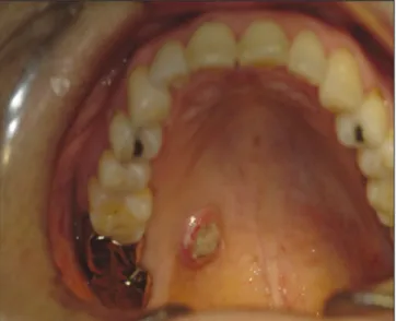
67세 여자환자로 2009년 3월 10일 본원에 처음 내원하셨 으며, 입천장에 생긴 무통성의 궤양성병소를 호소하였 다.(Fig. 1) 병소는 상악 구개부의 우측에 위치하고 있었으 며, 크기는 직경 1 cm 정도로 병소 중간 부위에 궤양이 있 었다. 궤양의 중심부는 흰색을, 주변은 붉은색을 띠고 있었 으며, 촉진할 때 약간의 통증을 호소하였다. 구강 외 특이 소견은 관찰되지 않았으며, 경부 림프절의 전이를 의심할
김 진 수700-705 대구시 중구 삼덕동2가188-1번지
경북대학교 치의학전문대학원 구강악안면외과학교실 Chin-Soo Kim
Department of Oral and Maxillofacial Surgery, School of dentistry, Kyungpook National University 188-1 Samduk-dong 2ga, Jung-gu, Daegu, 700-705, Korea TEL: +82-53-600-7562 FAX: +82-53-426-5365 E-mail: [email protected]
구개부에 발생한 다형성 저등급 선암종의 치험례
신영민
1∙최소영
1∙김진욱
1∙변기정
2∙김진수
11
경북대학교 치의학전문대학원 구강악안면외과학교실,
2울산대학교병원 치과
Polymorphous low-grade adenocarcinoma on hard palate: case report
Young-Min Shin
1, So-Young Choi
1, Jin-Wook Kim
1, Ki-Jung Byeon
2, Chin-Soo Kim
11
Department of Oral and Maxillofacial Surgery, School of Dentistry, Kyungpook National University, Daegu, Korea
2
Department of Dentistry, Ulsan University Hospital, Ulsan, Korea
Polymorphous low-grade adenocarcinomas (PLGA) are distinctive salivary gland neoplasms with a propensity to arise from the minor salivary glands.
The most frequent location of PLGA is the palate, even though other locations have been described. Previously used terms for PLGA include lobular carcinoma and terminal duct carcinoma.
Although the frequency of the tumor is unknown, the recognition of PLGA as an individual tumor has increased with the establishment of specific histopathological criteria characterizing the PLGA.
The first choice of treatment is a wide surgical excision including the subjacent bone if necessary. The prognosis is generally good and the recurrence rate ranges from 17% and 22%. Distant metastases is unusual (9%) but occur mainly in the regional lymph nodes.
This is a case report of a 67 year old female patient with PLGA who was treated with a wide excision by layers (2 stage) of the lesion including the surrounding bone. We present this case with a review of the relevant literature.
Key words:Polymorphous low-grade adenocarcinomas, Excision by layers
[paper submitted 2010. 9. 30 / revised 2011. 1. 12 / accepted 2011. 1. 28]
Abstract (J Korean Assoc Oral Maxillofac Surg 2011;37:72-6)
만한 경결감은 찾을 수 없었다.
이 병소는 5개월 전부터 생기기 시작하였고, 평상시에는 통증은 없었으나, 양치질하다가 가끔씩 피가 나고 불편감 을 느꼈다고 하였다. 병소의 크기 변화는 느끼지 못하고 계 시다가, 내원 1주일 전에 주위 치과의원에서 본원으로의 내원을 권유받아 내원하셨다. 환자의 체격 및 영양상태는 양호하며, 골격이상, 피부질환 등의 소견도 발견할 수 없었 으며, 복용하는 약물도 없었다. 혈액이화학적 검사에서의 수치들도 전부 정상범주에 속하였다. 가족력에서도 특이 한 사항을 발견할 수는 없었다.
술전 파노라마 및 경부 초음파검사에서 병소와 관련있는 특이한 소견은 없었으며, 골스캔에서도 구개골로의 침윤 을 의심할 만한 소견은 보이지 않았다. Paranasal sinus-com- puted tomography (PNS-CT)에서도 특이한 변화를 보이는 림프절 및 해부학적 구조는 없었고, positron emission
tomography-computed tomography (PET-CT)에서 우측 Level II 부위에서 전이가 의심되는 림프절이 관찰되었으나 원발 병소가 있는 부위에는 fluorodeoxyglucose (FDG) 섭취는 거 의 나타나지 않았다.(Fig. 2)
임상소견에서 악성 종양으로 잠정적 진단 후, 시행한 절 개생검술 결과에서 다형성 저등급 선암종으로 확진하였 다. 수술은 구강 내 접근을 통한 병소 주위의 구개부의 골 을 포함하는 광범위한 절제술을 시행하기로 결정하고, 구 개점막부의 결손이 2차 치유로 회복이 힘들다고 예상될 경 우 구개부회전피판술을 시행하기로 하였다.
2009년 5월 12일 비기관 삽관을 통해 전신마취 후 구개부 절개를 시작으로, 병소 경계에서 2 cm 거리를 두고 원형으 로 구개부 연조직의 절제 후 하방 구개골을 연조직 절제 경 계를 따라 2차적으로 절제하였다.(Figs. 3, 4) 절제 부위를 세척한 후 바셀린거즈를 넣고 상악에 레진 스플린트를 장
Fig. 1.Ulcerative lesion on right palate was showed at first visit. Fig. 2.PET-CT: without significant focal FDG uptake in right palate.(PET-CT: positron emission tomography-computed tomography, FDG: fluorodeoxyglucose)
Fig. 3.Excised mass of tumor and underlying bone. Fig. 4.Intraoral view of postoperation state.
착하여 치유를 유도하였다. 술후 10개월이 지난 현재 치유 양상은 양호하며, 다른 조직으로의 전이는 발견되지 않고 있다.(Fig. 5)
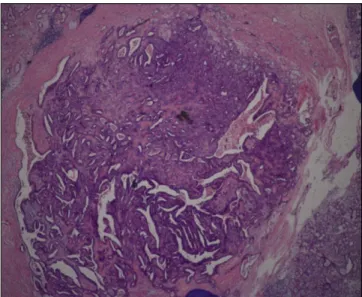
조직병리학적 소견으로는 원형 또는 계란형의 핵과 작은 핵소체를 가진 일정한 세포들의 군집들이 증식되어 있었 고, 세포질은 호산성을 띠고 있었다. 일부에선 adenoid나 solid한 형태를 띠고, 또 다른 일부에선 papillar, cribriform,
ductal 형태를 보였다. 신경 주위로의 침습양상이나, 골조
직으로의 침윤은 관찰할 수가 없었다.(Fig. 6)
Ⅲ. 고 찰
다형성 저등급 선암종은 소타액선에서 생기는 모든 종양 에서 7-11%를 차지하고, 악성 종양 중에 19-26%를 차지하 고 있으며, 구개부에서 가장 흔하게 발생한다
8. 하지만 입 술, 혀, 협부, 구강저 부위 등에서도 발생할 수 있으며, 아주 간혹 대타액선에서 발생하기도 한다
9-13. 수술 부위에서의 재발률은 9-33%정도이며, 경부 림프절로의 전이는 6-35%
로 알려져 있으나, 원위부로의 전이는 1% 이하로 보고되었 다
14. 매우 드물게 고등급 암종으로의 변형이 추적검사 기 간에 나타나기도 한다
15-17.
해부조직병리학적으로 이 병소는 저등급이며, solid, tra- becular, ductal, tubular, cribriform, cystic, papillary-cystic와 같은 다양한 성장양상을 보이나 전반적으로 일정한 세포 학적 양상을 나타낸다
8,18. 또한 신경 주위로의 침윤양상이 자주 보이나, 이 증례에서는 침윤양상이 보이지 않았다.
다형성 저등급 선암종의 진단은 일반적으로 어려운 편은 아니나, 다형성 선종(pleomorphic adenoma)와의 감별을 확 실히 해야한다. 이는 정확한 감별진단이 치료와 예후에 큰
영향을 미치기 때문이다. 다형성 선종과의 감별에서 신경 교원섬유산단백(glial fibrillary acid protein)이 유용하게 이 용되는데 이는 다형성 저등급 선암종에서는 이 단백과의 반응이 거의 나타나지 않으나 다형성 선종에는 반응이 나 타나기 때문이다
9,19. 또한 소타액선에서 생기는 다형성 선 종은 다형성 저등급 선암종과 달리 신경조직 주위로의 침 입이 관찰되지 않으며, 침윤적이지 않은 것으로 알려져 있 다. 감별진단이 필요한 다른 병소로는 선양낭성암종(ade- noid cystic carcinoma)이 있는데, 선양낭성암종의 세포는 다 형성 저등급 선암종에 비해 작고, 기저양(basaloid)이며, 높 은 핵세포질비를 보이며, 다염색성 핵과 투명한 세포질을 보인다.
다형성 저등급 선암종은 완전한 절제술이 필요하나 예후 는 일반적으로 양호한 편이며, 절개생검에서 확연하게 판 단하기는 쉽지 않으며, CT, magnetic resonance imaging (MRI) 등의 다른 방사선검사가 적절한 치료를 위해 필수적 이다. 다형성 저등급 선암종의 치료에 있어서 가장 적절한 초기 치료는 병소 경계의 정확한 평가 후에 국소적으로 광 범위하게 절제하는 것이다. 임상적 평가에서 림프조직으 로의 전이가 명확하지 않다면, 예방적 경부곽청술이 필요 없다. 이 증례에서는 병소 경계에서 2 cm 거리를 확보하여 연조직의 절제를 시행하였고, 하방 구개골을 연조직 절제 경계를 따라 2차적으로 절제하였다. 병소 자체가 크지 않 아 절제 후 2차 치유를 통한 충분한 연조직 회복이 가능하 리라 생각되어, 구개점막회전피판술은 시행하지 않았으 며, PET-CT에서 우측 경부에 전이가 의심되는 림프절이 존재한다 하였으나 PNS-CT 및 경부 초음파검사에서 특이 소견이 보이지 않아 예방적 경부곽청술은 시행하지 않았다.
구개부에 발생한 암종의 절제술을 시행할 경우 monobloc
Fig. 5.Ten months after surgery: operation site is unremarkable. Fig. 6.Histological overview.(H&E staining, original magnifi- cation 4×10)
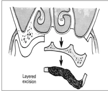
excision을 통해 수술을 하는데 이 경우 종양과 아래에 위치 한 골을 동시에 제거한다.(Fig. 7) 이 경우 상악동 점막과 비 점막골막의 보존이 어려워, 상악동 및 비강누공 등을 야기 할 수 있기에, 본 증례에선 종양 및 주위 점막을 우선 절제 한 후 하방에 위치한 골을 제거하는 2단계 절제술(excision in layer)을 시행하였다. 물론 선양낭성암종과 같은 종양에 서는 조직의 plane을 따라 침습적으로 자라는 특징이 있어 이러한 수술방법의 선택이 불가하나, 본 증례에서는 술전 에 시행한 PET-CT, 골스캔, PNS-CT에서 bone invasion 이 없는 것으로 판단하였으며, 수술 시에도 구개부의 점막제 거 후 구개골을 확인한 바, 골표면이 매끄러운 상태라 골로 의 종양의 침범이 없다고 진단하였기에 이런 방법을 선택 할 수 있었다고 생각되며, 이 방법을 통해 상악동과 비점막
을 용이하게 보존할 수 있었다
20.(Fig. 8) 수술 후 방사선검 사에서 상악동의 점막비후가 나타나기는 하였으나, 상악 동의 기능은 수술 전과 다르게 보이지 않았다.(Fig. 9)
현재 환자는 수술 후 경과관찰 중이며, 10개월이 지난 지 금까지 재발양상은 발견할 수 없었고, 수술 부위의 치유양 상은 매우 양호한 편이다. 경부에서 관찰되었던 전이가 의 심되는 림프절은 지금까지 큰 차이가 없으며 상악동의 기 능 또한 문제가 없었으며, 불편감도 호소하지 않고 있다.
Ⅳ. 결 론
본 증례에서는 구개부에 생긴 무통성의 궤양으로 내원한 67세 여자환자에서 임상, 방사선학적 소견 및 절개생검을 통해 구개부 우측에 발생한 다형성 저등급 선암종을 exci- sion in layer로 2단계로 절제함으로써 상악 및 비점막골막 을 보존하여 재발 및 상악동 누공 발생 등의 술후 합병증이 없는 양호한 결과를 얻었기에 문헌고찰과 함께 보고하는 바이다.
References
1.Furuse C, Tucci R, Machado de Sousa SO, Rodarte Carvalho Y, Cavalcanti de Arau′jo V. Comparative immunoprofile of poly- morphous low-grade adenocarcinoma and canalicular adenoma.
Ann Diagn Pathol 2003;7:278-80.
2.Freedman PD, Lumerman H. Lobular carcinoma of intraoral mi- nor salivary gland origin. Report of twelve cases. Oral Surg Oral Med Oral Pathol 1983;56:157-66.
3.Frierson HF Jr, Mills SE, Garland TA. Terminal duct carcinoma of minor salivary glands. A nonpapillary subtype of polymor- phous low-grade adenocarcinoma. Am J Clin Pathol 1985;84:8- 14.
4.Castle JT, Thompson LD, Frommelt RA, Wenig BM, Kessler HP. Polymorphous low grade adenocarcinoma: a clinicopatho- Fig. 7.Schematic view of monobloc excision. Fig. 8.Schematic view of excision by layers.
Fig. 9.Postoperative Water’s view.
logic study of 164 cases. Cancer 1999;86:207-19.
5.Neville BW, Damm DD, Allen CM, Bouquot JE. Oral and max- illofacial pathology. 2nd ed. Philadelphia: WB Saunders; 2002.
6.Clayton JR, Pogrel MA, Regezi JA. Simultaneous multifocal polymorphous low-grade adenocarcinoma. Report of two cases.
Oral Surg Oral Med Oral Pathol Oral Radiol Endod 1995;80:71- 7.
7.Kelsch RD, Bhuiya T, Fuchs A, Gentile P, Kahn MA, Fantasia JE. Polymorphous low-grade adenocarcinoma: flow cytometric, p53, and PCNA analysis. Oral Surg Oral Med Oral Pathol Oral Radiol Endod 1997;84:391-9.
8.Darling MR, Schneider JW, Phillips VM. Polymorphous low- grade adenocarcinoma and adenoid cystic carcinoma: a review and comparison of immunohistochemical markers. Oral Oncol 2002;38:641-5.
9.Curran AE, White DK, Damm DD, Murrah VA. Polymorphous low-grade adenocarcinoma versus pleomorphic adenoma of minor salivary glands: resolution of a diagnostic dilemma by immuno- histochemical analysis with glial fibrillary acidic protein. Oral Surg Oral Med Oral Pathol Oral Radiol Endod 2001;91:194-9.
10.Miliauskas JR. Polymorphous low-grade (terminal duct) adeno- carcinoma of the parotid gland. Histopathology 1991;19:555-7.
11.Haba R, Kobayashi S, Miki H, Hirakawa E, Saoo K, Iwai T, et al.
Polymorphous low-grade adenocarcinoma of submandibular gland origin. Acta Pathol Jpn 1993;43:774-8.
12.Blanchaert RH, Ord RA, Kumar D. Polymorphous low-grade adenocarcinoma of the sublingual gland. Int J Oral Maxillofac Surg 1998;27:115-7.
13.de Diego JI, Bernaldez R, Prim MP, Hardisson D. Polymorphous low-grade adenocarcinoma of the tongue. J Laryngol Otol 1996;
110:700-3.
14.Kumar M, Stivaros N, Barrett AW, Thomas GJ, Bounds G, Newman L. Polymorphous low-grade adenocarcinoma-a rare and aggressive entity in adolescence. Br J Oral Maxillofac Surg 2004;
42:195-9.
15.Mills SE, Garland TA, Allen MS Jr. Low-grade papillary adeno- carcinoma of palatal salivary gland origin. Am J Surg Pathol 1984;8:367-74.
16.Simpson RH, Pereira EM, Ribeiro AC, Abdulkadir A, Reis-Filho JS. Polymorphous low-grade adenocarcinoma of the salivary glands with transformation to high-grade carcinoma.
Histopathology 2002;41:250-9.
17.Pelkey TJ, Mills SE. Histologic transformation of polymorphous low-grade adenocarcinoma of salivary gland. Am J Clin Pathol 1999;111:785-91.
18.Gibbons D, Saboorian MH, Vuitch F, Gokaslan ST, Ashfaq R.
Fine-needle aspiration findings in patients with polymorphous low grade adenocarcinoma of the salivary glands. Cancer 1999;
87:31-6.
19.Gnepp DR, el-Mofty S. Polymorphous low-grade adenocarcino- ma: glial fibrillary acidic protein staining in the differential diag- nosis with cellular mixed tumors. Oral Surg Oral Med Oral Pathol Oral Radiol Endod 1997;83:691-5.
20.McGregor IA, McGregor FM. Cancer of the face and mouth:
pathology and management for surgeons. New York: Churchill Livingstone; 1986.