Received:July 7, 2015, Revised:July 29, 2015, Accepted:August 5, 2015
Corresponding to:Yong-Wook Park, Division of Rheumatology, Department of Internal Medicine, Chonnam National University Hospital, Chonnam National University Medical School, 42 Jebong-ro, Dong-gu, Gwangju 61469, Korea. E-mail:[email protected] pISSN: 2093-940X, eISSN: 2233-4718
Copyright ⓒ 2016 by The Korean College of Rheumatology. All rights reserved.
This is a Free Access article, which permits unrestricted non-commerical use, distribution, and reproduction in any medium, provided the original work is properly cited.
Clinical and Hematological Effects of Tocilizumab on Serum Hepcidin, Anemia Response and Disease Activity in Patients with Active Rheumatoid Arthritis
Ki-Jeong Park1, Hye-Mi Jin1, Young-Nan Cho1, Jeong-Hwa Kang1, Hyun-Ju Jung1, Ji-Hyoun Kang1, Ji-Eun Kim1, Yi-Rang Yim1, Jeong-Won Lee1, Kyung-Eun Lee1, Dong-Jin Park1, Tae-Jong Kim1, Shin-Seok Lee1, Seung-Jung Kee2, Yong-Wook Park1
1Division of Rheumatology, Department of Internal Medicine and 2Department Laboratory Medicine, Chonnam National University Hospital, Chonnam National University Medical School , Gwangju, Korea
Objective. The purpose of this study is to evaluate the clinical and hematological effects of tocilizumab in active rheumatoid arthritis (RA) patients. Methods. Fourteen patients with active RA were enrolled in this study. The patients received tocilizumab 8 mg/kg intravenously every four weeks for 6 months. Disease activity, anemia-related factors including serum hepcidin-25, and hematological parameters were monitored at baseline and at 1, 3, and 6 months after the initiation of treatment. Results.
Significant reductions in tender joint count, swollen joint count, visual analogue scale, erythrocyte sedimentation rate (ESR), and C-reactive (CRP) protein plus reductions in a 28-joint disease activity score were observed within one month after the first tocilizumab treatment. These effects lasted throughout the six-month study period. In addition, significant improvements in anemia-related factors such as hepcidin-25, ferritin, iron, hemoglobin, red blood cell counts and mean corpuscular volume were observed during the treatment period. Hematological parameters were improved with reductions in counts for leuko- cytes, monocytes, neutrophils, and platelets. The lymphocyte counts and their subset numbers were unchanged. Changes in hepcidin levels showed significant correlation with changes in CRP, ESR, ferritin, hemoglobin and counts for red blood cells, leukocytes, and neutrophils during the treatment period. Conclusion. This study demonstrates that tocilizumab significantly and meaningfully reduces disease burden in patients with active RA. In addition, tocilizumab diminishes the levels of in- flammatory anemia by inhibiting hepcidin production. These clinical data provide evidence of a favorable outcome from tocili- zumab in RA. (J Rheum Dis 2016;23:37-46)
Key Words. Rheumatoid arthritis, Tocilizumab, Hepcidins, Anemia, Disease activity
INTRODUCTION
Rheumatoid arthritis (RA) is an autoimmune disorder characterized by chronic inflammation that affects many organs and tissues, especially the synovial joints. The in- flammatory process induces synovitis, synovial hyper- plasia with neovascularization, and excessive synovial fluid buildup, which causes joint swelling, stiffness, and pain. Progressive RA leads to destruction of cartilage and bones in the joints [1,2]. RA patients may also manifest
multiple systemic symptoms such as fever, fatigue, ano- rexia, anemia, osteoporosis, weight loss, and muscle weakness [3]. In particular, anemia occurs in 30% to 60%
of RA patients and this rheumatoid anemia is a typical ex- ample of anemia of chronic disease (ACD) [3-5].
Currently, the etiology of RA is unclear, but certain proinflammatory cytokines, including tumor necrosis factor (TNF)-α, interleukin (IL)-1β, and IL-6 are known to play key roles in the pathogenesis of RA [6]. IL-6 is a multifaceted cytokine with various biological activities,
including regulation of the immune response, inflam- mation, and hematopoiesis [7]. Relevant to RA, IL-6 has a pivotal role in synovitis and osteoclast-mediated bone resorption [8,9]. IL-6 levels are found to be considerably increased in the serum and synovial fluid of RA patients and directly correlate with disease activity and in- flammation [10]. In addition, high levels of soluble IL-6 receptor (sIL-6R) have been shown to correlate with the degree of joint destruction [3]. IL-6 also induces hepcidin production during inflammation [11]. Hepcidin is known to be an iron regulatory peptide hormone produced in the liver and plays an important role in iron homeostasis and erythropoiesis [12]. Increases in hepcidin levels correlate with anemia in ACD, which implies that the regulation of hepcidin levels may be an important option for the treat- ment of chronic anemia [11].
Tocilizumab is a humanized monoclonal that inhibits IL-6R signaling by blocking IL-6 binding. This agent can diminish IL-6 triggered pathologic cascade and it also de- creases hepcidin-25 circulatory levels [13]. Previously to- cilizumab has been reported to improve anemia in mul- ti-centric Castleman’s disease [14] as well as joint swel- ling in animal model [15]. Additional studies showed that tocilizumab was more effective than a tested TNF-α in- hibitor in improving RA-related anemia and this was from inhibiting hepcidin production [16,17]. Thus, rela- tionships of IL-6R inhibition with acute phase reactants, anemia-related factors and clinical outcome need to be monitored in RA patients.
Accordingly the aim of this study was to assess the ef- fects of tocilizumab on disease activity, anemia-related factors including hepcidin, and hematological parameters during a six-month period in active RA patients.
MATERIALS AND METHODS
Subjects
The study cohort included 14 patients diagnosed as hav- ing RA (14 women; mean age±standard deviation [SD], 55.1±15.3 yr) according to the American College of Rheumatology/European League Against Rheumatism 2010 classification criteria for RA [18]. All subjects met the following criteria: moderate to severe active RA of more than six months duration prior to enrollment, in- adequate responses to more than one biological dis- ease-modifying anti-rheumatic drug, and recommenda- tion for treatment with tocilizumab by their attending physicians. The patients received tocilizumab 8 mg/kg in-
travenously once every four weeks. The study protocol was approved by the Institutional Review Board of Chonnam National University Hospital (CNUH-2013- 004), and written informed consent was obtained from all the participants in accordance with the Declaration of Helsinki.
Assessments
Tender joint count (TJC), swollen joint count (SJC), vis- ual analogue scale (VAS), erythrocyte sedimentation rate (ESR), C-reactive protein (CRP), 28-joint disease activity score (DAS28) were assessed at baseline and months 1, 3, and 6 during tocilizumab treatment. Serum samples were separated by centrifugation at 3,000 rpm and stored at −80oC until assayed. Serum hepcidin-25 and IL-6 were determined by enzyme-linked immunosorbent assay us- ing a hepcidin-25 bioactive assay (DRG International, Springfield, NJ, USA) and human IL-6 immunoassay (R&D Systems, Minneapolis, MN, USA). Serum eryth- ropoietin (EPO) levels were determined by chem- iluminescent immunoassay using Immulite 2,000 ana- lyzer (Siemens Healthcare Diagnostics, Salt Lake City, UT, USA). Other anemia-related factors and hematologic parameters were measured using standard laboratory assays.
Isolation of peripheral blood mononuclear cells and flow cytometry
Peripheral venous blood samples were collected in hep- arin-containing tubes, and peripheral blood mononuclear cells (PBMCs) were isolated by density-gradient cen- trifugation using Ficoll-Paque Plus solution (Amersham Biosciences, Uppsala, Sweden). T lymphocytes, CD4+ T cells, CD8+ T cells, B lymphocytes, natural killer (NK) cells, mucosal-associated invariant T (MAIT) cells, natu- ral killer T (NKT) cells were identified phenotypically as CD3+, CD3+CD4+, CD3+CD8+, CD3−CD19+, CD3−CD56+, CD3+TCRγδ−Vα7.2+CD161high and CD3+6B11+ cells, respectively, by flow cytometry as previously described [19-23]. The following monoclonal antibodies (mAbs) and reagents were used in this study:
fluorescein isothiocyanate (FITC)-conjugated anti-CD3, FITC-conjugated anti-CD4, FITC-conjugated anti-T cell re- ceptor (TCR) γδ, phycoerythrin (PE)-conjugated 6B11, PE-conjugated anti-CD56, allophycocyanin (APC)-con- jugated anti-CD8α, APC-conjugated anti-CD19, peri- dinin chlorophyll-αprotein-conjugated anti-CD3 and PE-Cy5-conjugated anti-CD161 (all from Becton
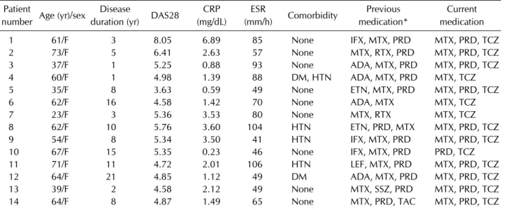
Table 1. Baseline clinical characteristics of the 14 patients with RA Patient
number Age (yr)/sex Disease
duration (yr) DAS28 CRP (mg/dL)
ESR
(mm/h) Comorbidity Previous medication*
Current medication
1 61/F 3 8.05 6.89 85 None IFX, MTX, PRD MTX, PRD, TCZ
2 73/F 5 6.41 2.63 57 None MTX, RTX, PRD MTX, PRD, TCZ
3 37/F 1 5.25 0.88 93 None ADA, MTX, PRD MTX, PRD, TCZ
4 60/F 1 4.98 1.39 88 DM, HTN ADA, MTX, PRD MTX, TCZ
5 35/F 8 3.63 0.59 49 None ETN, MTX, PRD MTX, PRD, TCZ
6 62/F 16 4.58 1.42 70 None ADA, MTX MTX, TCZ
7 23/F 3 5.36 3.53 80 None MTX, RTX MTX, TCZ
8 62/F 10 5.76 3.60 104 HTN ETN, PRD, MTX MTX, PRD, TCZ
9 54/F 8 5.34 3.50 41 HTN IFX, MTX, PRD MTX, PRD, TCZ
10 67/F 15 5.35 0.23 46 None IFX, MTX, PRD PRD, TCZ
11 71/F 11 4.72 2.01 106 HTN LEF, MTX, PRD MTX, PRD, TCZ
12 64/F 21 4.85 1.12 49 DM ADA, MTX, PRD MTX, PRD, TCZ
13 39/F 2 4.58 2.12 49 None MTX, SSZ, PRD MTX, PRD, TCZ
14 64/F 8 4.87 1.49 65 None MTX, PRD, TAC MTX, PRD, TCZ
ADA: adalimumab, CRP: C-reactive protein, DAS28: 28-joint disease activity score, DM: diabetes mellitus, ESR: erythrocyte sedimentation rate, ETN: etanercept, F: female, HTN: hypertension, IFX: infliximab, LEF: leflunomide, MTX: methotrexate, PRD:
prednisolone, RA: rheumatoid arthritis, RTX: rituximab, SSZ: sulfasalazine, TAC: tacrolimus, TCZ: tocilizumab. *Indicates recent medication used in RA patients 3 months before administration of tocilizumab.
Dickinson, San Diego, CA, USA); APC-conjugated anti- TCR Vα7.2 (BioLegend, San Diego, CA, USA); APC- Alexa Fluor 750-conjugated anti-CD3 (Beckman Coulter, Marseille, France). Cells were stained with the combina- tion of appropriate mAbs for 20 min at 4oC. Stained cells were analyzed on a Navios flow cytometer using Kaluza software (Beckman Coulter, Brea, CA, USA).
Statistical analysis
Wilcoxon’s signed rank test was used to compare changes in clinical parameters, hepcidin-25, anemia-re- lated factors and hematologic parameters during tocilizu- mab treatment period. Relationship between hepcidin levels and clinical parameters were examined using non- parametric Spearman rank sum correlation test. p-values less than 0.05 were considered statistically significant.
All statistical analyses were performed using SPSS ver.
18.0 software (SPSS, Chicago, IL, USA).
RESULTS
Patient characteristics
Fourteen RA patients were enrolled in this study and they were treated with tocilizumab during a six-month period.
The baseline clinical characteristics of the patients are summarized in Table 1. The mean values of clinical param- eters were as follows with mean±SD values: age
55.1±15.3 yr; disease duration 8.0±6.1 yr; hemoglobin (Hb) 11.1±1.1 g/dL; DAS28 5.27±1.03; ESR 70.1± 22.5 mm/h; and CRP 2.24±1.73 mg/dL (reference range: <0.3 mg/dL). According to the World Health Organization cri- teria for anemia (Hb levels of below 12.0 g/dL for women), 71.4% (10 of 14) RA patients who participated in this study were anemic. The patients had been previously treat- ed with: methotrexate (n=14); sulfasalazine (n=10); hy- droxychloroquine (n=8); leflunomide (n=6); tacrolimus (n=3); azathioprine (n=1); prednisolone (n=14); adali- mumab (n=13); etanercept (n=5); infliximab (n=5); and rituximab (n=5).
Improvement of disease activity in RA patients treated with tocilizumab during a six-month period
To evaluate the clinical effects of tocilizumab in active RA patients, TJC, SJC, VAS, ESR, CRP, and DAS28 were measured at baseline and months 1, 3, and 6 during tocili- zumab treatment. The median values of clinical parame- ters at baseline versus subsequent values at one month post tocilizumab were as follows (with p-values): 6.5 vs.
1.0 TJC (p<0.005); 7.5 vs. 3.0 SJC (p<0.005); 75.0 vs.
30.0 VAS (p<0.005); 67.5 vs. 10.0 mm/h ESR (p<
0.0005); 1.75 vs. 0.02 mg/dL CRP (p<0.0005); and 5.12 vs. 2.48 DAS28 (p<0.0005). One month after tocilizu- mab treatment, TJC, SJC, VAS, ESR, CRP and DAS28 were all significantly reduced compared with the base-
Figure 1. Clinical effects of tocilizumab in rheumatoid arthritis (RA) patients during six-month treatment period. Tocilizumab was administrated to 14 RA patients every four weeks. Disease activity was assessed at baseline and months 1, 3 and 6 during tocilizu- mab treatment. (A) Tender joint count (TJC). (B) Swollen joint count (SJC). (C) Visual analogue scale (VAS). (D) Erythrocyte sed- imentation rate (ESR). (E) C-reactive protein (CRP). (F) 28-joint disease activity score (DAS28). Symbols represent individual subjects. p-values were determined using Wilcoxon’s signed rank test. *p<0.005, **p<0.0005.
line. These clinical effects of tocilizumab lasted through- out the six-month period (Figure 1).
Effects of tocilizumab on hepcidin-25 and anemia- related factors in RA patients during a six-month period
To determine whether tocilizumab improves anemia in RA patients, anemia-related factors, such as serum levels of hepcidin-25, ferritin, iron, Hb and also red blood cell counts (RBCs), mean corpuscular volume (MCV), EPO and IL-6, were measured at baseline and months 1, 3, and 6 following start of tocilizumab treatment. The median values of anemia-related factors at baseline were as fol- lows: 20.3 ng/mL for serum hepcidin-25; 71.1 ng/mL for serum ferritin; 46.5 μg/dL for serum iron; 11.2 g/dL for Hb; 3.75×10⁶ cells/μL for RBC count; 92.4 fL for MCV;
15.7 mU/mL for EPO; and 13.4 pg/mL for IL-6. Six months after tocilizumab treatment, the median values of hepcidin-25, ferritin, iron, Hb, RBCs and MCV sig- nificantly changed as compared with the baseline.
However, no significant changes in EPO or IL-6 levels were found after tocilizumab treatment (Figure 2).
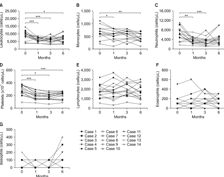
Effects of tocilizumab on hematological parame- ters in RA patients during a 6-month period
We investigated changes in hematological parameters in the 14 RA patients treated with tocilizumab during a six-month period. The median values of hematologic pa- rameters at baseline versus one month after tocilizumab treatment were 9,150 vs. 6,350 cells/μL (p<0.005) for leukocyte counts, 650 vs. 600 cells/μL (p<0.05) for monocyte counts, 6,300 vs. 3,550 cells/μL (p<0.01) for neutrophil counts, and 280×103 vs. 199×103 cells/μL (p<0.005) for platelet counts. One month after tocilizu- mab treatment, the median count values of leukocytes, monocytes, neutrophils and platelets were significantly reduced with respect to the baseline values. The tocilizu- mab effects lasted throughout the six-month period.
However, no significant changes in lymphocyte counts, eosinophil counts or basophil counts were found during
Figure 2. Effects of tocilizumab on hepcidin-25 and anemia-related factors in rheumatoid arthritis (RA) patients during the six-month treatment period. Tocilizumab was administrated to 14 RA patients every four weeks. Hepcidin-25 and anemia-related factors were measured at baseline and months 1, 3 and 6 during tocilizumab treatment. (A) Serum hepcidin-25. (B) Serum ferritin.
(C) Serum iron (Fe). (D) Hemoglobin (Hb). (E) Red blood cell counts (RBCs). (F) Mean corpuscular volume (MCV). (G) Erythropoietin (EPO). (H) Interleukin (IL)-6. Symbols represent individual subjects. p-values were determined using Wilcoxon’s signed rank test.
*p<0.05, **p<0.01, ***p<0.005, †p<0.001.
the six-month observation period (Figure 3).
To determine whether tocilizumab treatment influences proportions of lymphocyte subsets in peripheral blood, freshly isolated PBMCs from 14 RA patients treated with tocilizumab were stained with appropriate monoclonal antibodies, and then analyzed by flow cytometry. No sig- nificant changes in percentages of CD3+ T cells, CD4+
T cells, CD8+ T cells, B cells, NK cells, MAIT cells or NKT cells were found during the six-month observation period (Figure 4).
Relationships between serum hepcidin and clin- ical parameters in active RA patients
We monitored patient serum hepcidin levels at baseline
and during tocilizumab therapy with correlation levels calculated using Spearman’s coefficient. This was done in order to find significant correlations between hepcidin levels and other serum parameters and the subsequent changes following treatment with tocilizumab. Before tocilizumab therapy, serum hepcidin levels significantly correlated with serum ferritin and CRP levels (Spear- man’s correlation coefficient rs=0.657 [p<0.05] for fer- ritin; rs=0.547 [p<0.05] for CRP; Table 2). Moreover, a change from baseline in serum hepcidin level sig- nificantly correlated with changes from baseline in se- rum CRP, ESR, ferritin level, Hb level, leukocyte count, neutrophil count and RBC count throughout the treat- ment period (Spearman’s correlation coefficient rs=
Figure 3. Effects of tocilizumab on hematological parameters in rheumatoid arthritis (RA) patients during the six-month treatment period. Tocilizumab was administrated to 14 RA patients every four weeks. Hematologic parameters were measured at baseline and months 1, 3 and 6 during tocilizumab treatment. (A) Leukocyte count. (B) Monocyte counts. (C) Neutrophil counts. (D) Platelet counts. (E) Lymphocyte counts. (F) Eosinophil counts. (G) Basophil counts. Symbols represent individual subjects. p-values were determined using Wilcoxon’s signed rank test. *p<0.05, **p<0.01, ***p<0.005, †p<0.0005.
0.631 (p<0.0001); rs=0.351 (p<0.05); rs=0.499 (p
<0.005); rs=−0.434 (p<0.005); rs=0.404 (p<0.01);
rs=0.402 (p<0.01); and rs=−0.393 (p<0.05), re- spectively; Table 3). However, no significant correlations were found for changes of serum hepcidin level and DAS28, TJC, SJC, VAS, serum iron, MCV, EPO, IL-6 lev- els, lymphocyte count, monocyte count, eosinophil count, basophil count, or platelet count values from baseline to each time point during the treatment period (Table 3).
DISCUSSION
The present study is a first attempt to investigate clinical and hematological effects of tocilizumab in Korean pa-
tients with active RA. IL-6 has been reported to induce acute phase proteins, including hepcidin which is known as a key mediator of anemia of inflammation [24]. In sup- port of this mechanistic view, our data showed that tocili- zumab induced rapid and sustained reduction in hepcidin serum levels and subsequently improved other anemia- related factors as well, such as Hb, serum iron, MCV, and RBC count. In addition, tocilizumab was found to im- prove disease activity within one month after tocilizumab treatment and sustain this effect throughout a six-month period. However, lymphocytes and their subset levels were not affected by tocilizumab, which is in contrast to the notion that IL-6 differentiates T and B cells in RA [3].
The observation that tocilizumab improves disease ac- tivity has also been reported in Castleman disease [14],
Figure 4. Effects of tocilizumab on lymphocyte subsets in rheumatoid arthritis (RA) patients during the six-month treatment period.
Tocilizumab was administrated to 14 RA patients every four weeks. Percentages of lymphocyte subsets were measured at baseline and 6 months after tocilizumab treatment. Freshly isolated peripheral blood mononuclear cells (PBMCs) from 14 RA patients were stained with appropriate monoclonal antibodies and then analyzed by flow cytometry. (A) CD3+ T cells. (B) CD4+ T cells. (C) CD8+ T cells. (D) CD3−CD19+ B cells. (E) Natural killer (NK; CD3−CD56+) cells. (F) Mucosal associated invariant T (MAIT;
CD3+TCRγδ−Vα7.2+CD161high) cells. (G) Natural killer T (NKT; CD3+6B11+) cells. Symbols represent individual subjects. p-values were determined using Wilcoxon’s signed rank test.
systemic-onset juvenile idiopathic arthritis [25] and RA [16,17]. In the present study, we found that tocilizumab treatment resulted in a progressive decrease in TJC, SJC, VAS, ESR, CRP and DAS28 values in all active RA pa- tients during the treatment period, indicating that tocili- zumab induces early and sustained reductions in disease activity in RA patients. In a previous study, an early reduc- tion in CRP was observed within one week after admin- istration of tocilizumab [16]. In the present study, all the treated RA patients reached CRP normalization and DAS28 remission levels by 1 month and by 6 months, re- spectively, suggesting that CRP values, more early than DAS28, may reflect inflammation levels during the period
of tocilizumab therapy.
Hepcidin has emerged as a key regulator of iron homeo- stasis that is mainly regulated by IL-6 as part of the patho- genesis of ACD [3]. ACD is known as the most frequent cause of anemia in RA [17]. It can be postulated that IL-6 induced-hepcidin binds and degrades ferroportin, which in turn results in a decrease in iron export from enter- ocytes and macrophages into blood. As a consequence, serum iron decreases while iron stored within macro- phages increases, leading to elevated serum ferritin levels [26]. Interestingly, before tocilizumab therapy, serum hepcidin level in our RA patients showed a strong correla- tion with serum ferritin and CRP levels, possibly indicat-
Table 2. Spearman’s correlation coefficients for serum hep- cidin-25 with respect to clinical and laboratory parameters in 14 RA patients before tocilizumab therapy
Variable Correlation
coefficient (γs) p-value
CRP (mg/dL) 0.547 0.043
ESR (mm/h) 0.285 0.324
DAS28 0.244 0.400
Tender joint count 0.107 0.715 Swollen joint count 0.116 0.692
Visual analogue scale −0.109 0.712
Ferritin (ng/mL) 0.657 0.011
Iron (μg/dL) −0.029 0.923
Hemoglobin (g/dL) −0.150 0.610
Leukocyte (cells/μL) 0.257 0.375
Lymphocyte (cells/μL) 0.135 0.646
Monocyte (cells/μL) 0.049 0.868
Neutrophil (cells/μL) 0.081 0.782
Eosinophil (cells/μL) 0.290 0.315
Basophil (cells/μL) −0.140 0.633
Platelet (×103 cells/μL) −0.108 0.714 RBCs (×106 cells/μL) −0.152 0.605
MCV (fL) −0.099 0.737
EPO (mU/mL) −0.002 0.994
IL-6 (pg/mL) 0.160 0.584
CRP: C-reactive protein, DAS28: 28-joint disease activity score, EPO: erythropoietin, ESR: erythrocyte sedimentation rate, IL-6: interleukin-6, MCV: mean corpuscular volume, RA:
rheumatoid arthritis, RBC: red blood cell.
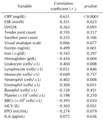
Table 3. Spearman’s correlation coefficients for changes from baseline in serum hepcidin-25 with respect to changes from baseline in clinical and laboratory parameters in 14 RA patients treated with tocilizumab over 6-month treatment
Variable Correlation
coefficient (γs) p-value
CRP (mg/dL) 0.631 <0.0001
ESR (mm/h) 0.351 0.023
DAS28 0.263 0.093
Tender joint count 0.195 0.217 Swollen joint count 0.253 0.106 Visual analogue scale 0.066 0.677
Ferritin (ng/mL) 0.499 0.001
Iron (μg/dL) −0.165 0.297
Hemoglobin (g/dL) −0.434 0.004
Leukocyte (cells/μL) 0.404 0.008
Lymphocyte (cells/μL) −0.031 0.846
Monocyte (cells/μL) −0.049 0.757
Neutrophil (cells/μL) 0.402 0.008
Eosinophil (cells/μL) 0.304 0.051
Basophil (cells/μL) −0.120 0.451
Platelet (×103 cells/μL) 0.198 0.210
RBCs (×106 cells/μL) −0.393 0.010
MCV (fL) −0.303 0.051
EPO (mU/mL) −0.274 0.079
IL-6 (pg/mL) 0.075 0.636
CRP: C-reactive protein, DAS28: 28-joint disease activity score, EPO: erythropoietin, ESR: erythrocyte sedimentation rate, IL-6: interleukin-6, MCV: mean corpuscular volume, RA:
rheumatoid arthritis, RBC: red blood cell.
ing a link between hepcidin levels and iron accumulation under inflammatory condition that bring about anemia in RA. Our results are consistent with those of previous studies in Castleman’s disease and RA [14,16,17].
Our longitudinal study demonstrated that blocking IL-6 pathway by tocilizumab induced rapid and sustained de- clines in serum hepcidin and ferritin levels that led to in- creases in gauge factors for anemia- iron levels, Hb con- centrations, RBC counts, and MCV values. A change from baseline in serum hepcidin level was observed to sig- nificantly correlate with changes in the above mentioned factors for anemia and these changes were maintained throughout the treatment period. These observations led us to speculate that improvement of anemia was due to improved iron utilization by a hepcidin level reduction.
Consistent with previous studies [16,17], these findings indicate that serum hepcidin may reflect inflammation and anemia, in particular during long-term tocilizumab therapy in RA patients. Previous studies have reported a significant positive correlation between serum hepcidin
levels and DAS28 score, suggesting that serum hepcidin could be a new surrogate biomarker of RA [17,27-29]. In the present study, however, no significant correlation was found between serum hepcidin level and disease activity as assessed by DAS28 score. This discrepancy may be due to the use of a different hepcidin kit from the previous studies and small sample size of our study.
Additional studies have reported that human IL-6 in- duced both thrombocytosis by increased thrombocytopoi- esis and leukocytosis by demargination of intravascular neutrophils and accelerated bone marrow release of the newly generated cells [30,31]. These studies suggested that IL-6 plays a key role in hematopoiesis [30,31]. We found that tocilizumab therapy induced rapid and sus- tained reductions in leukocyte, neutrophil, monocyte, and platelet counts throughout the six-month treatment peri- od, which is consistent with the previous findings [30,31].
In the present study, however, counts of lymphocyte, eosi- nophil, basophil, and lymphocyte subsets including T, B,
NK, NKT, and MAIT cells were not significantly changed during the six-month treatment period. There have been a number of reports that IL-6 may play an important role in the pathogenesis of autoimmunity via the development of antibody-producing plasma B cells and Th17 cells [32,33].
Indeed tocilizumab treatment was shown to decrease the frequency of circulating plasma cells in SLE patients [34].
We believe that further studies are needed to determine which lymphoid cell subsets could be specifically affected by blocking the IL-6 signaling pathway.
Our data revealed that no correlation exists between se- rum hepcidin and IL-6 levels before tocilizumab treat- ment and this is consistent with a number of previous studies [17,27]. One recent study, however, contrasted this as serum hepcidin levels showed a significant pos- itive correlation with IL-6 [16]. This controversy may be due to the presence of confounding regulatory factors of hepcidin production such as hypoxia, anemia, and iron deficiency all of which inhibit hepcidin synthesis [26,35].
A previous study has reported that serum IL-6 level in- creased after tocilizumab treatment in patients with Castleman’s disease and RA [36]. In our study, no overall changes in serum IL-6 levels were found, although the IL-6 levels were quite variable among the patients. The reason for this discrepancy is currently unclear, but one possible explanation is that tocilizumab is known to in- hibit IL-6R-mediated elimination of IL-6 and such varia- bility in serum IL-6 levels may reflect the patient-specific differences in production and degradation rates of IL-6 af- ter administration of tocilizumab [36].
CONCLUSION
In summary, our study demonstrates that tocilizumab reduces disease-activity in patients with active RA patients. In addition, tocilizumab improved inflamma- tory anemia by inhibiting hepcidin production. This clin- ical data provides additional evidence of an important role for IL-6 signaling in the pathogenesis of RA.
ACKNOWLEDGMENTS
This study was supported by a grant from the National Research Foundation of Korea funded by the Korean Go- vernment (#2013R1A2A2A01067956) and the Chon- nam National University Hospital Biomedical Research Institute (CRI13905-22.3, CRI14039-21 and CRI14039- 22).
CONFLICT OF INTEREST
No potential conflict of interest relevant to this article was reported.
REFERENCES
1. McInnes IB, Schett G. Cytokines in the pathogenesis of rheumatoid arthritis. Nat Rev Immunol 2007;7:429-42.
2. McInnes IB, Schett G. The pathogenesis of rheumatoid arthritis. N Engl J Med 2011;365:2205-19.
3. Hashizume M, Mihara M. The roles of interleukin-6 in the pathogenesis of rheumatoid arthritis. Arthritis 2011;2011:
765624.
4. Wilson A, Yu HT, Goodnough LT, Nissenson AR. Preva- lence and outcomes of anemia in rheumatoid arthritis: a sys- tematic review of the literature. Am J Med 2004;116 Suppl 7A:50S-7S.
5. Masson C. Rheumatoid anemia. Joint Bone Spine 2011;78:
131-7.
6. Voulgari PV, Kolios G, Papadopoulos GK, Katsaraki A, Seferiadis K, Drosos AA. Role of cytokines in the patho- genesis of anemia of chronic disease in rheumatoid arth- ritis. Clin Immunol 1999;92:153-60.
7. Romano M, Sironi M, Toniatti C, Polentarutti N, Fruscella P, Ghezzi P, et al. Role of IL-6 and its soluble receptor in in- duction of chemokines and leukocyte recruitment. Immuni- ty 1997;6:315-25.
8. Hashizume M, Hayakawa N, Mihara M. IL-6 trans-signal- ling directly induces RANKL on fibroblast-like synovial cells and is involved in RANKL induction by TNF-alpha and IL-17. Rheumatology 2008;47:1635-40.
9. Hashizume M, Hayakawa N, Suzuki M, Mihara M.
IL-6/sIL-6R trans-signalling, but not TNF-alpha induced angiogenesis in a HUVEC and synovial cell co-culture system. Rheumatol Int 2009;29:1449-54.
10. Madhok R, Crilly A, Watson J, Capell HA. Serum inter- leukin 6 levels in rheumatoid arthritis: correlations with clinical and laboratory indices of disease activity. Ann Rheum Dis 1993;52:232-4.
11. Nemeth E, Rivera S, Gabayan V, Keller C, Taudorf S, Pedersen BK, et al. IL-6 mediates hypoferremia of in- flammation by inducing the synthesis of the iron regulatory hormone hepcidin. J Clin Invest 2004;113:1271-6.
12. Roy C, Andrews NC. Anemia of inflammation: the hepcidin link. Curr Opin Hematol 2005;12:107-11.
13. Kawabata H, Tomosugi N, Kanda J, Tanaka Y, Yoshizaki K, Uchiyama T. Anti-interleukin 6 receptor antibody tocilizu- mab reduces the level of serum hepcidin in patients with multicentric Castleman's disease. Haematologica 2007;92:
857-8.
14. Song S, Tomosugi N, Kawabata H, Ishikawa T, Nishikawa T, Yoshizaki K. Down-regulation of hepcidin resulting from long-term treatment with an anti-IL-6 receptor antibody (tocilizumab) improves anemia of inflammation in multi- centric Castleman disease. Blood 2010;116:3627-34.
15. Hashizume M, Uchiyama Y, Horai N, Tomosugi N, Mihara M. Tocilizumab, a humanized anti-interleukin-6 receptor antibody, improved anemia in monkey arthritis by sup-
pressing IL-6-induced hepcidin production. Rheumatol Int 2010;30:917-23.
16. Isaacs JD, Harari O, Kobold U, Lee JS, Bernasconi C. Effect of tocilizumab on haematological markers implicates inter- leukin-6 signalling in the anaemia of rheumatoid arthritis.
Arthritis Res Ther 2013;15:R204.
17. Song SN, Iwahashi M, Tomosugi N, Uno K, Yamana J, Yamana S, et al. Comparative evaluation of the effects of treatment with tocilizumab and TNF-α inhibitors on se- rum hepcidin, anemia response and disease activity in rheu- matoid arthritis patients. Arthritis Res Ther 2013;15:R141.
18. Aletaha D, Neogi T, Silman AJ, Funovits J, Felson DT, Bingham CO 3rd, et al. 2010 Rheumatoid arthritis classi- fication criteria: an American College of Rheumatology/
European League Against Rheumatism collaborative initiative. Arthritis Rheum 2010;62:2569-81.
19. Park YW, Kee SJ, Cho YN, Lee EH, Lee HY, Kim EM, et al.
Impaired differentiation and cytotoxicity of natural killer cells in systemic lupus erythematosus. Arthritis Rheum 2009;60:1753-63.
20. Cho YN, Kee SJ, Lee SJ, Seo SR, Kim TJ, Lee SS, et al.
Numerical and functional deficiencies of natural killer T cells in systemic lupus erythematosus: their deficiency re- lated to disease activity. Rheumatology (Oxford) 2011;50:
1054-63.
21. Cho YN, Kee SJ, Kim TJ, Jin HM, Kim MJ, Jung HJ, et al.
Mucosal-associated invariant T cell deficiency in systemic lupus erythematosus. J Immunol 2014;193:3891-901.
22. Lee SJ, Cho YN, Kim TJ, Park SC, Park DJ, Jin HM, et al.
Natural killer T cell deficiency in active adult-onset Still's disease: correlation of deficiency of natural killer T cells with dysfunction of natural killer cells. Arthritis Rheum 2012;64:2868-77.
23. Jin HM, Kee SJ, Cho YN, Kang JH, Kim MJ, Jung HJ, et al.
Dysregulated osteoclastogenesis is related to natural killer T cell dysfunction in rheumatoid arthritis. Arthritis Rheu- matol 2015;67:2639-50.
24. Nemeth E, Valore EV, Territo M, Schiller G, Lichtenstein A, Ganz T. Hepcidin, a putative mediator of anemia of in- flammation, is a type II acute-phase protein. Blood 2003;
101:2461-3.
25. Yokota S, Miyamae T, Imagawa T, Iwata N, Katakura S, Mori M, et al. Therapeutic efficacy of humanized recombinant an- ti-interleukin-6 receptor antibody in children with sys- temic-onset juvenile idiopathic arthritis. Arthritis Rheum 2005;52:818-25.
26. van Santen S, van Dongen-Lases EC, de Vegt F, Laarakkers CM, van Riel PL, van Ede AE, et al. Hepcidin and hemoglo- bin content parameters in the diagnosis of iron deficiency in rheumatoid arthritis patients with anemia. Arthritis Rheum 2011;63:3672-80.
27. Koca SS, Isik A, Ustundag B, Metin K, Aksoy K. Serum pro-hepcidin levels in rheumatoid arthritis and systemic lu- pus erythematosus. Inflammation 2008;31:146-53.
28. Kim HR, Kim KW, Yoon SY, Kim SH, Lee SH. Serum pro-hepcidin could reflect disease activity in patients with rheumatoid arthritis. J Korean Med Sci 2010;25:348-52.
29. Sellam J, Kotti S, Fellahi S, Bastard JP, Meyer M, Lioté F, et al. Serum hepcidin level is not an independent surrogate bi- omarker of disease activity or of radiographic progression in rheumatoid arthritis: results from the ESPOIR cohort. Ann Rheum Dis 2013;72:312-4.
30. Maslak P, Nimer SD. The efficacy of IL-3, SCF, IL-6, and IL-11 in treating thrombocytopenia. Semin Hematol 1998;
35:253-60.
31. Suwa T, Hogg JC, English D, Van Eeden SF. Interleukin-6 induces demargination of intravascular neutrophils and shortens their transit in marrow. Am J Physiol Heart Circ Physiol 2000;279:H2954-60.
32. Muraguchi A, Hirano T, Tang B, Matsuda T, Horii Y, Nakajima K, et al. The essential role of B cell stimulatory fac- tor 2 (BSF-2/IL-6) for the terminal differentiation of B cells.
J Exp Med 1988;167:332-44.
33. Zhou L, Ivanov II, Spolski R, Min R, Shenderov K, Egawa T, et al. IL-6 programs T(H)-17 cell differentiation by promot- ing sequential engagement of the IL-21 and IL-23 pathways.
Nat Immunol 2007;8:967-74.
34. Illei GG, Shirota Y, Yarboro CH, Daruwalla J, Tackey E, Takada K, et al. Tocilizumab in systemic lupus eryth- ematosus: data on safety, preliminary efficacy, and impact on circulating plasma cells from an open-label phase I dos- age-escalation study. Arthritis Rheum 2010;62:542-52.
35. Nicolas G, Chauvet C, Viatte L, Danan JL, Bigard X, Devaux I, et al. The gene encoding the iron regulatory peptide hepci- din is regulated by anemia, hypoxia, and inflammation. J Clin Invest 2002;110:1037-44.
36. Nishimoto N, Terao K, Mima T, Nakahara H, Takagi N, Kakehi T. Mechanisms and pathologic significances in in- crease in serum interleukin-6 (IL-6) and soluble IL-6 re- ceptor after administration of an anti-IL-6 receptor anti- body, tocilizumab, in patients with rheumatoid arthritis and Castleman disease. Blood 2008;112:3959-64.