ORIGINAL ARTICLE
수술 전 위암조직에서의 Cadherin과 혈관내피 성장 인자 농도의 의의
유금혜, 심기남1, 정성애1, 유 권1, 주양희1, 이주호2
국립암센터 암예방검진센터, 이화여자대학교 의학전문대학원 내과학교실1, 외과학교실2
Significance of Preoperative Tissue Levels of Vascular-endothelial Cadherin, Liver-intestine Cadherin and Vascular Endothelial Growth Factor in Gastric Cancer
Kum Hei Ryu, Ki-Nam Shim1, Sung-Ae Jung1, Kwon Yoo1, Yang-Hee Joo1 and Joo Ho Lee2
Center for Cancer Prevention and Detection, National Cancer Center, Goyang, Departments of Internal Medicine1 and Surgery2, Ewha Medical Research Institute, Ewha Womans University School of Medicine, Seoul, Korea
Background/Aims: The aims of this study were to examine the expressions of endothelium specific VE-cadherin, intestine specific LI-cadherin, and vascular endothelial growth factor (VEGF), and to determine their relationships with the clinicopathological parameters of gastric cancer.
Methods: A total 47 patients with gastric cancer who underwent surgery were enrolled. Endoscopic biopsies were obtained from the cancer and normal mucosa, respectively. Using semiquantitative RT-PCR, the mRNA expression levels of VE-cadherin, LI-cadherin and VEGF were measured by tumor/normal (T/N) ratios. The protein expressions of VE-cadherin, LI-cadherin and VEGF were examined by Western blot and immunohistochemical stain in surgically resected tissues. The clinicopathological variables were reviewed and analyzed, retrospectively.
Results: Twenty two cases (46.8%) of VE-cadherin, 25 cases (53.2%) of LI-cadherin and 27 cases (51.1%) of VEGF mRNA expressions were overexpressed in gastric cancer compared to normal tissue. There was a tendency for T/N ratio of VE-cadherin mRNA to correlate with the lymphatic invasion (p=0.07) and the lymph node metastasis (p=0.099) in advanced gastric cancer.
The T/N ratio of LI-cadherin mRNA showed significant association with distant metastasis (p=0.031) and lymphatic invasion especially in advanced gastric cancer (p=0.023). There was a tendency for the T/N ratio of VEGF mRNA to correlate with the distant metastasis (p=0.073) in advanced gastric cancer.
Conclusions: As increased mRNA expression of LI-cadherin was associated with distant metastasis and lymphatic invasion especially in the biopsy specimen of advanced gastric cancer before surgery, it may provide useful preoperative information on tumor aggressiveness. (Korean J Gastroenterol 2012;60:229-241)
Key Words: Cadherin; Vascular endothelial growth factor; Stomach neoplasms
Received April 2, 2012. Revised May 15, 2012. Accepted May 17, 2012.
CC This is an open access article distributed under the terms of the Creative Commons Attribution Non-Commercial License (http://creativecommons.org/licenses/
by-nc/3.0) which permits unrestricted non-commercial use, distribution, and reproduction in any medium, provided the original work is properly cited.
교신저자: 심기남, 158-710, 서울시 양천구 목동 911-1, 이화여자대학교 의학전문대학원 내과학교실
Correspondence to: Ki-Nam Shim, Department of Internal Medicine, Ewha Womans University School of Medicine, 911-1 Mok-dong, Yangcheon-gu, Seoul 158-710, Korea. Tel: +82-2-2650-2632, Fax: +82-2-2655-2076, E-mail: [email protected]
Financial support: None. Conflict of interest: None.
INTRODUCTION
Gastric cancer is the most common cancer and the third most common cause of cancer-related death in Korea, ac- counting for 26,253 new cases in 2007 and 10,779 deaths
in 2006.1 The overall gastric cancer incidence is decreasing, possibly due to the decrease in Helicobacter pylori preva- lence associated with improved living standards. However, there are still many patients diagnosed with advanced stage disease, for which the prognosis is poor. In spite of intense in-
terest and extensive investigations, the prognosis for such cases has not improved significantly in the past several years.2 Therefore, early detection and curative resection of gastric cancer is still the most important issue.
The most frequently used tumor marker, for gastric cancer, is the carcinoembryonic antigen (CEA). However, this marker is elevated only in a modest proportion of advanced gastric cancer. It is essential to evaluate preoperative clinical stages, and the depth of tumor invasion to determine appropriate treatment modality and predict the prognosis of gastric can- cer, but no reliable methods or biologic markers that can pre- dict metastasis and vessel invasion have been found until now.2-4
Adhesion molecules such as the cadherins are considered important for the invasion and/or metastasis of tumors.
Cadherins are calcium dependent homophilic adhesion mol- ecules frequently associated with specific junctional struc- tures referred to as adherens junctions. Cadherins are ex- pressed in several types of tissues with some specificity:
E-cadherin is mostly present in epithelial cells, N-cadherin in the nervous system, smooth muscle cells, fibroblasts and en- dothelial cells, vascular-endothelial cadherin (VE-cadherin) is specific for the endothelium and related with vasculo- genesis, and liver-intestine cadherin (LI-cadherin) is found in the intestinal epithelium.5
The major transmembrane component of endothelial ad- herens junctions is VE-cadherin. VE-cadherin has been im- plicated in cell growth, migration, vasculogenesis and vas- cular remodeling.6,7 Angiogenesis is important in tumori- genesis, so the assessment of angiogenesis may provide sig- nificant information of cancer behavior.
LI-cadherin is solely expressed in the liver and intestine of the rat. Human LI-cadherin is exclusively found in the in- testinal epithelium and the stomach is also negative for LI-cadherin.8 LI-cadherin was recently found to be overex- pressed in gastric cancer and the expression of LI-cadherin was associated with intestinal metaplasia.3,8,9 Gastric can- cer is generally believed to develop from a multistep pro- gression from gastritis, intestinal metaplasia, dysplasia and subsequently to cancer.4 The expression of LI-cadherin may therefore occur during the steps of gastric carcinogenesis.
Vascular endothelial growth factor (VEGF) is a key regulator of angiogenesis and induces proliferation of the endothelium along with the formation of new capillary vessels that loosen
the tight adherens junctions and induce proliferation of the endothelium contributing to tubulogenesis and increased vascular permeability.10,11
Since there are few reports of VE-cadherin and LI-cadherin which can predict the characteristics of gastric cancer, we performed this study to examine the expression of VE-cad- herin, LI-cadherin and VEGF in gastric cancer and to evaluate the correlation between their expression and clinicopatho- logical parameters.
SUBJECTS AND METHODS
1. Materials
Tissue specimens from 47 patients with gastric cancer who were diagnosed and underwent a surgery at Ewha Womens University Mokdong Hospital, Seoul, Korea, from 2005 to 2008, were obtained by means of endoscopic biopsy. At least 4 pieces of endoscopic biopsies were taken from the cancer and macroscopically normal mucosa, at least 5 cm away from the cancer, respectively before defini- tive treatment. The collected specimens were frozen immedi- ately and stored at −70oC until extraction of mRNA to de- termine the levels of VE-cadherin, LI-cadherin and VEGF.
Patients had underwent a gastrectomy procedure in the de- partment of surgery. During the surgery, tissues of the cancer and normal mucosa were obtained. The collected tissues were frozen immediately and stored at −70oC for the protein analysis. The pathologic diagnoses for endoscopically biop- sied specimens were matched with those of the surgically re- sected tissues, and a consultant pathologist determined the pathologic staging. Preoperatively the stage was determined by endoscopic and computed tomographic findings. We de- termined the postoperative pathologic stage using the 6th edition of the International Union against Cancer (UICC)/
American Joint Committee on Cancer (AJCC) TNM classi- fication system. The clinicopathological data from 47 pa- tients who underwent a surgical procedure were reviewed.
Informed consents were obtained from all patients and the study was approved by the Institutional Review Board of Ewha Womans University Mokdong Hospital.
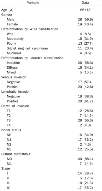
The patients were consisted of 28 men and 19 women ranging in age from 35 to 88 years (65±13 years [mean±
SD]). Histologically, 4 cases (8.5%) were well differentiated, 15 (31.9%) were moderately differentiated, 13 (27.7%) were
Table 1. Characteristics of the Patients
Variable Data
Age (yr) 65±13
Gender
Male 28 (59.6)
Female 19 (40.4)
Differentiation by WHO classification
Well 4 (8.5)
Moderately 15 (31.9)
Poorly 13 (27.7)
Signet ring cell carcinoma 11 (23.4)
Mucinous 4 (8.5)
Differentiation by Lauren's classification
Intestine 26 (55.3)
Diffuse 16 (34.1)
Mixed 5 (10.6)
Venous invasion
Negative 27 (57.4)
Positive 20 (42.6)
Lymphatic invasion
Negative 18 (38.3)
Positive 29 (61.7)
Depth of invasion
T1 12 (25.5)
T2 7 (14.9)
T3 26 (55.3)
T4 2 (4.3)
Nodal status
N0 16 (34.0)
N1 17 (36.2)
N2 2 (4.3)
N3 12 (25.5)
Distant metastasis
M0 40 (85.1)
M1 7 (14.9)
Stage
I 14 (29.7)
II 6 (12.8)
III 10 (21.3)
IV 17 (36.2)
Values are presented as mean±SD or n (%).
WHO, World Health Organization. Stages are determined by the 6th edition of International Union against Cancer/American Joint Committee on Cancer TNM classification System.
poorly differentiated, 11 (23.4%) were signet ring cell carci- noma, and 4 (8.5%) were mucinous type. Twenty six cases had intestinal type of differentiation and 16 (34.1%) were dif- fuse type according to Lauren's classification. Twenty cases (42.6%) had venous invasion and 29 cases (61.7%) had lym- phatic invasion. Of the 47 cancer cases, 12 (25.5%) were classified as T1, 7 (14.9%) were T2, 26 (55.3%) were T3, and 2 (4.3%) were T4; 16 cases (34.0%) were N0, 17 (36.2%) were N1, 2 (4.3%) were N2 and 12 (25.5%) were N3; 40 cases (85.1%) were M0 and 7 (14.9%) of M1. If we consider the TNM stage on the whole, 14 cases (29.8%) were in stage I, 6 (12.8%) were stage II, 10 (21.3%) were stage III and 17 cases (36.2%) were in stage IV (Table 1).
2. Semi-quantitative reverse transcript-polymerase chain reaction
1) RNA extraction
Total RNA was extracted from biopsy tissue using the easy-BLUETM total RNA extraction kit (Intron Biotechnology, Gyeonggido, Korea). Preperation of fresh tissues were added to 800 μL easy-BLUETM reagent and homogenized using a ho- mogenizer or equivalent. Vigorous vortex was applied in room temperature for 3 minutes. Then, 200 μL of chloroform was added and vortex was applied. After centrifuge the solution at 12,000 rpm (4oC) for 15 minutes, 400 μL of supernatant was transferred to an empty 1.5 mL tube. Then, 400 μL iso- propanol (2-propanol) was added, and mixed it well by invert- ing the tube 5-6 times. It was left for 5 minutes at room temperature. After centrifugating the solution at 12,000 rpm (4oC) for 10 minutes, remove the supernatant to obtain RNA pellet. Then, 1 mL of 75 % ethanol was added and the solution mixed well by inverting the tube 2-3 times. The mixtures were centrifuged for 5 minutes at 12,000 rpm (4oC). The super- natant discarded, and the remaining RNA pellet was dried.
RNA was dissolved using 20-50 μL of RNase, Dnase free wa- ter for storage at −70oC. The amount and purity of the ex- tracted RNA was quantitated by spectrophotometry.
2) cDNA synthesis (reverse transcription)
cDNA was synthesized with 2 μg of total RNA, oligodT primer. In a sterile RNase-free microcentrifuge tube, add 0.5 μg of oligodT primer and 2 μg RNA sample. The tube was heat- ed at 70oC for 5 minutes, and cooled immediately on ice.
Moloney Murine Leukemia Virus Reverse Transcriptase (M-MLV RT; Promega, Fitchburg, WI, USA) 200 unit, rRNasin
Ribonuclease inhibitor (Promega) 40 unit, 5×RT buffer and dNTP were added to the tube. The tube was gently mixed, in- cubated for 40 minutes at 42oC and heated for 5 minutes at 95oC. The cDNA was stored at −20oC.
3) Polymerase chain reaction amplification
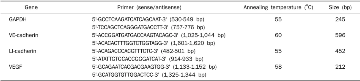
The primer sequences for amplification which were used are shown in Table 2. All primers were synthesized by Cosmo GeneTech (Seoul, Korea). The amplification reaction was car- ried out in a 20 μL of PCR mixture containing 20 μL of the syn-
Table 2. Primers of the Genes
Gene Primer (sense/antisense) Annealing temperature (oC) Size (bp)
GAPDH 5'-GCCTCAAGATCATCAGCAAT-3' (530-549 bp) 55 245
5'-TCCAGCTCAGGGATGACCTT-3' (757-776 bp)
VE-cadherin 5'-ACCGGATGATGACCAAGTACAGC-3' (1,025-1,044 bp) 60 596
5'-ACACACTTTGGTCTGGTAGG-3' (1,601-1,620 bp)
LI-cadherin 5'-ACAGACCCACGTTTCTC-3' (482-501 bp) 55 452
5'-ATATTGTGCACCGGGATCAT-3' (914-933 bp)
VEGF 5'-GCAGAATCACGACGAAGTGG-3' (1,133-1,152 bp) 58 212
5'-GCATGGTGTTGGACTCC-3' (1,325-1,344 bp)
GAPDH, glyceradlehyde 3-phosphate dehydrogenase; VE-cadherin, vascular-endothelial cadherin; LI-cadherin, liver-intestine cadherin; VEGF, vascular endothelial growth factor.
thesized cDNA solution, 4 μL of 5×polymerase reaction buf- fer, 200 μM of dNTP, 0.5 μM of each primer (sense and anti- sense) and 0.5 unit Taq polymerase (Promega). The PCR mix- ure was amplified using a GeneAmp PCR System 9600 (PERKIN-ELMER, Wellesley, MA, USA). Amplified products (10 μL) were identified by electrophoresis of PCR on 2 % agar- ose gel containing ethidium bromide and ultraviolet illumination. Forty seven endoscopic biopsy tissues were an- alyzed for mRNA expression. The expression of VE-cadherin, LI-cadherin and VEGF was evaluated using the tumor: normal (T/N) ratio for VE-cadheirn, LI-cadherin and VEGF. The results are expressed as the mean±SD for gastric cancer tissues.
3. Western blot analysis
Twenty four sets of cancerous and normal gastric tissue samples which were obtained by the surgical procedure for protein analysis were frozen immediately and kept at −70oC until use. The tissues were washed with 300 μL ice-cold phos- phate-buffered saline solution (PBS) and then washed into ly- sis buffer (ProprepTM, protein extraction solutionⓇ, Intron Biotechnology, Gyunggido, Korea). After incubation for 20 mi- nutes on ice, tissue lysates centrifuged at 12,000 rpm for 20 minutes at 4oC. The resulting supernatant was assayed for the protein concentration to ensure that a consistent amount of protein assay (BCA protein assay reagent A and BⓇ (50 : 1);
Pierce, Rockford, IL, USA)
After determination of protein concentrations, protein ex- tracts 50 μg were subjected to 10% sodium dodecyl sul- fate-polyacrylamide gel electrophoresis for 1.5 hours at 100 V room temperature. The fractionated proteins were trans- ferred overnight at 4oC onto the polyvinyl difluride membrane at 80 mA. Nonspecific binding of proteins was blocked by in- cubating the membranes in 5% skim milk in PBS containing
0.1% Tween 20 (PBSCT) for 120 minutes. The membranes were incubated with goat anti-VE-cadherin antibody, goat an- ti LI-cadherin and rabbit anti-VEGF antibody with the appro- priate dilution for overnight at 4oC, and then the membranes were rinsed three times with PBSCT at 5 minute intervals. The membranes were incubated with seondary antibody for 2 hours at room temperature. After the membranes were rinsed three times with PBST at 5 minute intervals, the blots were developed using the ECL western blotting detection sol- ution (ECLTM Western blotting detection reagents; Amersham Biosciences, Buckinghashire, UK). The blots were detected by LAS system (LAS-3000, Fugifilm, Seoul, Korea). The re- sults of western blotting were standardized with the density of glyceraldehyde 3-phosphate dehydrogenase (GAPDH).
4. Immunohistochemical study
Paraffin embedded 18 samples of gastric cancer tissue which were obtained by the surgical procedure were used for immunohistochemical study. Immunohistochemistry was performed through the biotin-streptavidin (B-SA) method us- ing the labelled streptavidin biotin (LSAB) kit (Dakocytomat- ion Inc., Carpinteria, CA, USA). The paraffinized tissues were sectioned 4 μm thickness and deparaffinized with xylene twice for 5 minutes. The tissues were rehydrated serially with 100%, 90%, 80% and 70% alcohol for 5 minutes and washed with distilled water. Antigen retrival condition included heat-induced epitope retrival in 10 nm/L citrate buffer (pH 6.0) for 7 minutes.
The endogenous peroxidase activity was blocked by in- cubation in 3 % hydrogen peroxide solution for 5 minutes. The tissue was incubated with the monoclonal anti-VE-cadhrin (SA-6452, dilution 1:200; Santa Cruz Biotechnology, Santa Cruz, CA, USA), anti-LI-cadherin (SC-6974, dilution 1:200;
Fig. 1. RT-PCR analysis of vascular-endothelial cadherin (VE-cad- herin) in gastric cancer tissue (C) and normal tissue (N). Re- presentative RT-PCR cases were shown. GAPDH, glyceraldehyde 3-phosphate dehydrogenase.
Fig. 2. Western blot analysis of vascular-endothelial cadherin (VE-cadherin) in gastric cancer tissue (C) and normal tissue (N).
Representative cases were shown. GAPDH, glyceraldehyde 3-phos- phate dehydrogenase.
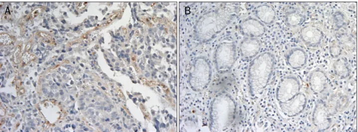
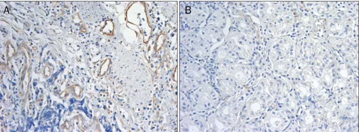
Fig. 3. Immunohistochemical staining with vascular-endothelial cadherin (VE-cadherin) (×200). (A) Immune staining was evident on vascular endothelial cells in gastric cancer tissues. (B) Immune reactivity of VE-cadherin was not detected in normal gastric tissues.
Santa Cruz Biotechnology), and anti-VEGF (SC-152, dilution 1:1,000; Santa Cruz Biotechnology) in a moist chamber for 2 hours at the room temperature. The negative control was incubated with diluent with background reducing compo- nents (Ready-to-use, Dakocytomation Inc.) instead of the pri- mary antibody. Subsequently, the tissues were incubated for 30 minutes with the biotinylated secondary antibody solution. The tissues were then incubated with streptavidin peroxidase solution for 30 minutes, and liquid DAB substrate chromogen solution was used as a substrate to yield the brown-colored reaction products. Hematoxylin staining was performed for counterstaining. The stained tissues were then photographed using the Olympus microscope (Olym- pus, Tokyo, Japan) with ColorView3 digital camera (Soft Imaging System, Gmbh, Münster, Germany). A cancer was re- garded as positive if any cancer cells showed immunostain- ing, whereas it was classified as negative if there was a com- plete absence of immunostaining in cancer cells.
5. Statistical analysis
STATA 10 (StataCorp, College Station, TX, USA) was used for the statistical analysis. Continuous variables were ex- pressed as means±SD. The association between clinico- pathological features and mRNA expressions of VE-cadherin, LI-cadherin and VEGF in cancer were studied by a non- parametric test. The correlations of the expression of VE-cad- herin, LI-cadherin and VEGF were analyzed by the Pearson correlation test. The data were considered significant if the p-value was less than 0.05.
RESULTS
1. Expression of VE-cadherin
Overexpression of mRNA of VE-cadherin was detected in 22 of 47 (46.8%) gastric cancer lesions. The mRNA ex- pression of VE-cadherin in gastric cancer and normal tissue were shown in Fig. 1. In western blot analysis, VE-cadherin
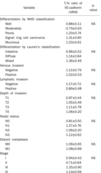
Table 3. Relationships between VE-cadherin mRNA Expression and Clinicopathological Characteristics
Variable
T/N ratio of VE-cadherin
mRNA
p- value
Differentiation by WHO classification
Well 0.88±0.11 NS
Moderately 0.79±0.63
Poorly 1.20±0.74 Signet ring cell carcinoma 1.31±0.60
Mucinous 1.20±0.61
Differentiation by Lauren’s classification
Intestine 0.96±0.52 NS
Diffuse 1.14±0.84
Mixed 1.36±0.49
Venous invasion
Negative 1.12±0.79 NS
Positive 1.02±0.53
Lymphatic invasion
Negative 1.17±0.72 NS
Positive 0.89±0.48
Depth of invasion
T1 0.97±0.44 NS
T2 1.05±0.49
T3 1.11±0.78
T4 1.06±0.20
Nodal status
N0 0.81±0.50 NS
N1 1.27±0.76
N2 1.06±0.20
N3 1.12±0.62
Distant metastasis
M0 1.06±0.65 NS
M1 1.08±0.69
Stage
I 0.94±0.43 NS
II 0.71±0.64
III 1.35±0.90
IV 1.13±0.59
Values are presented as mean±SD.
WHO, World Health Organization; VE-cadherin, vascular-endothelial cadherin; WHO, World Health Organization; NS, not significant.
Stages are determined by the 6th edition of International Union against cancer/American Joint Committee on Cancer TNM classi- fication System.
Table 4. Relationships between LI-cadherin mRNA Expression and Clinicopathological Characteristics
Variable
T/N ratio of LI-cadherin
mRNA p- value
Differentiation by WHO classification
Well 1.34±0.73 NS
Moderately 1.00±0.38 Poorly 1.19±0.61 Signet ring cell carcinoma 1.45±1.04
Mucinous 1.17±0.65
Differentiation by Lauren’s classification
Intestine 1.14±0.51 NS
Diffuse 1.07±0.44
Mixed 1.92±1.51
Venous invasion
Negative 1.18±0.46 NS
Positive 1.22±0.82
Lymphatic invasion
Negative 1.17±0.79 NS
Positive 1.03±0.44
Depth of invasion
T1 1.13±0.51 NS
T2 1.01±0.24
T3 1.25±0.83
T4 1.65±0.60
Nodal status
N0 1.05±0.46 0.082
N1 1.04±0.53
N2 1.65±0.60
N3 1.55±1.00
Distant metastasis
M0 1.13±0.69 0.031
M1 1.58±0.53
Stage
I 0.94±0.43 NS
II 0.71±0.64
III 1.35±0.90
IV 1.13±0.59
Values are presented as mean±SD.
LI-cadherin, liver-intestine cadherin; WHO, World Health Organiza- tion; NS, not significant. Stages are determined by the 6th edition of International Union against cancer/American Joint Com- mittee on Cancer TNM classification System.
proteins were expressed in 12 of 24 (50%) gastric cancer cas- es (Fig. 2). In immunohistochemical study, 12 of 18 (66.7%) cases showed positive immunostaining. Immunostaining was evident on the vascular endothelial cells in the gastric cancer tissues (Fig. 3).
There were no associations between the T/N ratio of VE-cadherin mRNA with differentiations by World Health Organization (WHO) classification and by Lauren's classi- fication, venous invasion, depth of invasion, nodal status,
and distant metastasis (Table 3).
There was a tendency for the T/N ratio of VE-cadherin mRNA to correlate with the lymphatic invasion (p=0.070) and lymph node metastasis (p=0.099) in advanced gastric can- cer (AGC).
2. Expression of LI-cadherin
LI-cadherin mRNA was overexpressed in 25 of 47 (53.2%) gastric cancer cases (Fig. 4). LI-cadherin proteins were ex-
Fig. 5. Western blot analysis of liver-intestine cadherin (LI-cadherin) in gastric cancer tissue (C) and normal tissue (N). LI-cadherin protein was detected in gastric cancer. Representative cases were shown.
GAPDH, glyceraldehyde 3-phosphate dehydrogenase.
Fig. 4. RT-PCR analysis of liver-intestine cadherin (LI-cadherin) in gastric cancer tissue (C) and normal tissue (N). LI-cadherin mRNA was detected in gastric cancer. Representative RT-PCR cases were shown. GAPDH, glyceraldehyde 3-phosphate dehydrogenase.
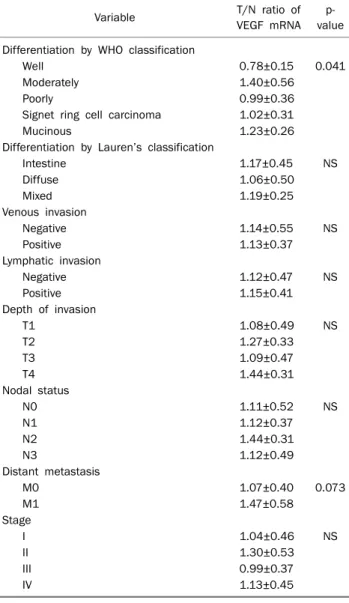
Table 5. Relationships between VEGF mRNA Expression and Clini- copathological Characteristics
Variable T/N ratio of
VEGF mRNA p- value Differentiation by WHO classification
Well 0.78±0.15 0.041
Moderately 1.40±0.56
Poorly 0.99±0.36 Signet ring cell carcinoma 1.02±0.31
Mucinous 1.23±0.26
Differentiation by Lauren’s classification
Intestine 1.17±0.45 NS
Diffuse 1.06±0.50
Mixed 1.19±0.25
Venous invasion
Negative 1.14±0.55 NS
Positive 1.13±0.37
Lymphatic invasion
Negative 1.12±0.47 NS
Positive 1.15±0.41
Depth of invasion
T1 1.08±0.49 NS
T2 1.27±0.33
T3 1.09±0.47
T4 1.44±0.31
Nodal status
N0 1.11±0.52 NS
N1 1.12±0.37
N2 1.44±0.31
N3 1.12±0.49
Distant metastasis
M0 1.07±0.40 0.073
M1 1.47±0.58
Stage
I 1.04±0.46 NS
II 1.30±0.53
III 0.99±0.37
IV 1.13±0.45
Values are presented as mean±SD.
VEGF, vascular endothelial growth factor; WHO, World Health Organization; NS, not significant. Stages are determined by the 6th edition of International Union against Cancer/American Joint Committee on Cancer TNM classification System.
pressed in 14 of 24 (58.3%) gastric cancer cases.
Representative examples of the Western blotting are shown in Fig. 5. In immunohistochemical study, 13 of 18 (72.2%) cases showed positive immunostaining. Immune reactivity of LI-cadherin in the mucosa of gastric cancer tissue was not- ed (Fig. 6).
There were no associations between the T/N ratio of LI-cadherin mRNA with differentiations by WHO classi- fication and by Lauren’s classification, venous invasion, and depth of invasion (Table 4).
There was a tendency for the T/N ratio of LI-cadherin mRNA to correlate with the nodal status (p=0.082). When nodal sta- tus was divided into two subgroups (N0/1 vs. N2/3), a sig- nificant difference was noted (1.05±0.49 vs. 1.56±0.93 [mean±SD]; p=0.013).
There was a significant relationship between the T/N ratio of LI-cadherin mRNA and distant metastasis (p=0.031) (Table 4). A significant relationship was also noted between the T/N ratio of LI-cadherin mRNA and lymphatic invasion es- pecially in AGC (p=0.023) (Table 6).
3. Expression of VEGF
Overexpression of the mRNA of VEGF in gastric cancer was observed in 27 of 47 cases (51.1%). Fig. 7 shows the mRNA expression of VEGF in gastric cancer and normal tissue. VEGF proteins were expressed in 14 of 24 (58.3%) gastric cancer specimens (Fig. 8). In immunohistochemical study, 10 of 18 (55.6%) cases showed positive immunostaining. Immuno- staining was evident in the vascular endothelial cells of the gastric cancer tissues (Fig. 9).
There were no associations between the T/N ratio of VEGF mRNA with differentiations by Lauren’s classification, ve- nous invasion, lymphatic invasion, depth of invasion and no- dal status (Table 5).
There was a tendency for the T/N ratio of VEGF mRNA to
Table 6. Relationships between VE-cadherin, LI-cadherin and VEGF mRNA Expression and Clinicopahtological Characteristices in Advanced Gastric Cancer
Variable (n) VE-cadherin LI-cadherin VEGF
T/N ratio p-value T/N ratio p-value T/N ratio p-value
Venous invasion
Negative (16) 1.05±0.59 NS 1.25±0.99 NS 1.14±0.26 NS
Positive (19) 1.14±0.80 1.02±0.46 1.16±0.56
Lymphatic invasion
Negative (7) 0.73±0.50 0.070 0.80±0.15 0.023 1.23±0.25 NS
Positive (28) 1.19±0.73 1.33±0.80 1.13±0.48
Lymph node metastasis
Negative (5) 0.54±0.58 0.099 0.88±0.15 NS 1.25±0.60 NS
Positive (30) 1.19±0.69 1.28±0.78 1.13±0.42
Distant metastasis
Negative (28) 1.10±0.72 NS 1.14±0.76 0.035 1.07±0.36 0.070
Positive (7) 1.08±0.70 1.58±0.53 1.47±0.59
Values are presented as mean±SD.
VE-cadherin, vascular-endothelial cadherin; LI-cadherin, liver-intestine cadherin; VEGF, vascular endothelial growth factor; NS, not significant.
Fig. 8. Western blot analysis of vascular endothelial growth factor (VEGF) in gastric cancer tissue (C) and normal tissue (N). VEGF protein was detected in gastric cancer. Representative cases were shown. GAPDH, glyceraldehyde 3-phosphate dehydrogenase.
Fig. 6. Immunohistochemical staining with liver-intestine cadherin (LI-cadherin). (A) Immune staining was evident on the mucosal cells in gastric cancer tissues (×100). (B) Immune reactivity of LI-cadherin was not detected in normal gastric tissues (×200).
Fig. 7. RT-PCR analysis of vascular endothelial growth factor (VEGF) in gastric cancer tissue (C) and normal tissue (N). VEGF mRNA was detected in gastric cancer. Representative RT-PCR cases were shown. GAPDH, glyceraldehyde 3-phosphate dehydrogenase.
correlate with distant metastasis (p=0.073) (Table 5). Also there was a tendency for the T/N ratio of VEGF mRNA to corre- late with distant metastasis in AGC (p=0.070) (Table 6).
4. Correlations with cadherins and VEGF
There were no significant correlations between the T/N ra- tios of VE-cadherin and VEGF (r=−0.187, p=0.207) (Fig. 10),
Fig. 9. Immunohistochemical staining with vascular endothelial growth factor (VEGF) (×200). (A) Immune staining was evident on vascular endothelial cells in gastric cancer tissues. (B) Immune reactivity of VEGF was not detected in normal gastric tissues.
Fig. 10. Correlation between vascular-endothelial cadherin (VE-cad- herin) and vascular endothelial growth factor (VEGF) mRNA expression. There was no significant relationship between VE-cad- herin and VEGF.
Fig. 11. Correlation between vascular-endothelial cadherin (VE-cad- herin) and liver-intestine cadherin (LI-cadherin) mRNA expression.
There was no significant relationship between VE-cadherin and LI-cadherin.
Fig. 12. Correlation between LI-cadherin and vascular endothelial growth factor (VEGF) mRNA expression. There was no significant relationship between LI-cadherin and VEGF.
between the T/N ratios of VE-cadherin and LI-cadherin (r=0.160, p=0.287) (Fig. 11) and between the T/N ratios of LI-cadherin and VEGF (r=0.067, p=0.655) (Fig. 12).
DISCUSSION
Cadherins play an important role in establishing adhe- rens-type junctions by mediating calcium-dependent cell to cell adhesion.12 Cadherins were first shown to support cell-cell contact and maintain tissue cohesion, and have been shown to be essential for development and post- embryonic life. In addition, cadherin can also act as cell-sig- naling receptors by regulating the location of β-catenin. β-cat-
enin is a distal component of the highly conserved Wnt path- way that governs cell survival, proliferation, and migration.13 The finding that cadherin cell-cell adhesion receptors are key regulators of tissue architecture during development and tis- sue homeostasis provides a good molecular evidence to link cell-cell adhesion, morphogenesis and cancer.14 They are divided into more than 10 subclasses that are distinct in their immunologic specificities and tissue distribution.15,16
The formation of new blood vessels (angiogenesis) and lymph vessels (lymphangiogenesis) significantly contribute to malignant growth and metastasis of solid tumors. The as- sessment of angiogenesis and lymphangiogenesis has emerged as a potentially useful prognostic and predictive factor in human malignancies.17 A recently characterized member of the cadherin family, VE-cadherin, also known as cadherin 5, is an endothelial cell-specific cadherin located at intercellular adherens junctions, where it is thought to play a role in the cohesion and organization of adherens junctions and in the control of permeability properties of vascular endothelium.7,18 VE-cadherin is important for tumor angio- genesis, and its expression is up-regulated in the vasculature of breast cancers.19 Blocking VE-cadherin function with mon- oclonal antibodies in mouse tumor models leads to the in- hibition of tumor angiogenesis and growth. VE-cadherin is crucial for vessel assembly and integrity during angio- genesis, and blocking its function with antibodies has been previously proposed as a promising therapeutic approach against tumor angiogenesis.5 VE-cadherin is considered to be an important agent that is indirectly responsible for tumor invasiveness due to its involvement in angiogenesis. Highly aggressive melanomas were found to express VE-cadherin on the surface of malignant cells and such expression is in- dispensable for melanoma to create embryonic-like vasculo- genic networks.20 The intensity or intracapillary extent of pos- itive immune staining for VE-cadherin in the capillary endo- thelium of hepatocellular carcinoma tissues was sig- nificantly associated with tumor size, capsular invasion and tumor cell differentiation.7 Elevated levels of VE-cadherin protein have been detected in patients with colon cancer, however no correlation was found in comparison between VE-cadherin in a levels and the clinicopathological features of colorectal cancer.20 This study showed for the first time that mRNA expression, protein expression and immune re- activity of VE-cadherin were noted 46.8%, 50.0% and 66.7%
in gastric cancer, respectively and that in AGC, there was a tendency for the T/N ratio of VE-cadherin mRNA to correlate with the lymphatic invasion (p=0.070) and lymph node meta- stasis (p=0.099) (Table 6). In esophageal cancer, VE-cadher- in gene expression showed significant correlations with other angiogenic and lymphangiogenic markers.17 Up to now there have been no published reports on VE-cadherin expression in gastric cancer. The report of esophageal cancer may sup- port our data and VE-cadherin may be a marker for lymphatic invasion and lymph node metastasis in AGC.
Gastric intestinal metaplasia, an intermediate step in Correa’s cascade of gastric carcinogenesis, is generally re- garded as a premalignant lesion.21 Gastric cancer is gen- erally believed to develop from a multistep progression of chronic gastritis, atrophic gastritis, intestinal metaplasia, dysplasia and subsequently to cancer. Epidemiological stud- ies suggest that patients with intestinal metaplasia have more than a 10-fold increased risk of developing gastric cancer.22 Intestinal metaplasia of the stomach represents an alteration of the gastric mucosa to epithelium with morpho- logical and biological characteristics of the intestine.
Intestinal metaplasia is found in approximately 20% of gas- tric biopsies and is more frequent in older patients.23 Intestine specific markers such as villin, sucrase-isomaltase, and aminopeptidase N are known to be expressed in in- testinal metaplastic cells and gastric adenocarcinomas while not observed in the normal gastric mucosa.24 LI-cad- herin, which was studied in gastric cancer expression, is an- other candidate for evaluating whether the gastric epi- thelium is undergoing neoplastic transformation. LI-cadher- in is a structurally unique member of the cadherin super- family.25 Whereas the so-called classic cadherins, such as E-, N- and P-cadherin, have five cadherin repeats within their ex- tracellular domain, LI-cadherin consists of seven cadherin repeats. LI-cadherin has only 21 amino acids in the cyto- plasm domain, although the classic cadherins have highly conserved cytoplasm domains with 150 to 160 amino acids.15,26 The expression of LI-cadherin differs by species. In the rat, LI-cadherin is expressed in the liver and intestinal epi- thelial cells, while in humans and the mouse, LI-cadherin is expressed in the intestinal epithelial cells, but not in the liver.
It has been reported that LI-cadherin is expressed in color- ectal and pancreatic cancers.27,28 In colorectal cancer, a re- duced LI-cadherin expression level was associated with a
high tumor grade, lymphatic invasion, lymph node meta- stasis and an advanced TNM stage.29 Recent studies have re- ported that LI-cadherin is overexpressed in gastric cancer and in gastric intestinal metaplasia.9,27 Dong et al.27 reported that immunohistochemistry showed LI-cadherin was mainly present on the mucosal cell membrane and absent in normal gastric tissues and the positive rate for LI-cadherin was 78.4%. Also in this study, immune reactivity of LI-cadherin was shown in the mucosal cells of gastric cancer tissues and positive rate of immunohistochemical stain was 72.2% and LI-cadherin mRNA was overexpressed in 53.2% gastric can- cer cases.
The evidence for the expression of LI-cadherin in gastric cancer continues to be debated. It was reported that there is a close association between the reduced expression of LI-cadherin and lymph node metastasis in gastric cancer.
LI-cadherin was expressed in the well-differentiated ad- enocarinoma cells but this expression was reduced in the de- differentiated adenocarcinoma cells. Therefore, the adhe- sion molecules associated with the invasion and metastasis of cancer tissues appear to depend on how little they are expressed.2 It was also reported that lymph node metastasis was associated with the overexpression of LI-cadherin.9,27 LI-cadherin is found to be complementary to the co-ex- pressed classical cadherins such as E-cadherin. LI-cadherin may be up-regulated in some situations where the classical cadherin-mediated adhesion is disrupted. It may be involved in the intermediate steps of gastric carcinogenesis. More ad- vanced disease may result in a greater change in the up-regu- lation of LI-cadherin. This may explain the significant associa- tion with lymph node metastasis.9,27 In this study, the T/N ra- tio of LI-cadherin was significantly correlated with distant metastasis and lymphatic invasion specially in AGC patients and there was a tendency for the T/N ratio of LI-cadherin mRNA to correlate with the nodal status. When nodal status was divided into two subgroups, local and distant nodal metastasis, (N0/1 vs. N2/3), mRNA expression of the distant nodal metastasis group was significantly higher than that of the local nodal metastasis group which is favored to the sec- ond opinion. As the exact adhesive function of LI-cadherin is still unknown, more studies are required to elucidate this further.
VEGF was first described as a potent vascular permeability factor secreted by tumor cells that stimulates a rapid and re-
versible increase in microvascular permeability without mast cell degranulation or endothelial cell damage. This tumor se- creted vascular permeability factor was later shown to be a highly selective and remarkably potent growth factor of endo- thelial cells. VEGF has been shown to promote the migration, growth and survival of endothelial cells and is essential for vasculogenesis and angiogenesis.30 In adults, deregulated expression of VEGF is involved in a variety of disease states, ranging from inflammation to cancer and metastasis.31 VEGF expression is rapidly increased in hypoxic tissues, providing a mechanism that can assist tissue reoxygenation by stim- ulating angiogenesis and induces the proliferation of endo- thelium, contributes to tubulogenesis and increases vas- cular permeability.10,30 VEGF has been shown to be a marker for a poor prognosis. In addition, VEGF expression correlates with the degree of gastric wall involvement and production of ascites and the peritoneal dissemination of gastric cancer.31,32 In this study, overexpression of VEGF mRNA showed a ten- dency to correlate with distant metastasis.
VEGF and VE-cadherin are required for efficient angio- genesis. A possible dependency of VEGF and VE-cadherin was suggested in an experimental model of nude mice with implanted human renal cell carcinoma in which antibodies against VEGF were shown to restrict tumor growth by de- creasing both VEGF and murine VE-cadherin mRNA expression.33 The mRNA level of VE-cadherin increases in the conditions of enhanced angiogenesis like pregnancy or can- cer before treatment. In such conditions, VEGF also increased. Apoptosis of endothelial cells causes gradual ex- pulsion of VE-cadherin for the cell surface. So VE-cadherin and VEGF seem to be independent markers of angiogenesis and separately reflect the dynamics of neovascularization.34 In colon cancer, VE-cadherin and VEGF were elevated in can- cer patients than in controls but there were no correlations between VE-cadherin and VEGF. These finding also suggest that VE-cadherin and VEGF seem to be independent markers of angiogenesis in colon cancer.20 Similarily in our study, there was no significant correlation between VE-cadherin and VEGF mRNA ratio in gastric cancer. And there were also no significant correlation between VE-cadherin and LI-cad- herin mRNA ratio and between LI-cadherin and VEGF mRNA ratio. These finding may suggest that VE-cadherin, LI-cadher- in, and VEGF seem to be independent markers for gastric cancer.
There were limitations in this study. First, the number of cancer specimens was relatively small. Only 47 gastric can- cer cases were available for RT-PCR, 24 cases for Western blot analysis, and 18 cases were available for immunohisto- chemistry. Because of the small number of cancer speci- mens and enrolling only the patient who underwent the surgi- cal procedure, the portion of histologically poorly differ- entiated adenocarcinoma and signet ring cell carcinoma group was relatively big in our study. Also, we have to consider the possible discrepancy between endoscopic forcep biop- sies and surgically resected specimen.
Secondly, the normal (noncancerous) gastric specimen, which were used in this study, were not confirmed normal by microscopically but suspected to be normal by macroscopi- cally and the specimens were from the gastric cancer pa- tients, not from normal healthy control. It is known that the non-cancerous mucosa surrounding gastric cancer is often affected by gastritis, atrophy and intestinal metaplasia, so the normal gastric specimen may not be really normal gastric mucosa.
In this study, LI-cadherin had significant relationships with distant metastasis and lymphatic invasion in advanced gas- tric cancer. VE-cadherin transcription trends to increase with lymphatic invasion and lymph node metastasis in advanced gastric cancer. VEGF transcription tend to increase when dis- tant metastasis was presented. As an RT-PCR assay can be performed easily with small amount of specimens, examina- tion of LI-cadherin in biopsy specimens, obtained before sur- gery, may provide useful preoperative informations on tumor aggressiveness.
REFERENCES
1. Ministry for Health Welfare and Family Affairs. Yearbook of health and welfare statistics (1993-2007). Seoul: Ministry of Health and Welfare (Korea), 2009.
2. Park SS, Kang SH, Park JM, et al. Expression of liver-intestine cadherin and its correlation with lymph node metastasis in gas- tric cancer: can it predict N stage preoperatively? Ann Surg Oncol 2007;14:94-99.
3. Grötzinger C, Kneifel J, Patschan D, et al. LI-cadherin: a marker of gastric metaplasia and neoplasia. Gut 2001;49:73-81.
4. Konturek PC, Konturek SJ, Brzozowski T. Gastric cancer and Helicobacter pylori infection. J Physiol Pharmacol 2006;57 (Suppl 3):51-65.
5. Cavallaro U, Liebner S, Dejana E. Endothelial cadherins and tu- mor angiogenesis. Exp Cell Res 2006;312:659-667.
6. Blaschuk OW, Devemy E. Cadherins as novel targets for anti-can- cer therapy. Eur J Pharmacol 2009;625:195-198.
7. Kato K, Takada T, Fukusato T. Expression of vascular endothe- lial-cadherin in human hepatocellular carcinoma tissues.
Hepatol Res 2007;37:444-453.
8. Dong WG, Yu QF, Xu Y, Fan LF. Li-cadherin is inversely correlated with galectin-3 expression in gastric cancer. Dig Dis Sci 2008;53:1811-1817.
9. Ko S, Chu KM, Luk JM, et al. Overexpression of LI-cadherin in gas- tric cancer is associated with lymph node metastasis. Biochem Biophys Res Commun 2004;319:562-568.
10. Carmeliet P, Collen D. Molecular basis of angiogenesis. Role of VEGF and VE-cadherin. Ann N Y Acad Sci 2000;902:249-262.
11. Lampugnani MG, Dejana E. Adherens junctions in endothelial cells regulate vessel maintenance and angiogenesis. Thromb Res 2007;120(Suppl 2):S1-S6.
12. George SJ, Dwivedi A. MMPs, cadherins, and cell proliferation.
Trends Cardiovasc Med 2004;14:100-105.
13. Brantjes H, Barker N, van Es J, Clevers H. TCF: Lady Justice cast- ing the final verdict on the outcome of Wnt signalling. Biol Chem 2002;383:255-261.
14. Takeichi M. Cadherin cell adhesion receptors as a morphoge- netic regulator. Science 1991;251:1451-1455.
15. Gessner R, Tauber R. Intestinal cell adhesion molecules.
Liver-intestine cadherin. Ann N Y Acad Sci 2000;915:136-143.
16. Yagi T, Takeichi M. Cadherin superfamily genes: functions, ge- nomic organization, and neurologic diversity. Genes Dev 2000;
14:1169-1180.
17. Loges S, Clausen H, Reichelt U, et al. Determination of micro- vessel density by quantitative real-time PCR in esophageal can- cer: correlation with histologic methods, angiogenic growth fac- tor expression, and lymph node metastasis. Clin Cancer Res 2007;13:76-80.
18. Wheelock MJ, Johnson KR. Cadherin-mediated cellular signa- ling. Curr Opin Cell Biol 2003;15:509-514.
19. Labelle M, Schnittler HJ, Aust DE, et al. Vascular endothelial cad- herin promotes breast cancer progression via transforming growth factor beta signaling. Cancer Res 2008;68:1388-1397.
20. Sulkowska M, Famulski W, Wincewicz A, et al. Levels of VE-cad- herin increase independently of VEGF in preoperative sera of pa- tients with colorectal cancer. Tumori 2006;92:67-71.
21. Ko S, Chu KM, Luk JM, et al. CDX2 co-localizes with liver-intestine cadherin in intestinal metaplasia and adenocarcinoma of the stomach. J Pathol 2005;205:615-622.
22. Leung WK, Sung JJ. Review article: intestinal metaplasia and gastric carcinogenesis. Aliment Pharmacol Ther 2002;16:
1209-1216.
23. Filipe MI, Potet F, Bogomoletz WV, et al. Incomplete sulphomu- cin-secreting intestinal metaplasia for gastric cancer. Prelimi- nary data from a prospective study from three centres. Gut 1985;26:1319-1326.
24. Osborn M, Mazzoleni G, Santini D, Marrano D, Martinelli G, Weber K. Villin, intestinal brush border hydrolases and keratin polypeptides in intestinal metaplasia and gastric cancer; an im- munohistologic study emphasizing the different degrees of in- testinal and gastric differentiation in signet ring cell carcinomas.
Virchows Arch A Pathol Anat Histopathol 1988;413:303-312.
25. Ito R, Oue N, Yoshida K, et al. Clinicopathological significant and prognostic influence of cadherin-17 expression in gastric cancer. Virchows Arch 2005;447:717-722.
26. Takamura M, Sakamoto M, Ino Y, et al. Expression of liver-intes- tine cadherin and its possible interaction with galectin-3 in duc- tal adenocarcinoma of the pancreas. Cancer Sci 2003;94:425- 430.
27. Dong W, Yu Q, Xu Y. Altered expression of a Li-cadherin in gastric cancer and intestinal metaplasia. Dig Dis Sci 2007;52:536-542.
28. Su MC, Yuan RH, Lin CY, Jeng YM. Cadherin-17 is a useful diag- nostic marker for adenocarcinomas of the digestive system.
Mod Pathol 2008;21:1379-1386.
29. Takamura M, Ichida T, Matsuda Y, et al. Reduced expression of liver-intestine cadherin is associated with progression and lymph node metastasis of human colorectal carcinoma. Cancer Lett 2004;212:253-259.
30. Gavard J, Gutkind JS. VEGF controls endothelial-cell perme-
ability by promoting the beta-arrestin-dependent endocytosis of VE-cadherin. Nat Cell Biol 2006;8:1223-1234.
31. Bazas VM, Lukyanova NY, Demash DV, Galakhin KO, Myasoedov DV. Relation between cell-to-cell adhesion and angiogenesis and clinico-morphological prognostic factors in patients with gastric cancer. Exp Oncol 2008;30:235-239.
32. Aoyagi K, Kouhuji K, Yano S, et al. VEGF significance in peritoneal recurrence from gastric cancer. Gastric Cancer 2005;8:155- 163.
33. Dagnaes-Hansen F, Rasmussen LM, Tilton R, Denner L, Flyvb- jerg A. A murine vascular endothelial growth factor antibody in- hibits in vivo growth of human Caki-I renal adenocarcinoma.
Anticancer Res 2003;23:1625-1630.
34. Rabascio C, Muratori E, Mancuso P, et al. Assessing tumor angio- genesis: increased circulating VE-cadherin RNA in patients with cancer indicates viability of circulating endothelial cells. Cancer Res 2004;64:4373-4377.