Introduction
Acute subdural hematomas (ASDH) are observed in one third of patients with severe traumatic brain injury.22) ASDH forms between the dura and arachnoid membranes usually due to tearing of bridging veins or arterial rupture.
Management of ASDH may vary from simple observation to different surgical evacuation technique. Two most fre-
quent surgical modalities are craniotomy (CO) and decom- pressive craniectomy (DC). CO procedure removes skull bone and subdural hematoma followed by replacement of original skull bone. DC also removes skull bone and hema- toma, but remains the bone unclosed for possible expansion of edematous brain tissue with or without an additional ex- pansile duroplasty.2)
Many studies have been reported showing effectiveness of DC for ASDH.1,5,6,10,17,18) However, not all patients show severe postoperative brain swelling after evacuation of he- matoma where theoretical benefit of DC is questionable. DC also carries disadvantage owing to lack of bone closure.2,9,10,16) The optimal surgical modality for ASDH still remains to be clarified.
The objective of this study is to analyze the surgical out- comes of CO and DC for evacuation of ASDH by comparing the preoperative clinical features, computed tomography (CT) images and postoperative complications which may
Craniotomy or Decompressive Craniectomy for Acute Subdural Hematomas: Surgical Selection and Clinical Outcome
Young Sub Kwon, MD1,2, Kook Hee Yang, MD, PhD1, and Yun Ho Lee, MD1
1Department of Neurosurgery, National Health Insurance Service Ilsan Hospital, Goyang, Korea
2Department of Neurosurgery, School of Medicine, Kangwon National University, Chuncheon, Korea
Objective: Craniotomy (CO) and decompressive craniectomy (DC) are two main surgical options for acute subdural he- matomas (ASDH). However, optimal selection of surgical modality is unclear and decision may vary with surgeon’s experi- ence. To clarify this point, we analyzed preoperative findings and surgical outcome of patients with ASDH treated with CO or DC.
Methods: From January 2010 to December 2014, data for 46 patients with ASDH who underwent CO or DC were retro- spectively reviewed. The demographic, clinical, imaging and clinical outcomes were analyzed and statistically compared.
Results: Twenty (43%) patients underwent CO and 26 (57%) patients received DC. In DC group, preoperative Glascow Coma Scale was lower (p=0.034), and more patient had non-reactive pupil (p=0.004). Computed tomography findings of DC group showed more frequent subarachnoid hemorrhage (p=0.003). Six month modified Rankin Scale showed favorable outcome in 60% of CO group and 23% of DC group (p=0.004). DC was done in patient with more unfavorable preoperative features (p=0.017). Patients with few unfavorable preoperative features (<6) had good outcome with CO (p<0.001).
Conclusion: In selective cases of few unfavorable clinical findings, CO may also be an effective surgical option for ASDH.
Although DC remains to be standard of surgical modality for patients with poor clinical status, CO can be an alternative con- sidering the possible complications of DC.
(Korean J Neurotrauma 2016;12(1):22-27) KEY WORDS: Hematoma, subdural, acute ㆍCraniotomy ㆍDecompressive craniectomy ㆍTreatment outcome.
Received: January 15, 2016 / Revised: February 11, 2016 Accepted: February 14, 2016
Address for correspondence: Yun Ho Lee, MD
Department of Neurosurgery, National Health Insurance Service Ilsan Hospital, 100 Ilsan-ro, Ilsandong-gu, Goyang 10444, Korea Tel: +82-31-900-0435, Fax: +82-31-900-0588
E-mail: [email protected]
cc This is an Open Access article distributed under the terms of Cre- ative Attributions Non-Commercial License (http://creativecommons.
org/licenses/by-nc/3.0/) which permits unrestricted noncommercial use, distribution, and reproduction in any medium, provided the original work is properly cited.
Korean J Neurotrauma 2016;12(1):22-27 http://dx.doi.org/10.13004/kjnt.2016.12.1.22
possibly aid in selection of optimal surgical modality.
Materials and Methods
We retrospectively reviewed 46 cases of ASDH surgical- ly treated with CO or DC in our hospital from January 2010 to December 2014. Demographic and preoperative medi- cal data were reviewed including age, sex, and presence of medical illness causing coagulopathy or use of antiplatelet agents. Preoperative data that may affect the surgical out- come were also collected such as time from trauma to sur- gery or time from clinical deterioration to surgery, preop- erative Glasgow Coma Scale (GCS), pupillary light reflex, and presence of major extracranial injury. Preoperative CT scans were analyzed for measurement of midline shift, pres- ence of intracerebral hemorrhage (ICH) or petechial hem- orrhage, obliteration of basal cistern and third ventricle, and presence of subarachnoid hemorrhage at basal cistern.
All of the patients underwent surgery for evacuation of ASDH through frontotemporoparietal CO of size 10×12 cm or larger. Decision for CO or DC was done by attending
neurosurgeon (Figure 1). Cases in which evacuation ASDH were not the main goal of surgery were excluded. Thus, evacuation of large traumatic ICH, decompression for ce- rebral swelling, and surgery other than frontotemporopa- rietal CO were excluded in this study.
Postoperative midline shift was measured from immedi- ate postoperative CT scan. Measurement of swelling above the bone flap was done for patients who underwent DC with CT scan taken within postoperative day 3 to 7 when maxi- mal brain swelling was observed. Imaginary line on absent bone flap was drawn and brain tissue above the imaginary line was measured (Figure 2). Medical records and CT scans were reviewed for patient who underwent cranioplasty. Post- operative outcome was recorded using modified Rankin Scale (mRS) 6 months after initial surgery. Outcome was defined good for patients with mRS score 1-3, and poor for patients with scores 4-6.
Preoperative clinical features were classified for further analysis where unfavorable feature was defined as age over 70 years, anticoagulation or antiplatelet use, time to surgery >4 hours, preoperative GCS <8, one or both non-
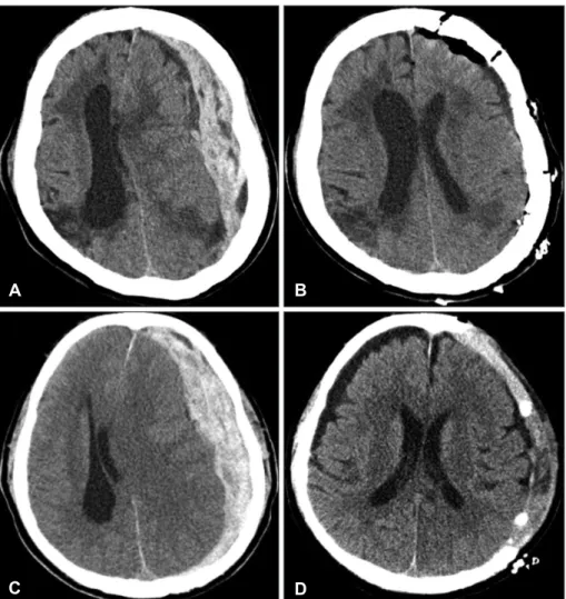
FIGURE 1. Preoperative (A) and postoperative (B) computed to- mography (CT) scan of 75-year- old female with acute subdural hematomas (ASDH) after trau- matic brain injury. She under- went craniotomy and evacuation of hematoma without remark- able postoperative brain swell- ing. Another case of 78-year-old male with ASDH (C, D) who un- derwent decompressive crani- ectomy. Preoperative (C) and postoperative (D) CT scan shows brain swelling, but removal of bone aids in control of raised in- tracranial pressure.
A B
C D
reactive pupil, and comorbid major extra-cranial injury. Pre- operative CT findings with ICH or petechial hemorrhage, obliterated basal cistern or 3rd ventricle, and presence of subarachnoid hemorrhage were also classified as unfavor- able preoperative feature.
Data was analyzed using Statistical Package for Social Sci- ences (SPSS) software for personal computers (SPSS ver. 21;
IBM Corp., Armonk, NY, USA). Unpaired Student’s t-test or Mann-Whitney test was used for continuous variables, and chi-squared test or Fisher’s exact test was used for cat- egorical variables. Probability value of less than 0.05 was considered as statistically significant.
Results
Demographic and preoperative clinical factors Forty six patients met the inclusion criteria of patient with ASDH who received either CO or DC. Twenty (43%) patients underwent CO, and 26 (57%) with DC. Mean age of CO group was 63.4 years, and DC was 65.5 years. Male was prevalent in both groups. Neither age nor gender distribu- tion showed significant difference between the groups.
Preoperative clinical data showed presence of coagulop- athy or use of antiplatelet in 5 of 20 (25%) patients of CO group and 12 of 26 (46%) patients of DC group. Time to sur- gery or clinical deterioration to surgery time was less than
FIGURE 2. Measurement of midline shift (A), and swelling above bone flap margin (B).
Imaginary line to absent bone flap was drawn congruent with measurement of contralateral hemisphere and brain tissue above the imaginary line was measured (B).
A B
TABLE 1. Patient demographics, preoperative clinical features, and preoperative computed tomography findings of craniotomy and decompressive craniectomy groups
CO (n=20) DC (n=26) p
Age Mean, years 63.4 (68)† 65.5 (68)† 0.515
<70 years 11 15
≥70 years 09 11
Gender M:F 12:8 16:10
Coagulopathy or antiplatelet use 05 (25%) 12 (46%) 0.676
Time to surgery ≤4 hours 11 (55%) 16 (62%) 0.655
>4 hours 09 (45%) 10 (38%)
GCS 8-12 13 (65%) 10 (38%) 0.034*
<8 07 (35%) 16 (62%)
Pupil reactivity Both reactive 12 (60%) 06 (23%) 0.004*
One reactive 06 (30%) 07 (27%)
None reactive 02 (10%) 13 (50%)
Major extracranial injury 02 (10%) 02 (8%) 0.783
Preoperative CT findings Mean midline shift (mm) 12.9 (13)† 13.3 (14)† 0.512 Presence of ICH or petechial hemorrhage 08 (40%) 13 (50%) 0.500 Obliterated basal cistern and 3rd ventricle 05 (25%) 13 (50%) 0.085
Subarachnoid hemorrhage 05 (25%) 18 (69%) 0.003*
*p<0.05, †median. CO: craniotomy, DC: decompressive craniectomy, M: male, F: female, GCS: Glasgow Coma Scale, CT:
computed tomography, ICH: intracerebral hemorrhage
4 hours in 11 of 20 (55%) patients in CO group and 16 of 26 (62%) patients in DC group. No statistical difference be- tween the two groups was significant in above preoperative clinical features. However, more patient in DC group had pre- operative GCS <8 (35% in CO vs. 62% in DC, p=0.034).
Preoperative pupillary reflex also showed more one or both non-reactive pupil in DC group (40% in CO vs. 77% in DC, p=0.004). Major combined extracranial injury were 2 pa- tients in both groups (Table 1).
Preoperative CT findings
Mean preoperative midline shift at preoperative CT were 12.9 mm in CO group and 13.3 mm in DC group (p=0.512).
Number of patients with ICH or petechial hemorrhage was 8 in CO group and 13 in DC group (40% in CO vs. 50% in DC, p=0.500). Obliteration of basal cistern and 3rd ventri- cle was 5 in CO group and 13 in DC group (25% in CO vs.
50% in DC, p=0.085). More patients showed preoperative subarachnoid hemorrhage in DC group (25% in CO vs. 69%
in DC, p=0.003).
Postoperative findings and patient outcome
Mean postoperative midline shift was larger in DC group (6.4 mm in CO vs. 9.1 mm in DC), but it was not statistical- ly significant (p=0.095). Reoperation was done in 4 of 20 (20%) patients in CO group which were due to recollection
of subdural hematoma in 2 patients and epidural hemor- rhage in 2 patients. In DC group, reoperation was done in 3 of 26 (12%) patients which were due to subgaleal hemato- ma in 1 patient and growth of traumatic ICH in 2 patients.
Cranioplasty was done in only 12 of 26 (46%) patients mainly due to patient condition. Six months postoperative mRS scores were less than 3 in 12 (60%), 4 & 5 in 7 (35%), and 6 in 1 (5%) of 20 patients in CO group. In DC group, mRS scores were less than 3 in 6 (23%), 4 & 5 in 7 (27%), and 6 in 13 (50%) of 26 patients. Difference of 6 months mRS scores between CO and DC groups were statistically significant (p=0.004) (Table 2).
Number of unfavorable preoperative features and clinical outcome
Mean number of unfavorable preoperative feature was 4.1 in CO group, and 5.8 in DC group (p=0.017) (Table 3).
In CO group, patient with preoperative adverse feature <6 showed good outcome in 13 patients and poor in 2 patients.
With preoperative adverse feature ≥6, no patient showed good outcome and 5 patients showed poor outcome (p<
0.001). In DC group, patient with preoperative adverse feature <6 showed good outcome in 4 patients and poor in 5 patients. With preoperative adverse feature ≥6, 2 pa- tient showed good outcome and 15 patients showed poor outcome (p=0.06).
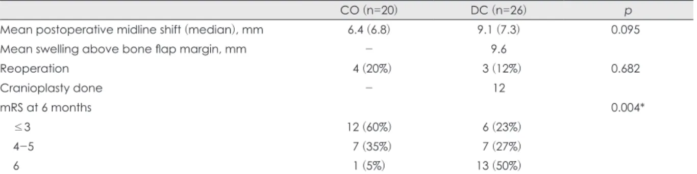
TABLE 2. Postoperative outcomes of CO and DC groups
CO (n=20) DC (n=26) p
Mean postoperative midline shift (median), mm 6.4 (6.8) 9.1 (7.3) 0.095
Mean swelling above bone flap margin, mm - 9.6
Reoperation 04 (20%) 03 (12%) 0.682
Cranioplasty done - 12
mRS at 6 months 0.004*
≤3 12 (60%) 06 (23%)
4-5 07 (35%) 07 (27%)
6 01 (5%)0 13 (50%)
*p<0.05. CO: craniotomy, DC: decompressive craniectomy, mRS: modified Rankin Scale TABLE 3. Number of unfavorable preoperative features and clinical outcome in CO and DC groups
CO (n=20) p DC (n=26) p
Mean number of unfavorable preoperative features 4.10 05.76 0.017*
Number of unfavorable preoperative features <0.001* 0.06
<6
Good 13 04
Poor 02 05
≥6
Good 00 02
Poor 05 15
Good: mRS score 1-3, Poor: mRS score 4-6. *p<0.05. CO: craniotomy, DC: decompressive craniectomy, mRS: modified Rankin Scale
Discussion
ASDH are present in approximately one third of patients with severe traumatic brain injury.22) Despite advances in emergency medical services and surgical techniques, ASDH remains one of the most lethal of all intracranial injuries.
Various surgical modality such as simple burr hole trephi- nation, CO, and DC are used for evacuation of ASDH. On the Brain Trauma Foundation guidelines published in 2006, they recommended that ASDH with thickness greater than 10 mm, or midline shifting greater than 5 mm on CT scan should be treated surgically.1) Mortality rates of ASDH rang- es from 55% to 79% even with surgical intervention of any modality.19)
Ransohoff et al.18) reported recovery rate of 40% for ASDH treated with hemicraniectomy followed by hema- toma removal. After the report, DC for ASDH has been recommended as a surgical modality of choice, and perform- ing DC in ASDH patients seemed to be attractive.1,4,7,8,13,14,20)
Girotto et al.7) reported DC technique of adequate size, ear- ly surgery, and GCS of 6 to 8 group would contribute signif- icantly to better outcome by reducing morbidity and mor- tality. Meier and Gräwe17) reported that DC benefits on overall outcome of patients with traumatic brain injury. The rationale behind performing DC lies in control of postoper- ative brain swelling and overwhelming intracranial hyper- tension, but little is known on the degree of postoperative swelling after evacuation of hematoma. Empirical decision for DC or CO is made by the neurosurgeon based on pa- tients’ clinical status and CT findings which may be con- founding unless brain swelling is noted intraoperatively af- ter evacuation of hematoma.
The analysis of postoperative brain swelling is difficult since the postoperative CT findings or intracranial pressure measurements will vary depending on closure or opening bone flap. Different trauma setting among patients makes randomized trial for CO and DC impossible and unethical.
Nevertheless, there were several retrospective series that compared the outcome of CO and DC.3,5,12,15,21) Woertgen et al.21) compared the surgical outcomes in ASDH which were not significantly different between CO and DC. They concluded that signs of herniation at presentation, and in- creasing age had most influence to patient outcome. So, preoperative clinical feature influenced most on outcome and DC does not seem to have a therapeutic advantage over CO in ASDH. More recent study by Chen et al.3) also report- ed similar results in 102 patients where DC group had high- er mortality rate which may be due to poorer preoperative clinical status. The study by Li et al.15) is notable where they
tried to diminish the effect of preoperative clinical status by using CRASH-CT prognostic model. Predicted outcome was calculated in 85 patients in a retrospective fashion. Fa- vorable outcomes were observed in 45% of CO versus 42%
of DC (p=0.83), but standardized morbidity ratio (observed/
expected unfavorable outcome) was 0.90 for CO group and 0.75 for DC group.
Our study showed poorer outcome in DC group com- pared with CO group (poor mRS 77%, 20 of 26 patients in DC group vs. 40%, 8 of 20 patients in CO group; p=0.004).
This results are may be due to more patients with low GCS score (GCS<8), unresponsive pupil, and comorbid CT le- sion in DC group. Our results carry similar selection bias that neurosurgeons tend to perform DC when patients’
preoperative clinical status is poor. To clarify this point, we counted on number of unfavorable features for each patient that may influence on poor outcome. On average, DC group had more adverse features than CO group, and thus poor outcome for DC group can be explained.
One notable finding is that in patients with few unfavor- able features (<6), good outcome (mRS less than 3) was achieved in majority of patients in CO group. However, similar results were not obtained in DC group with few un- favorable features. This implies that further stratification of unfavorable clinical features is needed which has larger im- pact on outcome. Nonetheless, it seems that some patients with few preoperative unfavorable features can benefit with CO without the need for bone removal.
Furthermore, various possible complications of DC need awareness of neurosurgeons. Subgaleal hemorrhage, her- niation through the cranial defect, subdural effusion, syn- drome of the trephined (sinking skin flap syndrome), and hydrocephalus were reported complications of DC.11,23) In our series, 1 patient underwent reoperation due to subgale- al hematoma and 2 patients had severe sinking of skin flap where difficulty was in cranioplasty resulted in complica- tion. DC also have disadvantage of requiring subsequent cranioplasty which harbor additional risk of complica- tion.2,11,16) Gooch et al.9) reported that immediate post-oper- ative complication rate of cranioplasty after DC was as high as 34% which were infection, wound breakdown, intracra- nial hemorrhage, and bone resorption. We also experienced complications of cranioplasty in our patients (4 of 12; epi- dural hematoma 2, infection 1, cerebrospinal fluid leakage 1) which interrupted patients’ recovery. In this context, there may be some advantage of CO in evacuation of ASDH.
However, this study is a retrospective single center study with small patient population. Limitations of selection bias hinder any conclusion on role of CO or DC for ASDH. We
think further investigation with larger patient population and carefully selected criteria is needed to clarify the opti- mal surgical modality for patient with ASDH.
Conclusion
In selective cases of few unfavorable clinical findings, CO may also be an effective surgical option for ASDH. Although DC remains to be standard of surgical modality for patients with poor clinical status, CO can be an alternative consid- ering the possible complications of DC. Controlled prospec- tive study with larger patient population is needed clarify this point.
■ The authors have no financial conflicts of interest.
REFERENCES
1) Bullock MR, Chesnut R, Ghajar J, Gordon D, Hartl R, Newell DW, et al. Surgical management of acute subdural hematomas.
Neurosurgery 58(3 Suppl):S16-S24; discussion Si-Siv, 2006 2) Chaturvedi J, Botta R, Prabhuraj AR, Shukla D, Bhat DI, Devi BI.
Complications of cranioplasty after decompressive craniectomy for traumatic brain injury. Br J Neurosurg 17:1-5, 2015
3) Chen SH, Chen Y, Fang WK, Huang DW, Huang KC, Tseng SH.
Comparison of craniotomy and decompressive craniectomy in severely head-injured patients with acute subdural hematoma. J Trauma 71:1632-1636, 2011
4) Chibbaro S, Marsella M, Romano A, Ippolito S, Benericetti E.
Combined internal uncusectomy and decompressive craniectomy for the treatment of severe closed head injury: experience with 80 cases. J Neurosurg 108:74-79, 2008
5) Coplin WM, Cullen NK, Policherla PN, Vinas FC, Wilseck JM, Zafonte RD, et al. Safety and feasibility of craniectomy with du- raplasty as the initial surgical intervention for severe traumatic brain injury. J Trauma 50:1050-1059, 2001
6) Girotto D, Ledić D, Bajek G, Jerković R, Dragicević S. Efficancy of decompressive craniectomy in treatment of severe brain injury at the Rijeka University Hospital Centre. Coll Antropol 35 Suppl 2:255-258, 2011
7) Girotto D, Ledić D, Daji V, Vujković Z, Mihelcić N. Neurosurgical procedure for treatment of traumatic subdural hematoma with severe brain injury: a single center matched-pair analysis. Coll Antropol 38:1255-1258, 2014
8) Godlewski B, Pawelczyk A, Pawelczyk T, Ceranowicz K, Wojdyn M, Radek M. Retrospective analysis of operative treatment of a series of 100 patients with subdural hematoma. Neurol Med Chir
(Tokyo) 53:26-33, 2013
9) Gooch MR, Gin GE, Kenning TJ, German JW. Complications of cranioplasty following decompressive craniectomy: analysis of 62 cases. Neurosurg Focus 26:E9, 2009
10) Honeybul S. Complications of decompressive craniectomy for head injury. J Clin Neurosci 17:430-435, 2010
11) Honeybul S, Ho KM. Decompressive craniectomy for severe trau- matic brain injury: the relationship between surgical complica- tions and the prediction of an unfavourable outcome. Injury 45:
1332-1339, 2014
12) Huang AP, Tu YK, Tsai YH, Chen YS, Hong WC, Yang CC, et al.
Decompressive craniectomy as the primary surgical intervention for hemorrhagic contusion. J Neurotrauma 25:1347-1354, 2008 13) Kalayci M, Aktunç E, Gül S, Hanci V, Edebali N, Cagavi F, et al.
Decompressive craniectomy for acute subdural haematoma: an overview of current prognostic factors and a discussion about some novel prognostic parametres. J Pak Med Assoc 63:38-49, 2013 14) Karibe H, Hayashi T, Hirano T, Kameyama M, Nakagawa A, Tom-
inaga T. Surgical management of traumatic acute subdural hema- toma in adults: a review. Neurol Med Chir (Tokyo) 54:887-894, 15) Li LM, Kolias AG, Guilfoyle MR, Timofeev I, Corteen EA, Pick-2014
ard JD, et al. Outcome following evacuation of acute subdural haematomas: a comparison of craniotomy with decompressive cra- niectomy. Acta Neurochir (Wien) 154:1555-1561, 2012 16) Liang ES, Tipper G, Hunt L, Gan PY. Cranioplasty outcomes and
associated complications: a single-centre observational study. Br J Neurosurg 30:122-127, 2016
17) Meier U, Gräwe A. The importance of decompressive craniecto- my for the management of severe head injuries. Acta Neurochir Suppl 86:367-371, 2003
18) Ransohoff J, Benjamin MV, Gage EL Jr, Epstein F. Hemicraniec- tomy in the management of acute subdural hematoma. J Neuro- surg 34:70-76, 1971
19) Servadei F. Prognostic factors in severely head injured adult patients with acute subdural haematoma’s. Acta Neurochir (Wien) 139:
279-285, 1997
20) Winter CD, Adamides A, Rosenfeld JV. The role of decompres- sive craniectomy in the management of traumatic brain injury: a critical review. J Clin Neurosci 12:619-623, 2005
21) Woertgen C, Rothoerl RD, Schebesch KM, Albert R. Comparison of craniotomy and craniectomy in patients with acute subdural haematoma. J Clin Neurosci 13:718-721, 2006
22) Yanaka K, Kamezaki T, Yamada T, Takano S, Meguro K, Nose T.
Acute subdural hematoma--prediction of outcome with a linear dis- criminant function. Neurol Med Chir (Tokyo) 33:552-558, 1993 23) Yang XF, Wen L, Shen F, Li G, Lou R, Liu WG, et al. Surgical com- plications secondary to decompressive craniectomy in patients with a head injury: a series of 108 consecutive cases. Acta Neuro- chir (Wien) 150:1241-1247; discussion 1248, 2008