서론
치과용 임플란트 치료의 성공적인 결과를 위한 중요한 기준 은 골조직과 임플란트 간의 강력한 골유착을 얻는 것이다. 안 정적인 골유착을 얻기 위한 조건으로 임플란트의 재료, 형태,
표면처리 등이 중요한 요인들로 고려된다.1구강 내 임플란트 의 재료로써 티타늄과 그 합금은 뛰어난 생체 적합성과 부식 저항성을 갖고 있어 현재 널리 사용되고 있다.2그러나, 티타늄 표면은 골유착 속도가 느리고 화학적으로 골과 직접 결합하지 않기 때문에,3골유착을 증진시키기 위하여 수많은 티타늄 표
마이크로그루브 및 열산화 복합 티타늄 표면의 골아세포분화 증진효과
서진호1∙이성복1∙안수진1∙박수정1∙이명현2∙이석원1*
1강동경희대학교치과병원(생체재료)보철과 경희대학교 치과대학, 치의학전문대학원 치과보철학교실
2한국세라믹기술원 그린세라믹본부
Effect of titanium surface microgrooves and thermal oxidation on in vitro osteoblast responses
Jin-Ho Seo1, Richard Leesungbok1, Su-Jin Ahn1, Su-Jung Park1, Myung-Hyun Lee2, Suk Won Lee1*
1Department of Biomaterials & Prosthodontics, Kyung Hee University Hospital at Gangdong, School of Dentistry, Kyung Hee University, Seoul, Republic of Korea
2Green Ceramics Division, Korea Institute of Ceramic Engineering and Technology, Seoul, Republic of Korea
Purpose: We aimed to investigate the effect of combined various microgrooves and thermal oxidation on the titanium (Ti) and to evaluate various in vitro responses of human periodontal ligament cells (PLCs). Materials and methods: Grade II titanium disks were fabricated. Microgrooves were applied on titanium discs to have 0/0 ㎛, 15/3.5 ㎛, 30/10 ㎛, and 60/10 ㎛ of respective width/depth by photolithography. Thermal oxidation was performed on the microgrooves of Ti substrata for 3 h at 700℃ in air. The exper- iments were divided into 3 groups: control group (ST), thermal oxidation group (ST/TO), and combined microgrooves and thermal oxidation group (Gr15-TO, Gr30-TO, Gr60- TO). Surface characterization was performed by field-emission scanning microscopy. Cell adhesion, osteoblastic differentiation, and mineralization were analyzed using the bromodeoxyurdine (BrdU), Alkaline phosphatase (ALP) activity, and extracellular calcium deposition assays, respectively. Statistical analysis was performed using the one- way analysis of variance and Pearson’s bivariate correlation analysis (SPSS Version 17.0). Results: In general, the combined microgrooves and thermal oxidation group (Gr15- TO, Gr30-TO, Gr60-TO) showed significantly higher levels compared with the control (ST) or thermal oxidation (ST-TO) groups in the BrdU expression, ALP activity, and extracellular calcium deposition. Gr60-TO group induced highest levels of cell adhesion and osteoblastic differentiation. Conclusion: Within the limitation of this study, we conclude that the Ti surface treatment using combined microgrooves and thermal oxidation is highly effective in inducing the cell adhesion andosteoblastic differentiation. The propose surface is also expected to be effective in inducing rapid and strong osseointegration of Ti oral implants. (J Korean Acad Prosthodont 2015;53:198-206)
Key words: Titanium; Microgrooves; Thermal oxidation; Osteoblastic differentiation
c cc
2015 The Korean Academy of Prosthodontics
This is an Open Access article distributed under the terms of the Creative Commons Attribution Non-Commercial License (http://creativecommons.org/licens- es/by-nc/3.0) which permits unrestricted non-commercial use, distribution, and reproduction in any medium, provided the original work is properly cited.
*Corresponding Author: Suk Won Lee
Department of Biomaterials & Prosthodontics, Kyung Hee University Hospital at Gangdong, Institute of Oral Biology, School of Dentistry, Kyung Hee University, 892 Dongnam-ro, Gangdong-gu, Seoul 134-727, Republic of Korea
+82 2 440 7519: e-mail, [email protected]
Article history: Received June 18, 2015 / Last Revision July 13, 2015 / Accepted July 15, 2015
※This research was supported by Basic Science Research Program through the National Research Foundation of Korea (NRF) funded by the Ministry of Education, Science and Technology (NRF-20100021268).
면처리 및 표면개질(surface modification)방법들이 개발되고 있다.4 특히 임플란트 표면에 인위적으로 적정한 거친 표면(roughed surface)을 부여 시 기계연마 표면(machined surface)에 비하여 효과 적으로 생체 내 임플란트골유착 증진을 얻을 수 있음이 보고 되었다.5임플란트에 거친 표면을 부여하는 SLA (sand blasting with large grit & acid-etched), Plasma spaying 같은 방법에서 calcium phosphate (Ca-P)와 같은 생활성 재료 및 생체모방적인 표면을 형 성하는 코팅 방법까지 임플란트 표면처리에 대한 다양한 연구 들이 현재까지 소개되고 있으며,6최근에는 비교적 저렴하고 간단한 티타늄 표면처리 방법인 열산화(thermal oxidation)가 소개 되었다.
티타늄과 그 합금은 산소와 강한 친화력을 갖고 있기 때문에 산소나 수분과 반응하여 2 - 5 nm 두께의 산화막을 형성한다.7 티타늄의 뛰어난 생체적합성 및 부식저항성은 이러한 안정된 부동태 티타늄 산화막을 통하여 얻어질 수 있다.8그러나, 자연 상태에서 산소와 반응하여 형성된 티타늄 산화막은 대부분 이 산화 티타늄(TiO2)이며 두께가 얇고 불안정하여 임플란트식립 시 쉽게 손상될 수 있다.9이처럼 불안정한 티타늄 산화막을 개 선하기 위하여 열산화법, 양극산화법, 플라즈마산화법, 전기 화학적 방법 등 다양한 피막 형성 방법들이 연구되고 있다.10그 중 열산화는 티타늄 표면에 상대적으로 두꺼운 산화막을 형성 하여 생체친화성 및 세포활성을 증진시키는 것으로 보고되고 있다.11실제로, 티타늄을 이용한 임플란트 제조과정에서 소결, 고온소독 등 다양한 열처리 공정이 이루어지며,12이러한 공정 중에 각기 다른 온도 환경에 따라 티타늄 산화막의 표면 특성 및 안정성은 달라지게 된다.13티타늄 산화막의 안정성 증진을 위한 적정한 열산화 처리 온도에 대한 여러 연구들에 따르면, 티타늄을 700℃에서 열산화 처리한 실험군이 다른 온도에서 열산화 처리한 실험군 등에 비해 골아세포접착 정도가 증진된 결과를 보였으며,14골아세포분화 및 골형성도 증진됨을 보였 다.15
마이크로 단위의 그루브가 부여된 표면성상은세포접착과 세포증식을 유도, 증가시킨다고 알려져 있다.16티타늄 표면에 포토리소그라피(photolithography)의 습성에칭기술(wet-etching technique)로 형성된 적절한 크기의 마이크로그루브는 인간치은 섬유아세포의 세포증식을 유발시킨다는 연구 결과가 보고되 었으며,17다양한 크기의 마이크로 및 서브 마이크로 단위의 표 면성상에 따라 골아세포의 분화의 정도가 달라짐이 확인되었 다.18마이크로/나노 단위 그루브가 형성된 티타늄 표면은 골아 세포의 활동을 향상시켰으며,19열산화 처리와 SLA (sandblast- ed/acid-etched)를 동반한 마이크로-나노 복합 표면은 골아세포 및 MG63세포의 활동성을 증가시켰다.20이에 따라, 마이크로-나 노 복합 표면은 골아세포의 분화 및 증식에 큰 영향을 끼친다 고 볼 수 있으며, 마이크로와 나노 각각의 특성을 살린 복합 임 플란트 표면이 빠르고 강력한 골유착을 유도할 수 있음을 시 사한다.
이전 연구들에서 적정한 마이크로그루브 크기와 열산화 처
리 온도는 규명이 시도되었지만, 마이크로그루브-열산화 복합 표면에서 골아세포분화를 촉진시키는 적정한 마이크로그루 브의 크기에 대한 연구는 이루어지지 않았다. 본 연구의 목적 은 다양한 크기의 마이크로그루브와 열산화 처리가 복합된 티 타늄 표면이 세포접착 및 골아세포분화에 미치는 영향을 조사 하여 치과용 임플란트 표면 적용에 가장 적합한 표면처리 조 건을 규명하는 것이다.
재료 및 방법 1. 재료
1) 시편의 제작
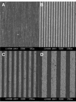
0.14 mm 두께의 grade-2 순수티타늄(commercially pure titanium, cp-Ti) 디스크(TSM-TECH Co., Ltd., Ulsan, Korea)를 silicon carbide (SiC) 로 Ra ≤ 0.1 ㎛의 표면 거칠기를 갖도록 기계적으로 연마한 (smooth titanium, ST), 이후 티타늄 표면을 포토리소그라피(pho- tolithography; MEMSware Inc.,Yangju, Gyeonggi, Korea)를 이용하여 마이크로그루브를 형성하였다. 기계적으로 연마된 티타늄 시 편의 표면에 UV-sensitive polymer (photo-resist, DTFR, Dongjin, Seoul, Korea)를 코팅시켰다. 코팅된 티타늄 시편 위에 마이크 로그루브의 모양이 형성된 Photo-mask를 위치시키고 이를 UV 광에 노출시켜, Photo-mask를 통과한 UV 광에 의해 티타늄 시편 의 표면에 마이크로그루브의 모양이 현상되도록 하였다. 티타 늄 시편의 마이크로그루브 모양은 폭/깊이 각각 0/0 ㎛, 15/3.5 ㎛, 30/10 ㎛ 및 60/10 ㎛로 각각 형성되었다(ST, Gr15, Gr30, Gr60, Fig. 1). 그루브가 부여됨에 따라 형성된 ridge의 크기는 각각의 그
Fig. 1. Field emission scanning electron microscopic images of (A) ST (×500), (B) Gr15 (×200), (C) Gr30 (×200) and (D) Gr60 (×200).
A B
C D
루브의 크기와 동일하였다. 코팅된 티타늄 시편을 용해액 (2.38% tetramethyl-ammonium hydroxide, TMAH, Dongjin, Seoul, Korea)에 담가 UV 광에 노출된 부위만 용해시켜 마이크로그루 브 모양을 현상하였고, photo-resist 코팅을 용해액(아세톤, 이소 프로필 알코올)으로 제거하였다. 마이크로그루브가 형성된 티 타늄 시편을 700℃에 3시간 동안 공기 중에 노출하여 열산화를 시행하였다(ST-TO, Gr15-TO, Gr30-TO, Gr60-TO). 준비된 모든 티타늄 시편은 실험 사용 전에 초음파 기계에 30분간 3회 세척 하고, 증류수에 3회 세척한 후, 상온에서 1일 건조시킨 후 사용 되었다.
2. 방법
1) 주사전자현미경(scanning electron microscopy, SEM) 관찰 Field emission scanning electron microscopy (S-800 FE-SEM�; HITACHI, Tokyo, Japan)를 통하여 표면을 관찰하였다.
2) 세포배양
교정치료 중인 환자에서 발치된 소구치의 치주인대로부터 얻어진 인간일차세포, 즉 인간치주인대세포(human periodontal ligament cells)를 배양하였다. 연구에 사용된 치주인대 조직은 강동경희대병원 IRB (Institutional Review Board)의 승인을 얻어 절 차에 따라 이용되었다. 발치된 소구치를 즉시 10% fetal bovine serum (FBS; Sigma-Aldrich, St. Louis, MO, USA)과 1% antibiotic/antimy- cotic solution (Invitrogen, Carlsbad, CA, USA)을 함유한 DMEM 배지 (Dulbecco’s modified Eagle’s medium, WelGene, Daegu, Korea)에 서 37℃, 5% CO2하에 보관하였다. 발치된 소구치의 치근 1/3부 위로부터 채취된 치주인대 조직은 1.1 units/ml dispose (Invitrogen, Carlsbad, CA, USA)와 264 units/ml collagenase (Invitrogen Invitrogen, Carlsbad, CA, USA)에서 37℃, 5% CO2하에 1시간 동안 처리되었 다. 준비된 조직은 10% fetal bovine serum (Sigma-Aldrich)과 1%
antibiotic/antimycotic solution (Invitrogen)을 함유한 DMEM 배지 (WelGene)에서 37℃, 5% CO2하에 배양되었다. 배양된 3 - 5 주기 세포가 본 연구에 사용되었다.
3) Bromdeoxyuridine 세포접착 실험
바닥을 제거한 배양접시에 표면처리된 지름 10 mm의 티타 늄 디스크를 실리콘 본딩제로 접착하여 ST, ST/TO, Gr15-TO, Gr30-TO, Gr60-TO의 5가지 대조군 및 실험군으로 이루어진 96- well 티타늄 시편들이 제작되었다. 인간치주인대세포는 96-well 티타늄 표본 바닥에 3×10³cell/ml 밀도로 접착되어, 37℃, 5% CO2 의 습도 하에 16시간 동안 인큐베이터 내에서 배양되었다. 1 ml 의 bromodeoxyurdine (BrdU) 표지 시약[1000 × conc., 인산완충용 액(phosphate buffered solution; Gibco BRL, Grand Island, NY, USA)에 희석된 10 nM 5-bromo-2-deoxyuridine, pH 7.4; Roche Diagnostics GmbH, Mannheim, Germany]을 각 well에 첨가하였고, 세포들은 37℃에서 2시간 동안 재배양되었다. 이러한 표지 기간 동안 pyrimidine 유
사체인 BrdU는 증식세포 DNA의 Thymidine 위치로 대체되어 들어가게 된다. 배지를 제거한 후 항체가 BrdU를 감지할 수 있 도록 200 ml의 FixDenat (Roche Diagnostics GmbH)를 첨가하여 30분 간 실온에서 세포를 고정시키고 DNA를 변성시킨다.
Fixdenat를 제거한 후 항-BrdU-POD (peroxidase에 결합된 monoclonal antibody; Roche Diagnostics GmbH) 부유액을 각 well마다 첨가하고 90분 간 실온에 보관하였다. 이 시간 동안 BrdU에 결합한 항- BrdU-POD가 새롭게 합성된 세포 DNA에 대체되어 들어가고 차후의 효소-기질반응에 의하여 이 면역 복합체가 감지되게 된다. 모든 군의 반응 산물들은 96-well plate에 옮겨져, microreader (ELISA analyzer; Bio-Rad, Hercules, CA, USA)를 이용하여 370 nm 에서 그 흡광도(optical density, OD)가 측정되었다.
4) 알칼리성 인산가수분해효소 활성 실험
인간치주인대세포의 골활성능을 확인하기 위해, 알칼리성 인산가수분해효소(alkaline phosphatase, ALP) 활성 레벨을 측정하 였다. 세포를 4×10⁵cell/ml의 밀도로 24-well 티타늄 표본에 위치 시키고 2일 동안 배양 후, 골활성배지[osteogenic media; DMEM 배 지(Dulbecco’s modified Eagle’s medium, WelGene, Daegu, Korea), 10%
FBS (Sigma-Aldrich), 50 ug/ml α-ascorbic acid (Sigma-Aldrich), 10 mM β-glycerophosphate (Sigma-Aldrich), 100 mM dexametasone (Sigma-Aldrich)]
에서 37℃, 5% CO2를 유지하여 7 및 14일 간 세포를 재배양하였 다. 배양된 세포는 인산완충용액(Gibco BRL)로 세척하고, 트립 신-EDTA 용액으로 처리하였으며, 0.1% Triton X-100 buffer로 용해 시키고, 얼음 안에서 초음파 처리하였다. ALP 존재 하에서 p- nitrophenol 생성물 수준의 흡광도는 microplate reader (ELISA ana- lyzer; Bio-Rad, Hercules, CA, USA)로 405 nm에서 측정되었다. 측정 치들을 표준 p-nitrophenol을 사용하여 비교하였고, 각 시점에서 티타늄 표본에서의 세포 수 차이를 반영하기 위해 총 단백질 양을 사용하여 보정하였다.
5) 세포외 칼슘 침착 실험
인간치주인대세포를 4×10⁵cell/ml의 밀도로 24-well 티타늄 대 조군과 실험군 표본에 위치시키고 2일 동안 배양하였다. 그 후 세포를 골활성배지에서 37℃, 5% CO2를 유지하여 21일 간 배양 하였다. 배양된 세포는 인산완충용액(Gibco BRL)로 세척되었 다. Calcium Liquicolor (Stanbio Laboratory, Boerne, TX, USA)를 사용 하여 시편 표면에 침착된 세포외 칼슘(extracellular calcium depo- sition) 양을 측정하였다. Microplate reader (Bio-Rad, Hercules)를 이용하여 650 nm에서 흡광도가 측정되였다.
6) 통계분석
BrdU 실험, 알칼리성 인산가수분해효소 활성 실험, 세포외 칼슘 침착 실험 등은 동시에 독립적으로 5회 시행되었으며, 평 균값과 표준편차를 계산하였다. ST, ST/TO, Gr15-TO, Gr30-TO, Gr60-TO 군 사이의 평균값을 비교하기 위하여 일요인분산분 석(one-way ANOVA)을 사용하였다. BrdU 실험, ALP 활성 실험 간
상관관계를 분석하기 위하여 피어슨 상관관계 분석을 시행하 였다. 모든 통계분석에는 SPSS (17.0) 소프트웨어 프로그램이 사 용되었다.
결과
1. 주사전자현미경 관찰
열산화 처리 전 다양한 마이크로그루브 크기에 따른 표면 성 상변화가 관찰되었다(Fig. 1). 대조군인 ST표면에 비하여 ST-TO 에서 티타늄 격자(grain)의 크리스탈화가 진행되었으며, 격자 의 크기는 수십 - 수백 nm로 형성되었음이 관찰되었다(Fig. 2). 이 는 열산화에 의한 나노 단위 표면 성상의 형성을 시사하는 것 으로써, 이러한 나노 성상이 인간치주인대세포의 접착과 골아 세포분화에 영향을 미쳤음을 알 수 있다.
2. 세포분화
1) Bromdeoxyuridine 세포접착 실험
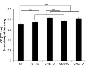
각 시편에 16시간 인간치주인대세포를 배양한 BrdU (Bromdeoxyuridine) assay를 통해 세포접착 정도를 확인하였다. 측 정된 데이터를 일요인분산분석 결과, Gr15-TO 군과 Gr60-TO 군 사이에는 유의성 있는 차이가 없었다(P>.001). 또한 Gr15-TO 와 Gr60-TO 군은 ST, ST/TO, Gr30-TO 등에 비하여 유의하게 증가 하였다(P<.001). 열산화 처리를 시행한 모든 실험군들 역시 ST 군에 비하여 유의하게 증가하였다(P<.001) (Table 1, Fig. 3).
2) 알칼리성 인산가수분해효소 활성 실험
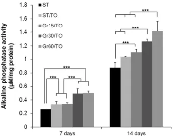
7일차에서 Gr30-TO와 Gr60-TO 군은 ST, ST/TO 및 Gr15-TO 군 등에 비해 유의하게 증가하였으며, ST/TO와 Gr15-TO 군은 대조 군인 ST에 비하여 유의하게 증가하였다(P<.001). 14일차 결과에 서는 Gr60/TO 군이 가장 높은 알칼리성 인산가수분해효소 활성 도를 보였으며 대조군과 모든 실험군들에 비해 유의하게 높았
Fig. 2. Field emission scanning electron microscopic images of the smooth tita- nium (ST) and thermally oxidized ST (ST-TO) groups at various magnifications.
(A) ST (×1,000), (B) ST (×50,000), (C) ST-TO (×1,000) , (D) ST-TO (×50,000).
Note that polished texture at nano- to submicro-scale widths appear in (A) and (B).
Fig. 3. Comparison result of the cell adhesion of human periodontal ligament cells on ST, ST-TO, Gr15-TO, Gr30-TO and Gr60-TO titanium substrata after 16 h of culture using bromodeoxyuridine assay. One-way ANOVA (n = 4).
***: significant difference (P<.001).
Table 1. Comparison of the cell adhesion of human periodontal ligament cells on titanium substrata with various surface topographies and chemistry after 16 h of culture by bromodeoxyuridine assay (optical density)
Titanium substrata with various surface topographies and chemistry
ST ST-TO Gr15-TO Gr30-TO Gr60-TO Sig.1)
n = 4 n = 4 n = 4 n = 4 n = 4
16 h 0.356 ± 0.005 0.376 ± 0.008 0.419 ± 0.009 0.389 ± 0.005 0.410 ± 0.010 <0.001
T2) a b c b c
1) Statistical significances were tested by one-way analysis of variance among groups.
2) The same letters indicate non-significant difference between groups based on Tukey's multiple comparison tests.
A B
C D
다(P<.001). 14일차에서 Gr15-TO, Gr30-TO, Gr60-TO 군은 ST 및 ST/TO 군과 비교 시 유의하게 증가하였다(P<.001) (Table 2, Fig. 4).
3. 세포광화
1) 세포외 칼슘 침착 실험
세포광화 결과를 확인하기 위하여 세포를 24일 간 배양한
후, extracellular calcium deposition assay 결과를 바탕으로 일요인분 산분석을 시행하였다. Gr60-TO의 평균 흡광도(optical density value, OD)가 대조군과 모든 실험군 등에 비하여 가장 높은 수치를 보 였으며 통계적으로 유의하게 높았다(P<.001). Gr15-TO와 Gr30-TO 군은 ST 및 ST/TO 군과 비교 시 유의하게 증가하였고, 모든 실 험군들은대조군과 비교하여 세포외 칼슘침착 결과는 유의하 게 증가하였다(P<.001) (Table 3, Fig. 5).
Table 2. Comparison of the alkaline phosphatase activity of human periodontal ligament cells on titanium substrata with various surface topographies and chemistry after 7 and 14 days osteogenic culture (μM/mg protein)
Titanium substrata with various surface topographies and chemistry
ST ST/TO Gr15-TO Gr30-TO Gr60-TO Sig.1)
n = 4 n = 4 n = 4 n = 4 n = 4
7 days 0.261 ± 0.006 0.337 ± 0.042 0.342 ± 0.018 0.493 ± 0.071 0.503 ± 0.027 <0.001
T2) a b b c c
14 days 0.878 ± 0.073 1.033 ± 0.010 1.107 ± 0.044 1.264 ± 0.034 1.417 ± 0.145 <0.001
T2) a a,b b,c c,d d
1) Statistical significances were tested by one-way analysis of variance among groups.
2) The same letters indicate non-significant difference between groups based on Tukey's multiple comparison tests.
Fig. 4. Comparison result of the alkaline phosphatase activity of human periodontal ligament cells on ST, ST-TO, Gr15-TO, Gr30-TO and Gr60-TO titanium substrata after 7 and 14 days of osteogenic culture. One-way ANOVA (n = 4).
***: significant difference (P<.001).
Fig. 5. Comparison result of the osteoblast differentiation of human periodontal ligament cells on ST, ST-TO, Gr15-TO, Gr30-TO and Gr60-TO titanium substrata after 24 days of osteogenic culture using extracellular calcium deposition assay.
One-way ANOVA (n = 4).
***: significant difference (P<.001).
Table 3. Comparison of the extracellular calcium deposition of human periodontal ligament cells on titanium substrata with various surface topographies and chemistry after 24 days of osteogenic culture by quantifying the calcium concentration per 24-well (μg/well)
Titanium substrata with various surface topographies and chemistry
ST ST/TO Gr15-TO Gr30-TO Gr60-TO Sig.1)
n = 4 n = 4 n = 4 n = 4 n = 4
24 days 2.365 ± 0.127 2.881 ± 0.133 3.273 ± 0.168 3.375 ± 0.170 3.842 ± 0.105 <0.001
T2) a b c c d
1) Statistical significances were tested by one-way analysis of variance among groups.
2) The same letters indicate non-significant difference between groups based on Tukey's multiple comparison tests.
4. 상관관계와 다중회기분석
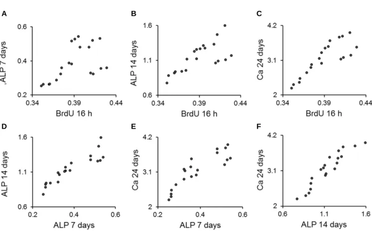
인간치주인대세포를 16 시간 배양 후 BrdU 실험 결과(BrdU 16 h); 인간치주인대세포를 7, 14일 골활성 배양 후 알칼리성 인산 가수분해효소 활성 실험 결과(ALP 7 days, ALP 14 days); 인간치주 인대세포를 24일 골활성 배양 후 세포외 칼슘 침착 실험 결과 (Ca 24 days) 등 4개의 실험 결과들을 사용하여 피어슨 상관관계 분석을 시행하였다. 그 결과, 각각의 모든 변수(BrdU 16 h, ALP 7 days, ALP 14 days, Ca 24 days)는 상호 간에 유의한 상관관계가 존 재하였다(P<.01) (Table 4, Fig. 6).
고찰
Bromdeoxyuridine 세포접착 실험은 세포성장 혹은 세포증식의 척도를 확인하기 위한 방법이다. 세포분열시에는 DNA합성이 필요하며, DNA합성은 세포분열 S phase에서 일어나게 된다. 이 때 DNA합성에 필요한 Thymidine을 대신하여 BrdU (bromod- eoxyurdine)를 사용하여 세포분열을 시킨 후, ELISA로 사용된 BrdU를 측정하여 세포의 성장 기준을 평가한다.21본 연구에서 는 16시간 세포 배양 후 BrdU 측정을 시행하여 세포들의 viability 정도를 확인하여 적용된 표면의 생체적합성을 평가하였다. 본 연구의 결과, 대조군에 비해 열산화 처리를 동반한 모든 실험 군 등은 BrdU 실험에서 유의성 있는 증가를 나타내었다(Table 1).
따라서 열산화를 통하여 티타늄 표면의 세포접착은 향상되었 고, 이에 따라 세포성장 및 세포증식 역시 증가되었을 것임을 유추할 수 있다. 알칼리성 인산가수분해효소 활성 및 세포외 칼슘침착 수치는 세포광화가 활성 될수록 증가되므로 골아세 포분화의 기준으로 평가된다.22본 연구의 알칼리성 인산가수 분해효소 및 세포외 칼슘침착 실험 결과를 살펴보면 열산화 처리된 모든 실험군들에서 대조군인 평활한 ST군과 비교하여 그 정도가 모두 유의하게 증가하였다(Table 2, Table 3). 이는 티타 늄 표면의 열산화 처리가 골아세포분화와 광화를 강력하게 촉
Fig. 6. Scatter-plot results from the Pearson's correlation analysis. Significant correlations were present for (A), (B), (C), (D), (E) and (F) (P<.01) (n = 20).
Table 4. Pearson correlation coefficients between the results of the bromodeoxyuridine assay, alkaline phosphatase activity and extracellular calcium deposition of human periodontal ligament cells
BrdU 16h ALP 7 days ALP 14 days
ALP 7 days 0.555**
ALP 14 days 0.713** 0.915**
Ca 24 days 0.844** 0.880** 0.941**
** Correlation is significant at the 0.01 level (2-tailed).
N = 20.
A B C
D E F
진함을 나타낸다. 티타늄 산화막의 안정성을 극대화하기 위한 적정 열산화 온도 조건 규명이 몇몇 연구들이 진행되었다. 티 타늄 표면에 700℃와 500℃에서 각각 열산화를 시행하였을 때, 700℃ 열산화 처리군의 세포접착이 증가되었고,14700℃ 열산 화 처리시 티타늄 표면에서 골아세포분화와 골형성이 증가된 결과가 보고되었다.15본 실험의 결과는 700℃의 적정한 온도조 건을 통한 열산화 처리한 티타늄 표면이 세포접착과 골광화를 증가시킨다는 다른 연구들과 상응한다고 볼 수 있다.23
본 연구에서 사용된 폭/깊이가 15/3.5 ㎛, 30/10 ㎛ 및 60/10 ㎛인 마이크로그루브들은, 이들 중 60/10 ㎛ 마이크로그루브 표면군 이 가장 높은 수준의 골아세포성숙(osteoblast maturation)을 유도 함을 보고한 이전 논문의 실험군들을 기준으로 선정되었다.24 본 연구에서, 마이크로그루브/열산화 동반 표면 실험군들인 Gr15-TO, Gr60-TO군이 대조군 혹은 ST-TO군과 비교하여 유의한 세포접착 증가를 유도하였으며(Table 1), 알칼리성 인산가수분 해효소, 세포외 칼슘침착 실험 등에서도 마이크로그루브/열산 화 동반 표면을 갖는 모든 실험군들이 ST와 ST-TO군과 비교하 여 유의한 골아세포분화 증가를 유도하였다(Table 2, Table 3).
이는 열산화를 동반한 마이크로그루브 티타늄 표면이 평활면 티타늄이나 열산화만 처리된 티타늄에 비해 세포접착, 골아세 포분화, 골광화 등 골유착에 필수적인 다양한 세포행동들을 종합적으로 향상시킴을 의미하며, 마이크로그루브 표면에 양 극산화를 통해 산화막 증가시킴으로써, 표면화학, 세포접착, 골아세포분화 및 유전자 발현 증진의 시너지 효과를 규명한 기존 연구 결과와 상응한다.25특히, Gr60-TO 실험군이 대조군 및 타 실험군들에 비해 세포접착, 골아세포분화, 골광화 등 모든 경우에서 가장 높은 수준의 증가를 나타낸 점은 본 연구의 핵 심 결과로 볼 수 있다. 이는 열산화가 동반된 60 ㎛ 폭과 10 ㎛ 깊 이의 마이크로그루브 표면 성상이 세포증식과 골활성 향상에 가장 효과적인 티타늄 표면을 유도함을 시사하며, 티타늄에 60/10 ㎛ 폭과 10 ㎛ 깊이의 마이크로그루브를 부여한 후 세포접 착 및 골아세포분화를 규명한 이전 연구들과 일부 상응하는 결과이다.26
피어슨상관관계분석을 통하여 BrdU 측정, 알칼리성 인산가 수분해효소 및 세포외 칼슘침착 실험들의 모든 결과들 간에 통계적으로 유의한 상관관계가 있음이 확인되었다(Table 4, Fig.
6). 특히, Brdu 16 h과 Ca 24 days 간의 높은 상관관계가 확인되었 으며, 이는 티타늄 표면의 세포접착과 골아세포분화/골광화 사이에 밀접한 관계가 존재함을 의미한다. 즉, 세포접착에 유 리한 표면이 활발한 세포증식을 유도하고, 이후 골아세포분화/
골강화까지 유효한 영향을 미친다는 점이 본 연구 결과에서 나타난 것이다. 본 연구를 통해 열산화 및 마이크로그루브 복 합 표면의 골아세포반응 촉진 효과가 확인되었으나, 더욱 구 체적인 최적의 열산화 온도 및 마이크로그루브 크기 조건을 규명하는 진전된 연구가 필요하다. 또한, 세포 수준 규명 이후 적절한 동물 모델을 이용한 성능 및 유효성 평가를 통하여 더 욱 구체적인 검증이 필요하다.
결론
본 연구에서 700℃ 열산화 처리 및 마이크로그루브 복합 티 타늄 표면의 세포접착 및 골활성 증진 능력이 규명되었다. 또 한, 60 ㎛ 폭과 10 ㎛ 깊이의 마이크로그루브에 열산화 처리한 복합 티타늄 표면이 가장 효과적으로 세포접착과 골활성을 증 진시킨다는 사실도 규명되었다. 향후 연구에서는 다양한 거칠 기를 가지는 단순 열산화 표면, 보다 다양한 크기의 단순 마이 크로그루브 표면 및 마이크로그루브-열산화 복합 표면에서의 비교를 통하여 마이크로그루브와 열산화 동반처리의 골아세 포 반응 유도 효과를 비교함과 동시에, 두 가지 표면 처리를 통 한 시너지 효과에 대한 추가적인 규명 또한 필요하리라 사료 된다. 이러한 향후 연구의 필요성에도 불구하고, 본 연구에서 규명된 적정한 마이크로그루브 크기와 열산화 처리 조건은 마 이크로그루브-열산화 복합 표면 티타늄 임플란트 개발의 기초 확립에 기여할 수 있을 것이다.
ORCID
Richard Leesungbok http://orcid.org/0000-0002-8381-723X Su-Jin Ahn http://orcid.org/0000-0003-2128-1561
Suk Won Lee http://orcid.org/0000-0003-2726-3567
References
1. Albrektsson T, Wennerberg A. Oral implant surfaces: Part 1--re- view focusing on topographic and chemical properties of different surfaces and in vivo responses to them. Int J Prosthodont 2004;
17:536-43.
2. Long M, Rack HJ. Titanium alloys in total joint replacement-- a materials science perspective. Biomaterials 1998;19:1621- 39.
3. Jansen JA, van de Waerden JP, Wolke JG, de Groot K. Histologic evaluation of the osseous adaptation to titanium and hydroxya- patite-coated titanium implants. J Biomed Mater Res 1991;25:973- 89.
4. Gaviria L, Salcido JP, Guda T, Ong JL. Current trends in dental implants. J Korean Assoc Oral Maxillofac Surg 2014;40:50-60.
5. Wong M, Eulenberger J, Schenk R, Hunziker E. Effect of surface topology on the osseointegration of implant materials in trabecular bone. J Biomed Mater Res 1995;29:1567-75.
6. Junker R, Dimakis A, Thoneick M, Jansen JA. Effects of implant surface coatings and composition on bone integration: a systematic review. Clin Oral Implants Res 2009;20:185-206.
7. Balasundaram G, Yao C, Webster TJ. TiO2nanotubes func- tionalized with regions of bone morphogenetic protein-2 increases osteoblast adhesion. J Biomed Mater Res A 2008;84:447-53.
8. Buch F, Albrektsson T, Herbst E. Direct current influence on bone formation in titanium implants. Biomaterials 1984;5:341-6.
9. Eisenbarth E, Velten D, Schenk-Meuser K, Linez P, Biehl V, Duschner H, Breme J, Hildebrand H. Interactions between
cells and titanium surfaces. Biomol Eng 2002;19:243-9.
10. Kataoka Y, Tamaki Y, Miyazaki T. Synergistic responses of su- perficial chemistry and micro topography of titanium created by wire-type electric discharge machining. Biomed Mater Eng 2011;21:113-21.
11. Feng B, Chen JY, Qi SK, He L, Zhao JZ, Zhang XD. Characteri- zation of surface oxide films on titanium and bioactivity. J Mater Sci 2002;13:457-64.
12. Raikar GN, Gregory JC, Ong JL, Lucas LC, Lemons JE, Kawahara D, Nakamura M. Surface characterization of titanium implants. J Vac Sci Technol 1995;13:2633-7.
13. Boyan BD, Hummert TW, Dean DD, Schwartz Z. Role of ma- terial surfaces in regulating bone and cartilage cell response.
Biomaterials 1996;17:137-46.
14. Garcl′a-Alonso MC, Saldaña L, Vallés G, Gonza′lez-Carrasco JL, Gonza′lez-Cabrero J, Martl′nez ME, Gil-Garay E, Munuera L. In vitro corrosion behaviour and osteoblast response of thermally oxidised Ti6Al4V alloy. Biomaterials 2003;24:19-26.
15. Saldaña L, Vilaboa N, Vallés G, Gonza′lez-Cabrero J, Munuera L. Osteoblast response to thermally oxidized Ti6Al4V alloy. J Biomed Mater Res A 2005;73:97-107.
16. den Braber ET, de Ruijter JE, Smits HT, Ginsel LA, von Recum AF, Jansen JA. Quantitative analysis of cell proliferation and ori- entation on substrata with uniform parallel surface micro- grooves. Biomaterials 1996;17:1093-9.
17. Lee SW, Kim SY, Rhyu IC, Chung WY, Leesungbok R, Lee KW.
Influence of microgroove dimension on cell behavior of human gingival fibroblasts cultured on titanium substrata. Clin Oral Implants Res 2009;20:56-66.
18. Zhao G, Zinger O, Schwartz Z, Wieland M, Landolt D, Boyan BD. Osteoblast-like cells are sensitive to submicron-scale surface structure. Clin Oral Implants Res 2006;17:258-64.
19. Zhao L, Mei S, Chu PK, Zhang Y, Wu Z. The influence of
hierarchical hybrid micro/nano-textured titanium surface with titania nanotubes on osteoblast functions. Biomaterials 2010;31:
5072-82.
20. Gittens RA, McLachlan T, Olivares-Navarrete R, Cai Y, Berner S, Tannenbaum R, Schwartz Z, Sandhage KH, Boyan BD.
The effects of combined micron-/submicron-scale surface roughness and nanoscale features on cell proliferation and dif- ferentiation. Biomaterials 2011;32:3395-403.
21. Gratzner HG. Monoclonal antibody to 5-bromo- and 5-iodo- deoxyuridine: A new reagent for detection of DNA replication.
Science 1982;218:474-5.
22. Owen TA, Aronow M, Shalhoub V, Barone LM, Wilming L, Tassinari MS, Kennedy MB, Pockwinse S, Lian JB, Stein GS.
Progressive development of the rat osteoblast phenotype in vitro: reciprocal relationships in expression of genes associated with osteoblast proliferation and differentiation during formation of the bone extracellular matrix. J Cell Physiol 1990;143:420-30.
23. Feng B, Weng J, Yang BC, Qu SX, Zhang XD. Characterization of surface oxide films on titanium and adhesion of osteoblast.
Biomaterials 2003;24:4663-70.
24. Lee MH, Oh N, Lee SW, Leesungbok R, Kim SE, Yun YP, Kang JH. Factors influencing osteoblast maturation on microgrooved titanium substrata. Biomaterials 2010;31:3804-15.
25. Lee MH, Kang JH, Lee SW. The effect of surface microgrooves and anodic oxidation on the surface characteristics of titanium and the osteogenic activity of human periodontal ligament cells.
Arch Oral Biol 2013;58:59-66.
26. Im BJ, Lee SW, Oh N, Lee MH, Kang JH, Leesungbok R, Lee SC, Ahn SJ, Park JS. Texture direction of combined mi- crogrooves and submicroscale topographies of titanium substrata influence adhesion, proliferation, and differentiation in human primary cells. Arch Oral Biol 2012;57:898-905.
마이크로그루브 및 열산화 복합 티타늄 표면의 골아세포분화 증진효과
서진호1∙이성복1∙안수진1∙박수정1∙이명현2∙이석원1*
1강동경희대학교치과병원(생체재료)보철과 경희대학교 치과대학, 치의학전문대학원 치과보철학교실
2한국세라믹기술원 그린세라믹본부
목적: 다양한 크기의 마이크로그루브가 형성된 티타늄 표면에 열산화 처리를 한 복합 표면의 표면특성을 규명하고, 인간치주인대세포 배양 시 표 면에 따른 다양한 세포행동들간 차이와 상관관계를 분석하고자 하였다.
재료 및 방법: Grade II 티타늄 디스크를 시편으로 제작하였다. 포토리소그라피를 이용하여 티타늄 시편의 마이크로그루브 크기를 폭/깊이 0/0 ㎛, 15/3.5 ㎛, 30/10 ㎛, 60/10 ㎛로 각각 형성하였다. 평활한 티타늄 표면인 대조군(ST)을 제외한 모든 실험군(ST/TO, Gr15-TO, Gr30-TO, Gr60-TO)에 700℃
에서 3시간동안 열산화 처리하고, 주사현미경 사진을 사용하여 표면특성을 평가하였다. 인간치주인대세포를 배양한 후 BrdU (Bromdeoxyuridine) 실 험, 알칼리성 인산가수분해효소 활성 실험, 세포외 칼슘 침착 실험을 통해 세포접착, 세포분화 및 골광화를 평가하였다. 통계분석으로는 일요인분 산분석과 피어슨상관관계분석(SPSS version 17.0)을 사용하였다.
결과: 열산화를 동반한 마이크로그루브가 형성된 실험군(Gr15-TO, Gr30-TO, Gr60-TO)들은 평활한 대조군(ST)과 단순 열산화 처리 실험군(ST-TO)에 비하여 BrdU 실험, 알칼리성 인산가수분해효소 활성 실험, 세포 외 칼슘 침착 실험 모두에서 유의하게 증가된 활성도를 나타내었다. 특히, Gr60-TO 군은 대조군 및 Gr15-TO, Gr30-TO, Gr60-TO 군 등에 비해 가장 증진된 세포접착 및 골아세포분화/골광화를 나타냈다.
결론: 본 연구의 한계 내에서, 열산화 처리 및 마이크로그루브 복합 티타늄 표면은 골아세포분화에 효과적 방법임이 확인되었다. 본 연구에서 규명 된 적정한 마이크로그루브 크기와 열산화 처리 조건은 마이크로그루브-열산화 복합 표면 티타늄 임플란트 개발의 기초 확립에 기여할 수 있을 것 이다. (대한치과보철학회지 2015;53:198-206)
주요단어: 티타늄; 마이크로그루브; 열산화; 세포접착; 골아세포분화
*교신저자: 이석원
134-727 서울시 강동구 동남로 892 (상일동 149) 경희대학교 치과대학, 치의학전문대 학원 치과보철학교실
02-440-7519: e-mail, [email protected]
원고접수일: 2015년 6월 18일 / 원고최종수정일: 2015년 7월 13일 / 원고채택일: 2015년 7월 15일
2015 대한치과보철학회
이 글은 크리에이티브 커먼즈 코리아 저작자표시-비영리 3.0 대한민국 라이선스에 따라 이용하실 수 있습니다.
c cc
※이 논문은 한국연구재단의 지원을 받아 수행된 기초연구사업임(NRF-20100021268).