• 검색 결과가 없습니다.

Whole Exome Sequencing in the Male Breast Cancer with Prolactinoma: A Case Report and Literature Review

N/A
N/A
Protected

Academic year: 2021

Share "Whole Exome Sequencing in the Male Breast Cancer with Prolactinoma: A Case Report and Literature Review"

Copied!
9
0
0

로드 중.... (전체 텍스트 보기)

전체 글

(1)

ABSTRACT

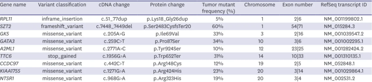
Male breast cancer (MBC) is rare and accounts for approximately 1% of all breast cancer cases worldwide. Previous studies have suggested that several factors significantly increase the risk of MBC. Prolactinoma has the highest incidence rate among patients with functional pituitary tumors. However, whether prolactinoma is involved in the onset and progression of breast cancer remains unclear. To date, there are only five case reports globally on MBC with concurrent prolactinoma. We hereby describe the first case of MBC with prolactinoma in China. We also explored the patient's genetic profile using whole exome sequencing. Our findings may help advance our understanding of the molecular pathogenesis of MBC. Further molecular analyses of such cases are warranted to improve auxiliary molecular diagnostic methods and targeted therapy for MBC.

Keywords: Breast neoplasms, male; Prolactinoma; Whole exome sequencing;

Pituitary neoplasms

INTRODUCTION

Male breast cancer (MBC) is a rare form of breast cancer, accounting for approximately 1%

and 1.4% of all breast cancer cases in Europe and the Americas, and China, respectively. Its incidence varies by race and region [1]. Consistent with other types of cancer, the risk of MBC increases with age. In contrast to female breast cancers, the age of MBC onset is delayed and follows a unimodal distribution. To date, prospective studies on MBC are scarce, and clinical trials on breast cancer treatments have often excluded male participants. In the past decade, great efforts have been made to improve our understanding of the biological characteristics of MBC and develop effective treatments. Previous studies have suggested that certain factors significantly increase the risk of MBC.

It has been reported that approximately 15%–20% of patients with MBC have a family history of breast or ovarian cancer, and approximately 10% of the patients have a genetic predisposition toward this disease. Indeed, BRCA2 is the most common gene mutation associated with MBC, and carriers have a lifetime risk of 5%–10%; in comparison, carriers of the BRCA1 mutation have a lifetime risk of 1%–5% [2]. In addition, Klinefelter's syndrome, which is a relatively common disease characterized by primary hypogonadism and low

Case Report

Received: Mar 12, 2020 Accepted: Jul 29, 2020 Correspondence to Donglin Luo

Department of Breast, Thyroid Surgery, Daping Hospital, Army Medical University, Chongqing 40042, China.

E-mail: luodonglin1967@163.com

© 2020 Korean Breast Cancer Society This is an Open Access article distributed under the terms of the Creative Commons Attribution Non-Commercial License (https://

creativecommons.org/licenses/by-nc/4.0/) which permits unrestricted non-commercial use, distribution, and reproduction in any medium, provided the original work is properly cited.

ORCID iDs Shuai Hao

https://orcid.org/0000-0003-4942-2215 Miao Huang

https://orcid.org/0000-0001-6828-8678 Wuguo Tian

https://orcid.org/0000-0001-6414-6808 Yi Chen

https://orcid.org/0000-0002-6919-5446 Jianjie Zhao

https://orcid.org/0000-0003-0950-9623 Donglin Luo

https://orcid.org/0000-0001-9329-0147 Funding

This study was funded by the Chonqing Science and Technology Bureau (No.2019QNXM027).

Conflict of Interest

The authors declare that they have no competing interests.

Shuai Hao

1

, Miao Huang

2

, Wuguo Tian

1

, Yi Chen

1

, Jianjie Zhao

1

, Donglin Luo

1

1

Department of Breast, Thyroid Surgery, Daping Hospital, Army Medical University, Chongqing, China

2

Nursing School, Chongqing Medical University, Chongqing, China

Whole Exome Sequencing in the Male Breast Cancer with Prolactinoma:

A Case Report and Literature Review

(2)

Author Contributions

Conceptualization: Hao S, Huang M, Tian W;

Funding acquisition: Tian W; Investigation:

Hao S, Tian W, Chen Y; Methodology: Huang M, Chen Y, Luo D; Project administration: Luo D; Supervision: Zhao J, Luo D; Validation: Zhao J, Luo D; Writing - original draft: Hao S; Writing - review & editing: Huang M.

testosterone levels, has been associated with increased risk of MBC. In addition to genetic factors, conditions related to elevated estrogen levels have also been associated with MBC, such as gynecomastia, liver disease, and obesity [3].

Prolactinoma (PRL) is the most common form of functional pituitary adenoma, accounting for 50% of all pituitary adenoma cases. Long-term excessive secretion of prolactin can lead to symptoms such as amenorrhea, galactorrhea and infertility in females. Besides, hyperprolactinemia can also cause loss of libido, erectile dysfunction with hypogonadism, and manifest as azoospermia in males. Previous studies have shown that prolactin may be involved in tumorigenesis by promoting cell proliferation, cell differentiation, as well as tumor angiogenesis and invasion [4]. However, whether prolactinoma is involved in the onset and progression of breast cancer remains unclear. To the best of our knowledge, only five cases of MBC with concurrent prolactinoma have currently been reported worldwide. In the present article, we report the first case of MBC with prolactinoma in China. We also explored the patient's genetic profile using whole exome sequencing (WES), with the aim to improve auxiliary molecular diagnostic methods and targeted therapy for MBC.

CASE REPORT

A 51-year-old male patient presented with a 4-year history of clear discharge from both nipples without an obvious cause, and a 3-year history of a right-sided breast lump, both of which had not been treated. As the lump had enlarged in the preceding months, the patient visited our hospital for consultation. Breast ultrasonography suggested a 3.4 × 1.7 cm sized, irregular and hypoechoic nodular lesion located near the right nipple with ill-defined borders and heterogeneous internal echoes. Breast magnetic resonance imaging (MRI) revealed a patchy, long T1 and T2 signal shadow located in the outer region of the right breast, the side toward the nipple was 0.8 cm and that toward the pectoralis major was 2 mm. The maximum section of the lesion was 3.2 × 1.5 cm in size; an enhanced scan showed an evident heterogeneous enhancement and irregular lesion shape. Small lymph nodes were detected bilaterally in the axillae, suggesting breast cancer (Figure 1). The patient underwent percutaneous biopsy of his right breast tumor under local anesthesia; pathological findings suggested a right- sided invasive breast cancer. A systemic physical examination, an enhanced computed tomography scan of the thorax and abdomen, and a whole-body bone scan showed no signs of abnormal metastasis. A coronal brain MRI scan suggested an enlarged pituitary; this was further confirmed by sagittal imaging, with the lesion measuring 1.2 × 0.9 cm, exhibiting heterogeneous signals and patchy long T1 and long T2 signals on the left side, suggesting a pituitary tumor (Figure 2). Hormone level testing revealed the following: prolactin 153 ng/mL, luteinizing hormone 4.21 mIU/mL, follicle-stimulating hormone 5.97 mIU/mL, progesterone 0.43 ng/mL, testosterone 4.24 ng/mL, and estradiol 43.2 pg/mL; these were consistent with a diagnosis of prolactinoma. After a multidisciplinary team (MDT) discussion on the subsequent treatment, the patient underwent a modified radical mastectomy (right side) under general anesthesia on June 5, 2019. The postoperative pathological findings revealed infiltrating ductal carcinoma, with massive degeneration and necrosis in the cancerous tissue.

Malignancy was not detected at the incision margins at the base or around the breast, and

metastasis was not found in the axillary lymph nodes (0/17). Immunohistochemistry showed

expressions of estrogen receptor (ER) (+++), progesterone receptor (PR) (+++), Her-2 (−),

and Ki-67 (90%) (Figure 3). The patient was started on a 6-cycle TEC chemotherapy regimen

(docetaxel 75 mg/m

2

+ epirubicin 75 mg/m

2

+ cyclophosphamide 500 mg/m

2

) on the ninth

(3)

day after surgery, which was followed by endocrinotherapy with oral tamoxifen at a dose of 20 mg/day. Oral bromocriptine at a dose of 7.5 mg/day was also administered to treat the prolactinoma. Follow-up outpatient visits were scheduled once every 3 months and included imaging and blood tests. An MRI re-examination of the pituitary on September 11, 2019 and December 19, 2019 showed no evident changes. Re-test of hormone levels showed prolactin levels of 110 ng/mL and 83 ng/mL on September 10, 2019 and December 18, 2019, respectively;

these were significantly reduced when compared with baseline measurements. To date, no recurrence of the tumor has been detected.

Figure 1. Findings of MRI of the breast.

Breast MRI showing a space-occupying mass in the outer region of the right breast, measuring approximately 3.2

× 1.5 cm, 0.8 cm side towards the nipple and 2 mm side towards the pectoralis major.

MRI = magnetic resonance imaging.

A B

Figure 2. Findings of MRI of the brain.

(A) Coronal MRI and (B) sagittal MRI showing an enlarged pituitary, measuring 1.2 × 0.9 cm, with heterogeneous signals and patchy long T1 and long T2 signals displayed on the left side.

MRI = magnetic resonance imaging.

(4)

Given that the molecular characteristics of this type of a tumor have not yet been reported, we performed WES using the Illumina's NovoSeq platform to explore the tumor-associated molecular changes. Paraffin-embedded tumor tissue specimens were used for the WES (Genetron Health, Beijing, China) assay. All experiments and bioinformatic analyses were performed according to the standard procedures. The WES assay detected nine somatic mutations (Table 1). Signaling pathway enrichment (Figure 4) and Gene Ontology (GO) functional analyses (Figure 5) revealed that these genes were primarily enriched in the Kyoto Encyclopedia of Genes and Genomes (KEGG) signaling pathway of inflammatory bowel disease (IBD). GO functional analyses demonstrated that the tumor was enriched for the regulation of secretion by cells, cell surface receptor signaling pathway involved in cell-cell signaling, cell morphogenesis involved in neuron differentiation, axon development, organic hydroxy compound metabolic process, axonogenesis, positive regulation of secretion, positive regulation of secretion by cell, small molecule catabolic process, and the organic hydroxy compound catabolic process. In addition, the enrichment of these functions was consistent with the patient's phenotype.

This case report was approved by the Institutional Review Board of Daping Hospital, Army Medical University (IRB No. 2020[43]), and the study protocol adhered to the principles of the Declaration of Helsinki. Written informed consent was obtained from the patient.

A B

Figure 3. Histopathological characteristics of breast cancer.

(A, B) Histology of the resected mastectomy specimen showing an infiltrating ductal carcinoma of the breast (hematoxylin and eosin stain, magnification ×200 and ×400, respectively).

Table 1. Tissue mutation profiling results of the patient

Gene name Variant classification cDNA change Protein change Tumor mutant

frequency (%) Chromosome Exon number RefSeq transcript ID

RPL11 inframe_insertion c.51_77dup p.Lys18_Gly26dup 5% 1 2|6 NM_001199802.1

SZT2 frameshift_variant c.7448_7449del p.Ser2483CysfsTer20 60% 1 54|71 NM_015284.3

GK5 missense_variant c.205A>G p.Ile69Val 33% 3 2|16 NM_001039547.2

GATA3 missense_variant c.259C>T p.Pro87Ser 34% 10 3|6 NM_001002295.1

A2ML1 missense_variant c.2771A>C p.Tyr924Ser 10% 12 23|25 NM_001282424.2

TTC6 stop_gained c.1956G>A p.Trp652Ter 31% 14 10|33 NM_001310135.1

CCDC97 missense_variant c.442C>T p.Arg148Cys 12% 19 2|5 NM_052848.1

KIAA1755 missense_variant c.1271G>A p.Arg424His 23% 20 3|14 NM_001029864.1

NTSR1 missense_variant c.968G>A p.Arg323His 19% 20 3|4 NM_002531.2

(5)

DISCUSSION

To date, the etiology of MBC remains unclear. A previous study has reported

hyperestrogenism induced by endocrine disorders to be a major risk factor. However, MBC may also be associated with obesity, liver disease, radiation exposure, and family history,

Rich ratio

20 15

10 5

KEGG

Ribosome

Description

Neuroactive ligand-receptor interaction

IBD

Calcium signaling pathway

p value

0 0.01 0.02 0.03 0.04 0.05

Size 0

Figure 4. Signaling pathway enrichment of genes.

Genes primarily enriched in the KEGG signaling pathway of inflammatory bowel disease.

IBD = inflammatory bowel disease; KEGG = Kyoto Encyclopedia of Genes and Genomes.

Rich ratio 7.5 5.0 2.5

GO

Small molecule catabolic process

Description

Cell surface receptor signaling pathway involved in cell-cell signaling Organic hydroxy compound metabolic process

Axonogenesis Regulation of secretion by cell Positive regulation of secretion by cell

Positive regulation of secretion

Cell morphogenesis involved in neuron differentiation Organic hydroxy compound catabolic process

Axon development

p value

0 0.025 0.050 0.075 0.100

Size 0 0.25 0.50 0.75 1.00

Figure 5. GO functional analyses of these genes.

GO functional analyses showing enrichment in the regulation of secretion by cells, cell surface receptor signaling pathway involved in cell-cell signaling, cell morphogenesis involved in neuron differentiation, axon development, organic hydroxy compound metabolic process, axonogenesis, positive regulation of secretion, positive regulation of secretion by cell, small molecule catabolic process, and in the organic hydroxy compound catabolic process.

GO = Gene Ontology.

(6)

among others [3]. The majority of currently available evidence on MBC is derived from retrospective analyses of case reports presented over the past decades, where patients were treated with regimens and protocols established from studies on female breast cancer. At the time of writing, many aspects of MBC remain unknown.

The influence of prolactin on breast cancer is unclear, and different studies on prolactin levels in patients with breast cancer have revealed conflicting results. Some researchers have noted that prolactin levels in patients with breast cancer are not significantly higher than those in control groups. For instance, Dekkers et al. [5] have reported that the standardized mortality ratio among 1,342 patients with breast cancer receiving treatments for hyperprolactinemia was 1.07 (95% confidence interval [CI], 0.5–2.03), suggesting that patients receiving treatment for hyperprolactinemia were not at an increased risk of breast cancer. However, a 10-year study involving 1,400 cases has shown that prolactin was closely associated with the occurrence of breast cancer, especially in postmenopausal women (relative risk [RR], 1.37; p <

0.05) and ER-positive patients (RR, 1.28; 95% CI, 1.07–1.54; p = 0.003) [6].

Hyperprolactinemia (HPRL) is a common clinical syndrome with physiological,

pharmacological, and pathological causes, with prolactinoma being the most commonly seen pathological cause. Prolactinoma has the highest incidence rate among patients with functional pituitary tumors [4].

To date, there are only five case reports on concurrent prolactinoma and MBC (Table 2) [7-11], and no such case has been reported in China. In all of the previously reported cases, HPRL occurred just before or at the time of tumor diagnosis, suggesting that prolactin may play a role in the occurrence of breast cancer. Three of the previously reported cases presented with gynecomastia, one of them had a palpable breast lump, whereas the remaining cases involved bloody nipple discharge. All of them presented with abnormally high levels of prolactin.

In one of these, there was a significant time interval between the diagnosis of pituitary prolactinoma and breast cancer, while no such significance was observed in the remaining two cases. In the present case, the pituitary tumor was detected via brain MRI after the diagnosis of breast cancer, and a diagnosis of prolactinoma was confirmed by abnormally high prolactin level. Nevertheless, given that the first incidence of nipple discharge occurred without an obvious cause 4 years before admission, the early onset of prolactinoma cannot be excluded. Therefore, regular breast examinations for patients with prolactinoma may help with the identification of early-stage breast cancer.

Among the 5 previously reported cases, three had bilateral breast cancer and 2 had a unilateral tumor; 4 presented with invasive ductal carcinoma and the remaining only had ductal carcinoma in situ. In the present case, an invasive ductal carcinoma was confirmed by the immunohistochemical expression of ER+, consistent with the previously reported cases. In addition, to the best of our knowledge, this is the first study to examine the Table 2. Prolactinoma associated with breast cancer in male patients

Author Year Age Type Side Symptom Interval * Treatment

Olsson et al. [7] 1984 48 IDC Bilateral Gynecomastia R-26/L-36 S, DXT

Haga et al. [8] 1993 68 IDC L, unilateral Palpable breast lump 0 B

Volm et al. [9] 1997 70 IDC Bilateral Gynecomastia 8 S, DXT

Forloni et al. [10] 2001 45 L-IDC, R-DCIS Bilateral Gynecomastia 0 B

Mallawaarachchi et al. [11] 2011 56 DCIS R, unilateral Bloody nipple discharge 3 B

* Interval, between the diagnosis of prolactinoma and breast cancer.

IDC = invasive ductal carcinoma; DCIS = ductal carcinoma in situ; L = left; R = right; S = pituitary surgery; DXT = radiotherapy; B = bromocriptine.

(7)

molecular characteristics of this tumor type. Nine mutant genes were found through the WES (Genetron Health) assay of paraffin-embedded tumor tissue specimens. This mutational landscape was compared with those of breast cancer hosted by The Cancer Genome Atlas repository. Figure 6 shows the mutational landscape of 938 breast cancer tissues stratified by sex along with the mutation status of our case listed on the left. Among the nine somatic mutants, GATA3 appears as one of the most recurrent mutants in breast cancer. Most somatic mutants in GATA3 are frameshift mutations which may cause the loss of function.

In addition, these mutations are enriched in the zinc finger 1 (ZnF1) and zinc finger 2 (ZnF2) domains which are responsible for maintaining stability and DNA binding. Although GATA3 p.Pro87Ser detected in our case falls outside of known hotspots, it is predicted by the scale-invariant feature transform algorithm as a possibly damaging mutation. The exact functional consequence of this mutant requires further investigation. Considering the high allele frequency (34%) in our case, it is possible that the GATA3 mutation served as an early oncogenic event. Previous studies have suggested that GATA3 functions in the differentiation of luminal epithelial cells [12]. More specifically, the lack of normal GATA3 expression drives tumorigenesis through promoting the epithelial-mesenchymal transition (EMT), a known characteristic in the progression of various cancers. The protein encoded by the GATA3 gene belongs to the GATA3 family of transcription factors, which is involved in the development of breast cancer through the regulation of different target genes involved in various pathways such as epithelial-mesenchymal transformation, metastasis and tumor differentiation [12].

GATA3 is frequently mutated in breast cancer, and the mutation mostly occurs in exons 5 and 6. Approximately 41% (27/66) of the code shift mutation in GATA3 was predicted to lead to early generation of the stop codon, and the encoded truncated protein products were stable and expressed in the tumor [13]. Association of GATA3 with good prognosis has been reported in female breast cancers but not in MBCs. It is possible that the mutant is not

4,000 2,000

0 0 100 200 300

34%

33%

19%

14%

13%

12%

2.0%

1.0%

1.0%

0.4%

0.4%

0.3%

0.3%

0.1%

TP53 PIK3CA TTN CDH1 GATA3 MUC16 SZT2 A2ML1 KIAA1755 GK5 NTSR1 CCDC97 TTC6 RPL11

Case Gender Male Female

frameshift_variant stop_gained

Multi_Hit missense_variant splice_variant

inframeshift_variant Figure 6. Mutational landscape of breast cancer.

Mutational landscape of 938 breast tumor samples from TCGA. The left cell indicates the somatic mutants detected in our case. The upper penal shows somatic mutations with high recurrence (> 10%) in breast cancer.

The lower penal shows the rest of the somatic mutants in our case. Colors in the panel indicate different mutation types. The left bar represents the occurrence of corresponding mutants in males and females.

TCGA = The Cancer Genome Atlas.

(8)

clinically important, but the functional consequence of the mutant is important. Due to the rarity of MBCs with prolactinoma, further in-depth studies are required to better elucidate the clinical implications of GATA3 mutation in males [14]. Further pathway enrichment and GO functional analyses revealed that these genes were mainly enriched in the KEGG signaling pathway of IBD. A previous study has suggested that IBD significantly correlated with breast cancer [15]. Furthermore, this functional enrichment profile was consistent with the phenotype of the present patient. This finding may help advance our understanding of the molecular pathogenesis of breast cancer. Further molecular analyses of such cases are warranted, with an aim to develop targeted therapies suitable for MBC patients.

With the development in science and technology, research on prolactinoma has advanced surgery, medication, radiation, and other treatment methods that reduce tumor size, restore normal levels of prolactin, maintain pituitary and other neural functions, prevent tumor recurrence, and improve patients' quality of life. Drug therapies are the most effective treatment for prolactinoma. Dopamine receptor agonists have allowed for restoration of normal prolactin levels and tumor size reduction in most patients [4]. Among the previously reported cases, 2 patients had a giant pituitary adenoma, each of which was removed surgically; the other three patients were treated with oral drug therapies and their prolactin levels were well controlled. Owing to the smaller pituitary adenoma in the present case, he was treated with bromocriptine at a dose of 7.5 mg/day, which significantly reduced his prolactin level at 3 and 6 months after treatment. During the ongoing follow-up, no disease progression has been detected.

Here, we report the first case of MBC with concurrent prolactinoma in China. Based on the present case and literature review findings, prolactin may play a role in the occurrence of breast cancer. We advocate careful and regular examinations of patients with prolactinoma, as well as clinical assessment and imaging tests for populations at high risk of breast cancer, as these may aid in the early detection and prompt treatment of new breast cancer patients.

However, as this is the only currently available report involving analysis of the patient's molecular data, future studies on larger samples are required to create a detailed molecular atlas of breast cancer with prolactinoma to facilitate the development of molecular diagnostic method and targeted therapy.

ACKNOWLEDGMENTS

We would like to thank Xiajie Wei (Genetron Health Co. Ltd., Beijing, China) and Hongling Yuan (Genetron Health Co. Ltd., Beijing China) for data processing and bioinformatics analysis.

REFERENCES

1. Chen W, Zheng R, Baade PD, Zhang S, Zeng H, Bray F, et al. Cancer statistics in China, 2015. CA Cancer J Clin 2016;66:115-32.

PUBMED | CROSSREF

2. Silvestri V, Barrowdale D, Mulligan AM, Neuhausen SL, Fox S, Karlan BY, et al.. Male breast cancer in BRCA1 and BRCA2 mutation carriers: pathology data from the Consortium of Investigators of Modifiers of BRCA1/2. Breast Cancer Res 2016;18:15.

PUBMED | CROSSREF

(9)

3. Giordano SH. Breast cancer in men. N Engl J Med 2018;378:2311-20.

PUBMED | CROSSREF

4. Romijn JA. Hyperprolactinemia and prolactinoma. Handb Clin Neurol 2014;124:185-95.

PUBMED | CROSSREF

5. Dekkers OM, Romijn JA, de Boer A, Vandenbroucke JP. The risk for breast cancer is not evidently increased in women with hyperprolactinemia. Pituitary 2010;13:195-8.

PUBMED | CROSSREF

6. Tworoger SS, Eliassen AH, Zhang X, Qian J, Sluss PM, Rosner BA, et al. A 20-year prospective study of plasma prolactin as a risk marker of breast cancer development. Cancer Res 2013;73:4810-9.

PUBMED | CROSSREF

7. Olsson H, Alm P, Kristoffersson U, Landin-Olsson M. Hypophyseal tumor and gynecomastia preceding bilateral breast cancer development in a man. Cancer 1984;53:1974-7.

PUBMED | CROSSREF

8. Haga S, Watanabe O, Shimizu T, Iida T, Imamura H, Kajiwara T, et al. Breast cancer in a male patient with prolactinoma. Surg Today 1993;23:251-5.

PUBMED | CROSSREF

9. Volm MD, Talamonti MS, Thangavelu M, Gradishar WK. Pituitary adenoma and bilateral male breast cancer: an unusual association. J Surg Oncol 1997;64:74-8.

PUBMED | CROSSREF

10. Forloni F, Giovilli M, Pecis C, Bortolani E, Preziosi A, Barzaghi ME, et al. Pituitary prolactin-secreting macroadenoma combined with bilateral breast cancer in a 45-year-old male. J Endocrinol Invest 2001;24:454-9.

PUBMED | CROSSREF

11. Mallawaarachchi CM, Ivanova S, Shorthouse A, Shousha S, Sinnett D. A rare case of male breast ductal carcinoma in-situ associated with prolactinoma. BMJ Case Rep 2011;2011:bcr0120091421.

PUBMED | CROSSREF

12. Du F, Yuan P, Wang T, Zhao J, Zhao Z, Luo Y, et al. The significance and therapeutic potential of GATA3 expression and mutation in breast cancer: a systematic review. Med Res Rev 2015;35:1300-15.

PUBMED | CROSSREF

13. Mair B, Konopka T, Kerzendorfer C, Sleiman K, Salic S, Serra V, et al. Gain- and loss-of-function mutations in the breast cancer gene GATA3 result in differential drug sensitivity. PLoS Genet 2016;12:e1006279.

PUBMED | CROSSREF

14. Gonzalez RS, Wang J, Kraus T, Sullivan H, Adams AL, Cohen C. GATA-3 expression in male and female breast cancers: comparison of clinicopathologic parameters and prognostic relevance. Hum Pathol 2013;44:1065-70.

PUBMED | CROSSREF

15. Tsai MS, Chen HP, Hung CM, Lee PH, Lin CL, Kao CH. Hospitalization for inflammatory bowel disease is associated with increased risk of breast cancer: a nationwide cohort study of an Asian population. Ann Surg Oncol 2015;22:1996-2002.

PUBMED | CROSSREF

수치

Figure 1. Findings of MRI of the breast.
Table 1. Tissue mutation profiling results of the patient
Figure 4. Signaling pathway enrichment of genes.

참조

관련 문서

 Often found connected to other molecules on the outsides of cells --- cellular recognition, cell signaling, cell

ƒ Monosaccharide may be present in the form of Monosaccharide may be present in the form of a linear or ring structure. ƒ In solution it is in the form of

These results suggest that the bilobalide inhibits the cell proliferation and induces the apoptotic cell death in FaDu human pharyngeal squamous cell carcinoma via both

ƒ The amount of a compound per unit cell mass (or cell The amount of a compound per unit cell mass (or cell volume).

Taken together, these results suggested that latex containing the ficin inhibited the cell growth and induced apoptosis by caspase and Bcl-2 family signaling pathway in

In the present study, consistent to that report, up-regulation of VEGF mRNA was observed in the colon cancer cell with the acquired resistance to 5-FU and this

The PIG3 was interacted with the CTSB in human prostate cancer cell line PC3, which was verified by co-immunoprecipitation analysis using specific

In this study, if the cell potential increases in voltaic cell environment and the effect of charge increases with applied external voltage, it promote s phase change