Introduction
Recovery of normal knee range of motion (ROM) is among the primary goals of total knee arthroplasty (TKA) since knee ROM is an important clinical indicator of knee joint function that can be used as a basis for the assessment of the success of TKA1-4). Most daily living activities require 90o–120o knee ROM2,4); how- ever, kneeling, squatting, and sitting cross legged, which necessi- tate high flexion of the knee joint over 120o, are also required for
daily living activities in some cultural, religious, or occupational environment2,3,5,6). Among the various factors influencing postop- erative knee joint ROM, implant design plays an important role, and the high-flexion TKA implant design was developed to en- able more than 120o postoperative flexion of the knee joint1-3,6-8). Until recently, several manufacturers have introduced various de- signs of high-flexion TKA implants, and a number of researches regarding the difference between the conventional implant design and high-flexion implant design have been performed. However, the reality is there are as many studies reporting insignificant dif- ferences between the implant designs9-13) as those reporting high- er flexion after implantation of the high-flexion TKA designs14-17). We compared the minimum 5-year follow-up results at our insti- tution with the results of the previous papers. Although a number of studies reported good results of high-flexion TKA, few reports assessed the results after years of repetitive high-flexion. The pur- pose of this study is to analyze the survival rate of NexGen LPS- Flex (Zimmer Inc., Warsaw, IN, USA) high-flexion implant at minimum 5-year follow-up based on radiological measurements
High-Flexion Total Knee Arthroplasty Using NexGen LPS-Flex System: Minimum 5-year Follow-up Results
Seung Joon Rhee, MD, Sung Min Hong, MD, and Jeung Tak Suh, MD
Department of Orthopaedic Surgery, Pusan National University Hospital, Busan, Korea
Purpose: This study is to report clinical and radiological results of high-flexion total knee arthroplasty (TKA) using NexGen LPS-flex system at a minimum 5-year follow-up, and to analyze the implant survivorship based on the results.
Materials and Methods: A total of 80 patients (118 knees) who underwent patellar preserving TKA using NexGen LPS-flex implant between February 2007 and February 2008 and could be followed for minimum 5 years were reviewed. The range of motion (ROM), hip-knee-ankle angle, Knee Society Knee score (KSKS), and Knee Society Function score (KSFS) were assessed preoperatively and at the last follow-up and analyzed.
Implant position of the femoral and tibial components on the immediate postoperative and last follow-up X-rays were compared.
Results: The mean ROM was 110.2o±14.5o (range, 60o to 140o) preoperatively and 132.4o±5.2o (range, 90o to 145o) at the last follow-up. KSKS was 36.9o±6.4o preoperatively and 94.2o±3.2o at the last follow-up. KSFS was 30.5o±5.7o preoperatively and 93.7o±4.1o at the last follow-up. There was no statistically significant change in the implant position measured as α, β, γ, and δ angles at the last follow-up compared to the immediate postoperative values. Radiolucent lines were observed in 13 knees (11%) on the last follow-up X-rays. Revision TKA was performed due to aseptic implant loosening in 1 knee (0.84%), and the survival rate at the 5th postoperative year was 99.2%.
Conclusions: The clinical and radiological outcomes of high-flexion TKA using NexGen LPS-Flex implant design were satisfactory with 99.2%
implant survival rate after 5 years of protected activities of daily living.
Keywords: Knee, Arthroplasty, Prosthesis design, Survival analylsis pISSN 2234-0726 · eISSN 2234-2451
Knee Surgery & Related Research
Received February 10, 2015; Revised April 19, 2015;
Accepted June 2, 2015
Correspondence to: Jeung Tak Suh, MD
Department of Orthopaedic Surgery, Pusan National University Hospital, 179 Gudeok-ro, Seo-gu, Busan 49241, Korea
Tel: +82-51-240-7248, Fax: +82-51-247-8395 E-mail: [email protected]
156
This is an Open Access article distributed under the terms of the Creative Commons Attribution Non-Commercial License (http://creativecommons.org/licenses/by-nc/4.0/) which permits unrestricted non-commercial use, distribution, and reproduction in any medium, provided the original work is properly cited.
Copyright © 2015 KOREAN KNEE SOCIETY www.jksrr.org
and revision rates.
Materials and Methods
1. Materials
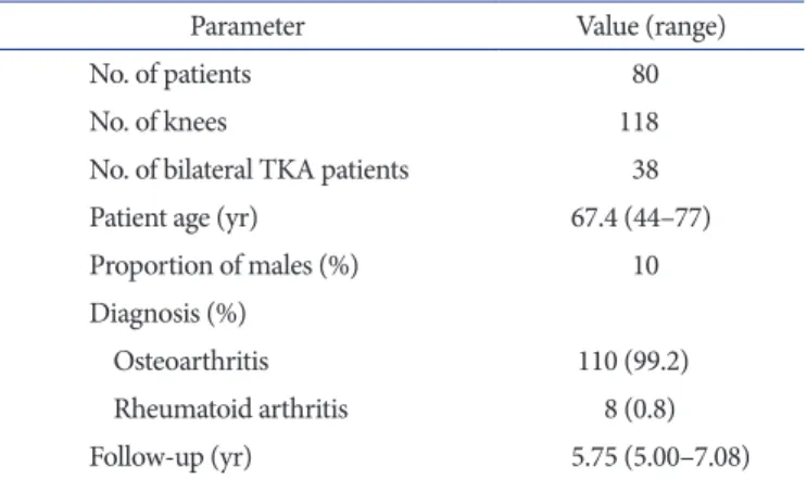
During the period from February 2007 to February 2008, a single surgeon operated TKA using NexGen LPS-Flex implant on 143 knees of 127 patients at our institution. Among them, 118 knees of 80 patients which could be followed for minimum 5 years (range, 5 to 7 years and 1 month; mean, 5 years and 9 months) were enrolled and their clinical and radiological data were analyzed. Patients with more than 15o of preoperative varus alignment or more than 30o of flexion contracture were excluded from the enrollment. There were 8 males (8 knees) and 72 fe- males (110 knees), and their age at the time of TKA was a mean of 67.4 years (range, 44 to 77 years). The indication for surgery was osteoarthritis in 73 patents (110 knees) and rheumatoid ar- thritis in 7 patients (8 knees). Unilateral surgery was done in 42 patients (42 knees) and staged bilateral surgery was done in 38 patients (76 knees) (Table 1).
2. Surgical Techniques and Rehabilitation
Midline skin incision and medial parapatellar approach were applied in all of the cases. Cruciate ligaments were resected as the implant design directs, and both the femoral and tibial com- ponents were fixed with bone cement. Bone cement was spread on the cut surface of the tibia and femur, and also on the implant itself. We cemented and implanted tibial implant first with 6–7 times off impaction using manufactured impactor and then removed excessive cement. After implantation of the tibial com- ponent was completed, the same routine was performed with the femoral implant. Just one mix of the cement was used for the
whole course of cementing. Then, the trial insert was inserted and maintained in position until the heat from the consolidating cement completely dissipated to secure balanced extension gap and to prevent the implant lift-off by the cement volume incre- ment during the consolidation phase. Patellar resurfacing was not done in any case, but osteophyte removal and micro-drilling were done only in the cases with predictable poor patellofemoral tracking due to their severe degeneration.
Quadriceps muscle strengthening exercise was begun on the day of operation. Passive and active ROM exercises were begun on the 3rd postoperative day after the removal of drain. Full weight-bearing ambulation was permitted from the 1st postop- erative week. Patients were encouraged to gain their maximum ROM until 2 to 3 months after TKA; however, we did not rec- ommend squatting, kneeling, or sitting cross legged. Patients were instructed to avoid weight-bearing high-flexion activities as much as possible; however, they were encouraged to gain the ability to perform high-flexion.
3. Clinical and Radiological Evaluation
On clinical evaluation, Knee Society Knee score (KSKS) and Knee Society Function score (KSFS) assessed preoperatively and at the 5th postoperative year follow-up visit were compared. The roentgenographic evaluation and scoring system of the American Knee Society was used to measure and compare the preoperative radiography and postoperative 5-year radiography. Flexion angle of the knee joint was measured by comparing the angle between the longitudinal midline of the femur and tibia in full extension and full flexion positions on the sagittal plane radiograph. As for the change in the implant position, femoral component angle in coronal plane (α angle), tibial component angle in coronal plane (β angle), femoral component angle in sagittal plane (γ angle), and tibial component angle in sagittal plane (δ angle) were measured radiographically. Changes in the α, β, γ, and δ angles between the immediate postoperative and 5-year postoperative follow-up were assessed. Bone-cement interface was set to 7 zones in coro- nal plane view of the femoral component and another 7 zones in coronal plane view of the tibial component, and 3 zones in sagittal plane. Thickness of radiolucent lines in 0.5 mm increments were measured in each zone. The sum of the scores in all of the zones was regarded as insignificant if it was less than 5 and highly suspi- cious of failure if the score was over 9 regardless of clinical symp- toms. Scores between 5 and 9 were regarded as an indication for serial follow-up as to the progression of implant loosening. Gross displacement or tilting of the implant was regarded as implant fixation failure regardless of the presence of a radiolucent line.
Table 1. Patient Demographics
Parameter Value (range)
No. of patients 80
No. of knees 118
No. of bilateral TKA patients 38
Patient age (yr) 67.4 (44–77)
Proportion of males (%) 10
Diagnosis (%)
Osteoarthritis 110 (99.2)
Rheumatoid arthritis 8 (0.8)
Follow-up (yr) 5.75 (5.00–7.08)
TKA: total knee arthroplasty
4. Survival Analysis
Kaplan-Meier survival analysis was done based on the 1-year follow-up results. The 1-year maintenance of the implant in its original location and in positions suggesting possibly good main- tenance of the implant for a longer period was defined as annual success. Patient who were in their 5th postoperative year follow- up period were not included in this analysis, and deaths due to diseases or follow-up loss cases were excluded. Implant removal, revision, or infection was defined as failure.
5. Statistical Analysis
Independent t-test was performed to compare clinical and ra- diological results of preoperative and last follow-up assessments using SPSS ver. 18.0 (SPSS Inc., Chicago, IL, USA). Calculation of survival rates and standard deviations was also done using SPSS ver. 18.0. Log-rank test was used to determine the relationship between the survival rate and gender and causative diseases.
Results
1. Clinical Results
The mean knee flexion angle increased from a mean of 110.2o±14.5o (range, 60o to 140o) preoperatively to a mean of 132.4o±5.2o (range, 90o to 145o) at the last follow-up. The mean knee flexion contracture decreased from 7.8o (range, 0o to 25o) preoperatively to 1.3o (range, 0o to 10o) at the last follow-up.
The KSKS increased from a mean of 36.9±6.4 preoperatively to a mean of 94.2±3.2 at the last follow-up. The KSFS increased from a mean of 30.5±5.7 preoperatively to a mean of 93.7±4.12 at the last follow-up (Table 2). Most patients were satisfied with the improvement in pain: 68 patients (100 knees, 85%) out of 80 patients reported no pain or intermittent slight pain at the last follow-up visit.
2. Radiological Results
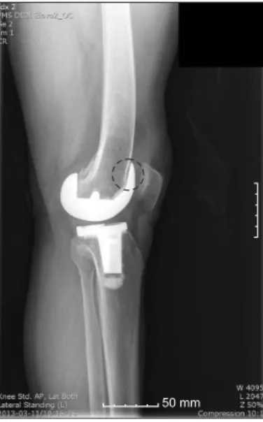
Mechanical axis of the lower limb changed from a mean of 10.2o±2.8o varus (range, 23o varus to 7o valgus) preoperatively to a mean of 3.2o±0.6o valgus (range, 4o varus to 7o valgus) postop- eratively. As for the implant position, the mean values of α, β, γ, and δ angles were 96.2o±1.5o (range, 94o to 99o), 92.4o±1.1o (range, 88o to 94o), 3.2o±1.2o (range, 0o to 5o), and 85.6o±2.4o (range, 80o to 90o), respectively, immediately after surgery and 95.7o±1.7o (range, 93o to 98o), 92.9o±1.9o (range, 89o to 96o), 3.2o±1.2o (range, 0o to 5o), and 85.6o±2.4o (range, 80o to 90o), respectively, at the last follow-up. There was no significant change in the position and alignment of implant between the immediate postoperative and last follow-up measurements (Table 3). Radiolucent lines were observed in 13 knees (11%) out of 118 knees during the 5-year follow-up. All of the radiolucent lines were in the femoral com- ponent: 4 were in zone 1 (anterior surface) and 9 were in zone 4 (posterior surface) (Figs. 1 and 2). The mean score of radiolucent lines in the femoral component was 0.5 (Table 4). However, there
Table 2. Changes in the ROM, HKAA, KSKS, and KSFS between the Preoperative and Last Follow-up Assessments
Variable Preoperative Last follow-up Mean gain p-value ROM (o) 110.2±14.5 132.4±5.2 52.2 0.015
HKAA (o) 10.2±2.8 –3.2±0.6 –13 0.002
KSKS 36.9±6.4 94.2±3.2 57.3 0.023
KSFS 30.5±5.7 93.7±4.1 63.2 0.011
Values are presented as mean±standard deviation.
ROM: range of motion, HKAA: hip-knee-ankle angle, KSKS: Knee Society Knee score, KSFS: Knee Society Function score.
Table 3. Changes of Component Alignment Angle between the Preoperative and Last Follow-up Assessments
Angle Preoperative (o) Last follow-up (o) p-value
α angle 96.2±1.5 95.7±1.7 0.861
β angle 92.4±1.1 92.9±1.9 0.332
γ angle 3.2±0.3 3.2±1.2 0.457
δ angle 85.3±1.9 85.6±2.4 0.182
Values are presented as mean±standard deviation.
Fig. 1. Follow-up roentgenogram of a 65-year-old female patient show- ing a radiolucent line between the anterior flange of the femur and the femoral implant (zone 1).
50 mm
was no statistically significant relationship between the existence of radiolucent lines and postoperative ROM (p=0.901) and KSKS (p=0.321).
3. Survival Analysis
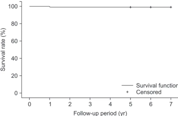
Removal of the implant due to loosening or revision operation was done in 1 knee (0.84%). The overall survival rate calculated using the Kaplan-Meier method was 99.2% (Fig. 3), and there was no statistically significant relationship between the survival rate and the gender (p=0.121) and causative diseases (p=0.257).
4. Complications
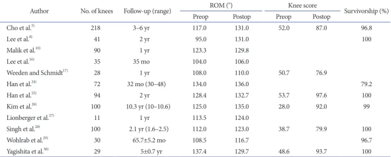
There were 2 cases of delayed wound healing and 1 case of revi- sion due to aseptic loosening of the implant (Fig. 4). The 2 knees with delayed wound healing had past history of diabetes mellitus and their wounds were healed 3 weeks after the operation. Asep- tic loosening occurred in the femoral component in the patient who had rheumatoid arthritis.
Discussion
Recently, vigorous research efforts have been made on implant designs, especially high-flexion designs, due to the increasing number of TKA even among young patients and the growing expectation on functional outcomes based on the improvement of implant durability. High-flexion after TKA is generally defined as greater than 125o of knee joint flexion18). Compared with the conventional implant designs that allow maximum 120o knee flexion, high-flexion designs theoretically enable more than 135o knee joint flexion. Although the preoperative knee joint ROM is among the most important factors influencing high-flexion after TKA19), maximum flexion angle could only be achieved with the Fig. 2. Follow-up roentgenogram of a 70-year-old female patient show-
ing a radiolucent line between the posterior cut surface of the femur and the posterior condyle of the femoral implant (zone 4).
Table 4. Incidence and Zone of Radiolucency Parameter Incidence
(%) Score Zone of radiolucency (cases) Femoral component 13 (11) 0.5 1 (4), 4 (9)
Tibial component 0
Fig. 3. Kaplan-Meier survival analysis: 99.2% survival rate at 5 years after surgery.
Survivalrate(%)
0 80 60 40 20 0
Follow-up period (yr) 100
7
1 2 3 4 5 6
Survival function Censored
Fig. 4. A 74-year-old female patient was diagnosed with aseptic loosen- ing and eventually underwent revision.
aid of an improved implant design.
There are several design characteristics of high-flexion implant designs for improved dynamic conditions compared to the con- ventional designs20). Increased posterior condylar offset makes the posterior condylar curve similar to the round shape, which enables femoral rollback in the higher flexion angle without impinging of the tibial component16,21,22). Trochlear groove of the implant is elongated to avoid patella being entrapped in the intercondylar space with the longer distal contact surface of the patella and femoral components under the high-flexion circum- stances. Anterior lip of the tibial polyethylene insert is beveled to avoid irritating patella and patellar tendon in the high-flexion state. Cam-post mechanism is also modified to increase the jump distance while preventing implant dislocation. Such changes in the implant design necessitate several changes in the surgical techniques as well. For example, 2–3 mm more resection of the posterior condyle is required to insert an implant with greater posterior offset. Sufficient exposure of the posterior joint capsule enhances removal of the posterior osteophytes impinging during high-flexion and enables concise balancing of the flexion gap21). Meticulous resection of the tissues in the intercondylar space must be done for the well-operating high cam-post mechanism.
Individualized implantation of a tibial component with a proper posterior slope and slightly posterior placement of the compo- nent to prevent flexion limitation caused by anterior sloping or anterior positioning of the tibial component are also required.
Joint line elevation should be minimized to avoid patellar baja, which causes flexion limitation23). On the other hand, it is also
true that there are studies reporting increased chances of aseptic loosening, polyethylene insert wear, patellar insufficiency, and instability related to the characteristics of high-flexion implant designs3,5,6,24).
During our research on the NexGen LPS-Flex single implant design, we could find 9 previous literatures regarding the same implant design5,8,12,13,15,24-30) (Table 5). However, only 3 studies fol- lowed a relatively large group of more than 100 cases, and only 2 reports were based on more than 5-year follow-up. There was only 1 report which followed over 100 cases for more than 5 years. Under such circumstances, our minimum 5-year follow-up research on 118 knees with the NexGen LPS-Flex high-flexion implant design provides valuable and rare data. Our results showed a mean 22.2o increase in the knee joint flexion angle from 110.2o±14.5o (range, 60o to 140o) preoperatively to 132.4o±5.2o (range, 90o to 145o) at the last follow-up, which was the highest flexion angle compared to that in the abovementioned research- es, suggesting excellent improvement. KSKS was improved from 36.9±6.4 preoperatively to 94.2±3.2 at the last follow-up, and it was similar to the results of conventional literatures ranging from 76.9 to 97.6. In particular, in our study, radiological assessment on implant position and radiolucent lines was carried out to iden- tify the presence of aseptic loosening and osteolysis. There was no significant change in the implant position from preoperative period to the last follow-up. Radiolucent lines were observed in 13 knees (11%) and all of which were in the femoral component, but none of them met the criteria for implant failure. The exis- tence of radiolucent lines showed no statistical correlation with
Table 5. Summary of Published Clinical Outcomes of Total Knee Arthroplasty Using the NexGen LPS-Flex Knee System
Author No. of knees Follow-up (range) ROM (o) Knee score
Survivorship (%)
Preop Postop Preop Postop
Cho et al.5) 218 3–6 yr 117.0 131.0 52.0 87.0 96.8
Lee et al.8) 41 2 yr 95.0 131.0 100
Malik et al.10) 90 1 yr 123.3 129.8
Lee et al.16) 35 35 mo 104.0 106.0
Weeden and Schmidt17) 28 1 yr 108.0 110.0 50.7 76.9
Han et al.24) 72 32 mo (30–48) 134.0 136.0 79.2
Han et al.25) 94 2 yr 128.4 132.7 53.7 97.6 100
Kim et al.26) 100 10.3 yr (10–10.6) 125.0 135.0 28.0 92.0 99
Lionberger et al.27) 11 1 yr 113.5 124.0
Singh et al.28) 100 2.1 yr (1.6–2.5) 112.0 123.0 38.7 79.9 100
Wohlrab et al.29) 30 65.7±5.2 mo 108.5 116.7 96.7
Yagishita et al.30) 29 5±0.7 yr 137.4 129.7 48.6 93.7 100
ROM: range of motion, Preop: preoperative, Postop: postoperative.
ROM or KSKS. In addition, the patients who showed radiolucent lines in the follow-up radiography reported they could perform more squatting and kneeling during daily living activities than they were recommended to do. Compared to the study of Han et al.24), which reports high incidence of radiolucent lines (38%) and revision surgery (21%) at a mean of 23-month follow-up in the patients with the same NexGen LPS-Flex implant, our results showed 11% incidence of radiolucent lines and revision surgery was required in 1 knee due to aseptic loosening. Han et al.24) al- lowed patients to do weight-bearing high flexion activities as tol- erated. On the contrary, we advised patients not to squat or kneel while encouraging them to recover the ability to squat and kneel.
We suspect that the postoperative patient education and activity restriction could have a profound influence on the difference of the study results. As for the survival analysis, there are literatures calculating survival rates merely based on the ratio of the total number of cases to the number of revision surgery, ignoring the timing of revision surgery or the influence of drop-outs during the research period. In contrast, the 99.2% survival rate in our study was calculated from the Kaplan-Meier survival analysis fac- toring in the influence of drop-out cases as well.
Until recently, numerous research and a few meta-analysis stud- ies have been carried out regarding TKA implants. However, it is difficult to draw a convincing conclusion from a simple compara- tive study of the results due to the diverse operative techniques of individual surgeons and differences in the measurement meth- odology. The limitation of our study lies in the fact that it is not a comparative study, simply providing clinical results and the mid- term survival rate of a single implant design. Therefore, we be- lieve it is necessary to conduct a long-term comparative study on the conventional implant design vs. high-flexion design to ana- lyze practical influence on the results of TKA in future research.
Conclusions
High-flexion TKA using NexGen LPS-Flex implant design yielded satisfactory clinical and radiological outcomes with 99.2%
implant survival rate after 5 years of protected activities of daily living.
Conflict of Interest
No potential conflict of interest relevant to this article was re- ported.
References
1. Anouchi YS, McShane M, Kelly F Jr, Elting J, Stiehl J. Range of motion in total knee replacement. Clin Orthop Relat Res.
1996;(331):87-92.
2. Endres S. High-flexion versus conventional total knee ar- throplasty: a 5-year study. J Orthop Surg (Hong Kong). 2011;
19:226-9.
3. Huddleston JI, Scarborough DM, Goldvasser D, Freiberg AA, Malchau H. 2009 Marshall Urist Young Investigator Award: how often do patients with high-flex total knee ar- throplasty use high flexion? Clin Orthop Relat Res. 2009;
467:1898-906.
4. Miner AL, Lingard EA, Wright EA, Sledge CB, Katz JN;
Kinemax Outcomes Group. Knee range of motion after total knee arthroplasty: how important is this as an outcome mea- sure? J Arthroplasty. 2003;18:286-94.
5. Cho SD, Youm YS, Park KB. Three- to six-year follow-up results after high-flexion total knee arthroplasty: can we al- low passive deep knee bending? Knee Surg Sports Traumatol Arthrosc. 2011;19:899-903.
6. Moon YW, Seo JG, Chang MJ, Yang JH, Jang SW. Minimum five-year follow-up results of single-radius, high-flex posteri- or-stabilized TKA. Orthopedics. 2010;33.
7. Bauman RD, Johnson DR, Menge TJ, Kim RH, Dennis DA.
Can a high-flexion total knee arthroplasty relieve pain and restore function without premature failure? Clin Orthop Relat Res. 2012;470:150-8.
8. Lee BS, Kim JM, Lee SJ, Jung KH, Lee DH, Cha EJ, Bin SI.
High-flexion total knee arthroplasty improves flexion of stiff knees. Knee Surg Sports Traumatol Arthrosc. 2011;19:936-42.
9. Kim YH, Sohn KS, Kim JS. Range of motion of standard and high-flexion posterior stabilized total knee prostheses: a prospective, randomized study. J Bone Joint Surg Am. 2005;
87:1470-5.
10. Malik A, Salas A, Ben Ari J, Ma Y, Gonzalez Della Valle A.
Range of motion and function are similar in patients under- going TKA with posterior stabilised and high-flexion inserts.
Int Orthop. 2010;34:965-72.
11. McCalden RW, MacDonald SJ, Bourne RB, Marr JT. A randomized controlled trial comparing “high-flex” vs “stan- dard” posterior cruciate substituting polyethylene tibial inserts in total knee arthroplasty. J Arthroplasty. 2009;24(6 Suppl):33-8.
12. Ng FY, Wong HL, Yau WP, Chiu KY, Tang WM. Comparison of range of motion after standard and high-flexion posterior
stabilised total knee replacement. Int Orthop. 2008;32:795-8.
13. Nutton RW, van der Linden ML, Rowe PJ, Gaston P, Wade FA. A prospective randomised double-blind study of func- tional outcome and range of flexion following total knee replacement with the NexGen standard and high flexion components. J Bone Joint Surg Br. 2008;90:37-42.
14. Argenson JN, Komistek RD, Mahfouz M, Walker SA, Auba- niac JM, Dennis DA. A high flexion total knee arthroplasty design replicates healthy knee motion. Clin Orthop Relat Res. 2004;(428):174-9.
15. Bin SI, Nam TS. Early results of high-flex total knee arthro- plasty: comparison study at 1 year after surgery. Knee Surg Sports Traumatol Arthrosc. 2007;15:350-5.
16. Li G, Most E, Sultan PG, Schule S, Zayontz S, Park SE, Rubash HE. Knee kinematics with a high-flexion posterior stabilized total knee prosthesis: an in vitro robotic experi- mental investigation. J Bone Joint Surg Am. 2004;86:1721-9.
17. Weeden SH, Schmidt R. A randomized, prospective study of primary total knee components designed for increased flex- ion. J Arthroplasty. 2007;22:349-52.
18. Long WJ, Scuderi GR. High-flexion total knee arthroplasty. J Arthroplasty. 2008;23(7 Suppl):6-10.
19. Ritter MA, Harty LD, Davis KE, Meding JB, Berend ME.
Predicting range of motion after total knee arthroplasty.
Clustering, log-linear regression, and regression tree analy- sis. J Bone Joint Surg Am. 2003;85:1278-85.
20. Argenson JN, Scuderi GR, Komistek RD, Scott WN, Kelly MA, Aubaniac JM. In vivo kinematic evaluation and design considerations related to high flexion in total knee arthro- plasty. J Biomech. 2005;38:277-84.
21. Goldstein WM, Raab DJ, Gleason TF, Branson JJ, Berland K.
Why posterior cruciate-retaining and substituting total knee replacements have similar ranges of motion. The importance of posterior condylar offset and cleanout of posterior condy- lar space. J Bone Joint Surg Am. 2006;88 Suppl 4:182-8.
22. Massin P, Gournay A. Optimization of the posterior condy- lar offset, tibial slope, and condylar roll-back in total knee arthroplasty. J Arthroplasty. 2006;21:889-96.
23. Figgie HE 3rd, Goldberg VM, Heiple KG, Moller HS 3rd, Gordon NH. The influence of tibial-patellofemoral location on function of the knee in patients with the posterior stabi- lized condylar knee prosthesis. J Bone Joint Surg Am. 1986;
68:1035-40.
24. Han HS, Kang SB, Yoon KS. High incidence of loosening of the femoral component in legacy posterior stabilised-flex to- tal knee replacement. J Bone Joint Surg Br. 2007;89:1457-61.
25. Han CW, Yang IH, Lee WS, Park KK, Han CD. Evaluation of postoperative range of motion and functional outcomes after cruciate-retaining and posterior-stabilized high-flexion total knee arthroplasty. Yonsei Med J. 2012;53:794-800.
26. Kim YH, Park JW, Kim JS. High-flexion total knee arthro- plasty: survivorship and prevalence of osteolysis: results after a minimum of ten years of follow-up. J Bone Joint Surg Am.
2012;94:1378-84.
27. Lionberger DR, Eggers MD, Brewer KE, Fang L. Improved knee flexion following high-flexion total knee arthroplasty. J Orthop Surg Res. 2012;7:22.
28. Singh H, Mittal V, Nadkarni B, Agarwal S, Gulati D. Gender- specific high-flexion knee prosthesis in Indian women: a prospective randomised study. J Orthop Surg (Hong Kong).
2012;20:153-6.
29. Wohlrab D, Hube R, Zeh A, Hein W. Clinical and radio- logical results of high flex total knee arthroplasty: a 5 year follow-up. Arch Orthop Trauma Surg. 2009;129:21-4.
30. Yagishita K, Muneta T, Ju YJ, Morito T, Yamazaki J, Sekiya I.
High-flex posterior cruciate-retaining vs posterior cruciate- substituting designs in simultaneous bilateral total knee ar- throplasty: a prospective, randomized study. J Arthroplasty.
2012;27:36.