Abstract
Purpose: The aim of this study is to investigate changes in the marginal bone level where a scalloped implant was placed as well as to evaluate the influencing factors, such as time of placement or area of placement.
Materials and Methods: The participants of this study included patients who received a scalloped implant from April 2011 to October 2012 and visited the Department of Prosthodontics, Gangnam Severance Dental Hospital after over 12 months. The survival rate and changes of mesial and distal marginal bone levels were calculated. In addition, the factors influencing the marginal bone level were evaluated according to the time of placement, area of placement and time. Subsequently, a statistic analysis was performed using an independent t-test and a linear mixed model. The significant level was 95%.
Results: Thirty-five patients with 43 scalloped implants were included in this retrospective study. Changes of marginal bone level were 0.34 mm mesially and 0.29 mm distally. There were no statistically significant differences among all of the influencing factors, except for time.
Conclusion: In conclusion, the present study suggests that the use of scalloped implant can be a predictable treatment option even in areas where mild dehiscence exists on the buccal plate.
Key Words: marginal bone level, scalloped implant, survival rate
물결 형태 임플란트의 변연골 변화에 관한 후향적 연구
최석근, 권주현, 김선재, 한종현
연세대학교 치과대학 강남세브란스 치과전문병원 치과보철과
Peri-Implant Bone Level Changes of Scalloped Implant: A Retrospective Radiographic Study
Seok-Geun Choi, Joo-Hyun Kwon, Sun-Jai Kim, Chong-Hyun Han
Department of Prosthodontics, Gangnam Severance Dental Hospital, College of Dentistry, Yonsei University, Seoul, Korea ISSN 1229-5418 Implantology 2014; 18(1): 2~10
Reprint requests: Chong-Hyun Han
Department of Prosthodontics, Gangnam Severance Dental Hospital, 211, Eonju-ro, Gangnam-gu, Seoul 135-720, Korea
Tel: 82-2-2019-1340, Fax: 82-2-2019-4808 E-mail: [email protected]
Received for publication: March 12, 2014 Accepted for publication: March 15, 2014
교신저자: 한종현
(135-720) 서울시 강남구 언주로 211 강남세브란스 치과전문병원 치과보철과 Tel: 82-2-2019-1340, Fax: 82-2-2019-4808 E-mail: [email protected]
원고접수일: 2014년 3월 12일 게재확정일: 2014년 3월 15일
Copyright © 2014. The Korean Academy of Oral & Maxillofacial Implantology
This is an Open Access article distributed under the terms of the Creative Commons Attribution Non-Commercial License (http://creativecommons.org/licenses/by-nc/3.0/) which permits
협설측과 근원심측 플랫폼의 높이가 다른 최초의 상용 화된 물결 형태 임플란트는 연속된 치아 상실부위에 임 플란트 수복 시, 임플란트 사이 소주(interproximal bone peak)의 흡수를 방지하여 연조직 심미성을 향상시키는 것을 고유목적으로 개발되었으나9, 협설측 플랫폼 높이 가 근원심측보다 치근단 방향으로 오목하게 위치되므로 인접면 변연골에 유리골이 형성되는 것을 방지하는 부가 적인 효과까지 기대할 수 있었다. 그러나 실제 임상에 적 용한 결과, 기존의 편평한 플랫폼 임플란트에 비해 오히려 인접면 변연골 흡수량이 더 큰 결과를 보였는데, 임플란트 상부에 미세나사산의 부재와 활택한 표면의 치은 관통부 위(transmucosal part)가 그 원인으로 추정되었다10-12.
치아를 발거한 후에는 협측골이 설측골보다 흡수가 많
다13,14는 원리에 근거하여, 근원심 및 설측에 비해 협측
플랫폼이 1.5~1.7 mm 치근단 방향으로 경사진 형태의 새로운 임플란트가 소개되었다. 이렇게 경사진 플랫폼 형태를 가지는 임플란트는 치은 관통부위 없이, 변연골 과 접촉하는 임플란트 상부에는 수평방향으로 주행하는 미세나사산이 부여된 디자인을 가진다. 보철물 장착 후 1 년간 관찰한 연구에서 근원심 변연골 변화량은 약 0.54±1.29 mm라고 보고되어 플랫폼이 편평하지 않은 임플란트의 가능성을 제시하였다15.
잔존 치조골의 흡수가 많이 진행되어 골의 폭이 매우 좁은 경우, 임플란트 식립을 위한 골삭제 후 협측뿐 아니 라 설측 치조골 수준도 인접면 치조골에 비해 치근단으 로 오목한 형태를 가지게 된다. 이런 문제를 해결하기 위 해 처음 상용화되었던 물결 모양 임플란트의 형태를 완 전히 개선하여 골삭제 후 남은 물결 모양의 치조골 형태
치의학에서 임플란트를 이용한 보철치료는 장기 간의 임상 연구에서 높은 성공률을 보이고 있 으며 다양한 형태로 발전되고 있다1. 임플란트 치료의 장기적인 성공을 판단하는 객관적인 기준으로 임 플란트 주위 변연골 수준, 임플란트 주위 연조직 상태, 그리고 보철물의 상태가 흔히 이용된다. 이 중 안정적으 로 유지되는 임플란트 주위 변연골 수준은 임플란트 치료 의 장기적인 성공을 위해 필수적이며2, 임플란트 주위 연 조직의 높이에도 큰 영향을 미치므로3,4, 결과적으로 임플 란트 치료의 심미성에 결정적인 영향을 미친다고 할 수 있다5-7.
임플란트 보철물에 기능하중이 가해질 때 임플란트를 통해 변연골에 작용하는 응력은 변연골의 흡수에 관여하 는 요소 중 하나로 알려져 있는데, 너무 과도한 응력이 작용하는 경우뿐 아니라 너무 작은 응력이 가해지는 경 우에도 골흡수가 일어날 수 있다고 하였다8. 그러므로 임 플란트 변연골에 적절한 양의 응력이 임플란트를 통해 전달된다면 변연골 흡수를 최소화하여 장기적으로 안정 적인 연조직 수준을 유지할 수 있다.
대부분의 임플란트 플랫폼(platform)은 협설, 근원심 높이가 동일한 편평한 형태이다. 이에 반해 치조골 폭경 이 넓지 않은 경우에는 치아 발거 후 즉시식립을 하는 경 우나 지연식립을 하는 경우라도 골삭제 후 협설(순구개) 측 변연골이 인접면 변연골의 높이보다 치근단 방향으로 낮게 위치하는 오목한 물결(scallop) 형태를 가지게 된다.
이 경우 임플란트의 플랫폼을 협측(순측) 변연골 높이에 맞춰 식립하면 인접면 부위에서는 임플란트 플랫폼이 변 연골보다 치근단 방향으로 깊게 위치하므로 인접면 치조
Original Article
이 부여된 임플란트가 개발되었다(Fig. 1). Park 등16은 다양한 형태의 물결 모양 임플란트를 비교한 동물 실험 에서 미세나사산이 수평방향으로 주행하는 것보다 물결 모양 외형과 평행하게 폐곡선 형태로 주행하는 임플란트 에서 더 우수한 골 반응을 보였다고 하였고, Choi 등17은 미세나사산을 가진 물결 형태 임플란트가 기존의 편평한 플랫폼 형태의 임플란트에 비해 인접면 변연골에 전달하 는 응력이 적고, 따라서 인접면 변연골을 보존하는 데 효 과적이라고 보고하였다.
현재까지 협설측이 근원심보다 낮은 물결 형태의 플랫 폼 임플란트를 협설측 치조정 높이에 맞춰 식립한 후 변 연골 변화를 관찰한 임상연구는 전혀 보고된 바 없다. 이 번 후향적 연구의 목적은 물결 모양 외형과 평행하게 미 세나사산이 주행하는 물결 모양 플랫폼 임플란트를 골삭 제 후 협설측 변연골이 근원심측보다 치근단 방향으로 오목하게 위치하는 치아 상실 부위에 적용한 뒤, 수복 후 1년 이상의 관찰기간 동안 변연골의 변화를 방사선적으 로 분석하여 물결 모양 임플란트의 임상효과를 평가하는 것이다.
II
연구재료 및 방법2011년 4월부터 2012년 10월까지 강남세브란스 치과전 문병원 보철과에 내원하여 물결 모양 임플란트 치료를 받은 환자를 대상으로 하였다. 다음에 해당하는 환자는 연구대상에서 제외하였다.
(1) 조절되지 않는 고혈압 또는 당뇨가 있는 환자 (2) 악골 내 악성 종양의 병력이 있는 환자
(3) 그 외에 중대한 전신적 질환의 병력이 있는 환자 (4) 알코올 남용의 과거력이 있거나 현재 남용하고 있
는 환자
(5) 방사선 치료의 병력이 있는 환자
연구대상에 포함된 환자 중 술 후 내원 기간이 최소 12 개월 이상인 환자 35명, 43개 임플란트를 대상으로 하였 다. 식립한 임플란트는 물결 형태의 임플란트(Tae- geukplant®; Warantec, Seoul, Korea)를 사용하였으며 근원심측은 400 μm, 순설측은 300 μm 간격으로 닫힌 형태의 미세나사산을 가지고 있고, 가늘어지는 고정체 형태의 내측 연결형 구조이다(Fig. 1). 증례에 따라 직경 은 3.8, 4.3, 5.3 mm, 길이는 8.5, 10.0, 11.5 mm를 각각 사용하였다(Table 1).
임플란트는 발치 후 즉시 식립하거나 발치 후 3개월의 치유기간을 거친 후 식립하였다. 발치 또는 임플란트 수
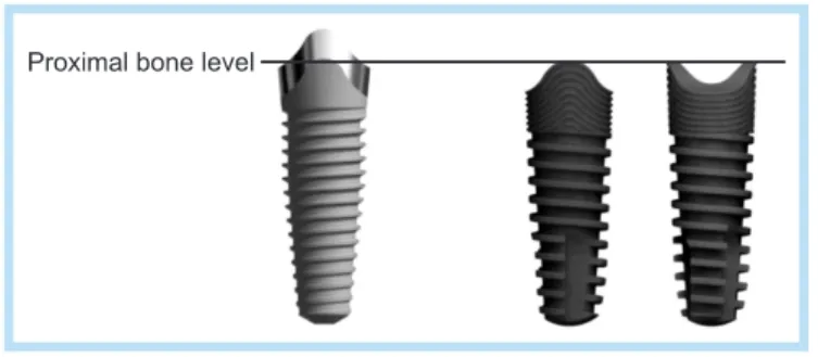
Fig. 1. The comparison between the first commercial scalloped implant (left: NobelPerfectTM; Nobel Biocare) and newly introduced scalloped implant (right: Taegeukplant®; Warantec). The smooth-rough surface border of NobelPerfectTM follows the alveolar bony scallop, whereas scallop platform of Taegeukplant® follows the alveolar bony scallop.
Seok-Geun Choi et al. : Peri-Implant Bone Level Changes of Scalloped Implant: A Retrospective Radiographic Study. Implantology 2014
Table 1. Used implants
Length (mm) Diameter (mm) 3.8 4.3 5.3 8.5
10.0 11.5
0 4 0
0 9 23
1 2 4
Seok-Geun Choi et al. : Peri-Implant Bone Level Changes of Scalloped Implant: A Retrospective Radiographic Study. Implantology 2014
를 마취하였다. 발치 후 즉시식립하는 경우 최대한 변연 골에 외상을 가하지 않도록 발치하였으며, 이후 순/협측 치은을 전층 박리하여 순/협측 변연골을 노출시켰다. 이 후 임플란트의 순/협측 부위를 변연골의 높이와 같게 식 립하였다. 이 결과 임플란트의 인접면 최상 부위가 즉시 식립하는 경우 인접면 변연골의 최상방점에서 1 mm 하 방, 발치와가 치유된 후에 식립하는 경우 인접면 변연골 의 최상방점에서 0.5 mm 하방에 위치하였다. 임플란트 는 순/협측으로 임플란트 외면으로부터 순/협측 골판 (labial/buccal plate) 외면까지 거리가 2 mm 이상이 되도 록 식립하였으며 초기 고정력은 모든 고정체에서 35 Ncm 이상을 나타냈다.
전치부의 경우 일체형 지대주를, 구치부의 경우 치유 지대주를 연결하고 흡수성 봉합사(Vicryl 4/0; Johnson
& Johnson Medical NV, St-Stevens-Woluwe, Belgium) 를 이용하여 치은을 봉합하였다. 다음날 수술 부위를 드 레싱하면서 전치부의 경우에만 임시치관을 장착하였고, 1주일 후 봉합사를 제거하였다.
고정체 식립 3개월 후 일체형 지대주를 고정체에 연결 하고(구치부), 최종 인상을 채득하여 금속도재관으로 최 종 보철물을 제작하였다. 고정체 식립 4개월 후 최종 보 철물을 장착하고 치근단 방사선 사진을 촬영하였다. 이 후 최종 보철물 장착 시점을 기준으로 3개월, 6개월, 1 년, 2년 후 치근단 방사선 사진을 촬영하였다.
촬영한 치근단 방사선 사진상에서 영상 분석 프로그램 (Image J; National Institute of Health, Bethesda, MD, USA)을 이용하여 고정체 플랫폼의 인접면 최상방점 (implant shoulder, IS)을 기준으로 첫 번째 골-임플란트
한 통계분석 프로그램(IBM SPSS Statistics version 20.0; IBM Co., Armonk, NY, USA)을 사용하여 근심측 과 원심측의 인접면 변연골 변화량의 비교는 독립변수 t-검정을 이용하였으며, 식립 부위(상/하악, 전치부/구 치부)나 식립 시기(즉시식립/치유 후 식립), 시간 등에 따 른 영향을 알아보기 위해 선형혼합모형(linear mixed model)을 이용하였다. 유의수준은 두 가지 통계분석 모 두 95%로 설정하였다(p=0.05).
III
연구결과환자수는 35명, 남녀 비율은 각각 12명, 23명이었으며 환자의 평균연령은 61.2세(25~84세)였다. 평균 추적관
Fig. 2. Implant shoulder-bone-implant contact (IS-BIC).
Original Article
찰 기간은 14.2개월(12~23개월)이었으며, 누적 생존율은 100%였다.
43개 임플란트의 근심측 평균 변화량은 0.34 mm, 원 심측 평균 변화량은 0.29 mm였으며, 두 부위 사이에 유 의한 차이는 보이지 않았다(p>0.05) (Table 2).
식립한 부위에 따른 평균 변화량은 전치부의 경우 근 심측 0.41 mm, 원심측 0.34 mm, 구치부의 경우 근심측 0.29 mm, 원심측 0.25 mm로 나타났다. 두 부위 사이에 유의한 차이는 없었다(p>0.05) (Table 3).
식립한 악궁에 따른 평균 변화량은 상악의 경우 근심 측 0.38 mm, 원심측 0.27 mm, 하악의 경우 근심측 0.24 mm, 원심측 0.32 mm로 나타났으며, 마찬가지로 상악과 하악 사이에 유의한 차이는 없었다(p> 0.05) (Table 4).
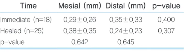
식립한 시기에 따른 평균 변화량은 즉시식립의 경우
근심측 0.29 mm, 원심측 0.35 mm, 치유 후 식립의 경우 근심측 0.38 mm, 원심측 0.24 mm로 나타났다. 마찬가
Table 2. Overall mean change of proximal marginal bone
Mesial (mm) Distal (mm) p-value Overall (n=43) 0.34±0.31 0.29±0.28 0.512 No statistically significant difference between two sites (p>0.05).
Values are presented as mean±standard deviation.
Seok-Geun Choi et al. : Peri-Implant Bone Level Changes of Scalloped Implant: A Retrospective Radiographic Study. Implantology 2014
Table 3. Mean change of proximal marginal bone with placement position
Position Mesial (mm) Distal (mm) p-value Anterior (n=24)
Posterior (n=19) p-value
0.41±0.35 0.29±0.24
0.207
0.34±0.32 0.25±0.22
0.255
0.238 0.682
No statistically significant difference among each sites (p>0.05).
Values are presented as mean±standard deviation.
Seok-Geun Choi et al. : Peri-Implant Bone Level Changes of Scalloped Implant: A Retrospective Radiographic Study. Implantology 2014
Table 4. Mean change of proximal marginal bone with jaw
Jaw Mesial (mm) Distal (mm) p-value Maxilla (n=30)
Mandible (n=13) p-value
0.38±0.35 0.24±0.21
0.802
0.27±0.26 0.32±0.31
0.754
0.195 0.548
No statistically significant difference among each sites (p>0.05).
Values are presented as mean±standard deviation.
Seok-Geun Choi et al. : Peri-Implant Bone Level Changes of Scalloped Implant: A Retrospective Radiographic Study. Implantology 2014
Table 5. Mean change of proximal marginal bone with placement time
Time Mesial (mm) Distal (mm) p-value Immediate (n=18)
Healed (n=25) p-value
0.29±0.26 0.38±0.35
0.642
0.35±0.33 0.24±0.23
0.645
0.400 0.307
No statistically significant difference among each sites (p>0.05).
Values are presented as mean±standard deviation.
Seok-Geun Choi et al. : Peri-Implant Bone Level Changes of Scalloped Implant: A Retrospective Radiographic Study. Implantology 2014
Table 6. Mean change of proximal marginal bone with period
Period (mo) Mesial (mm) Distal (mm) p-value 6
12 p-value
0.18±0.15 0.30±0.26
0.001
0.17±0.15 0.26±0.23
0.001
0.741 0.842
Statistically significant difference between 6 months and 12 months (p<0.05).
Values are presented as mean±standard deviation.
Seok-Geun Choi et al. : Peri-Implant Bone Level Changes of Scalloped Implant: A Retrospective Radiographic Study. Implantology 2014
지로 유의한 차이는 없었다(p>0.05) (Table 5).
식립 후 시간에 따른 평균 변화량은 근심측과 원심측 모두 최종 보철물 장착 시점을 기준으로 6개월에서 12개 월이 되는 시점에서 유의한 차이를 나타내기 시작했다 (p<0.05) (Table 6, Fig. 3).
IV
총괄 및 고찰본 연구에서는 물결 모양 임플란트를 식립하여 인접면 변연골의 변화량을 측정하고, 이를 토대로 물결 모양 임 플란트의 인접변 변연골 유지능력을 평가하였다.
변연골의 소실에 대한 여러 연구결과 중에 교합력과 변연골 소실 사이의 상관관계는 임상 연구18에서 입증되 었으며 이를 뒷받침하는 연구19도 보고되었다. 교합력은 보철물과 지대주, 고정체를 따라 치조골로 전달되며, 특 히 피질골-임플란트 계면에 높은 응력이 집중된다20.
Prendergast와 Huiskes21는 유한 요소 연구에서 변연골 에 가해지는 과부하는 미세손상을 초래하고 골개형(bone remodeling)에 의해 미세균열이 치유되는 것보다 미세손
수 있다고 하였다.
Frost8는 피질골에 스트레인이 4,000 με 이상 발생하 면 미세손상과 골개형 사이의 불균형으로 인해 비가역적 인 골소실이 일어나며 100 με 이하의 스트레인이 발생 할 때에는 불활동으로 인한 골흡수가 일어날 수 있다고 하였다. 따라서 변연골에 응력을 분산시켜 적절한 범위 의 스트레인을 발생시키는 것이 골조직의 항상성 유지에 중요하다.
기존의 연구들18,22-25은 대부분 피질골에 집중되는 과부 하로 인한 골흡수에 초점을 맞추어 왔다. 내측 연결형 임 플란트의 경우 지대주와 고정체 사이의 긴밀한 접촉으로 외측 연결형 임플란트에 비해 고정체 내부를 따라 응력 이 넓게 분산된다26. 그러므로 과도한 응력으로 인한 골 손상은 임플란트 디자인의 개선으로 어느 정도 극복이 가능해졌다.
임상적으로 전치부는 치조정의 형태가 구치부에 비해 편평하지 않고 볼록하거나 경사진 경우가 많으며 피질골 의 두께도 얇다. 이러한 부위에서 기존의 편평한 플랫폼 형태를 가진 임플란트는 고정체를 노출시키지 않기 위해 인접면이 치조정보다 깊게(subcrestally) 식립되어야 하 는데(Fig. 4), 이후 임플란트와 접촉하지 않은 인접면의
Original Article
NobelPerfectTM (Nobel Biocare, Kloten, Switzerland) 임플란트는 내측 연결형 구조와 변연골의 형태에 맞춘 임플란트 경부 디자인을 갖추고 있으나 상부 미세나사산 의 부재, 활택한 표면, 치은 관통부위로 인한 식립 깊이 문제 등으로 인해 좋지 않은 임상 결과를 나타냈다10-12.
이후 임플란트 디자인의 연구와 개발이 거듭되었고 Park 등16과 Choi 등17은 미세나사산을 부여한 물결 형태 임플란트의 인접면 변연골 보존 가능성을 보고하였다.
본 연구에서 사용된 물결 형태 임플란트인 Tae- geukplant®는 platform-switching 형태의 임플란트-지 대주 연결구조를 갖추고 있으며 닫힌 형태의 미세나사산 을 가지고 있다.
이 연구에 사용된 Taegeukplant®는 기존의 Nobel- PerfectTM와 달리 최종 보철물 장착 후 골증가(bone gain) 양상을 나타냈다. 구체적으로 식립한 부위(상/하악, 전 치부/구치부)나 식립한 시기(즉시식립/발치와 치유 후 식립) 등에 있어서는 인접면 변연골의 변화량에 유의한 차이를 나타내지 않았다. 하지만 임플란트를 식립하고 시간이 지남에 따라서 유의한 차이를 보이기 시작했다 (Fig. 3). 이는 임플란트 플랫폼 높이까지 부여한 미세나 사선과 거친 표면, 그리고 잘록한 형태의 임플란트-지대
주 연결구조로 인한 것이라 생각한다.
또한 응력분산 측면에서 기존의 유한요소 분석 결과27 처럼 인접면 변연골에 너무 적거나 많지 않은 적절한 수 준의 생리적 부하가 가해지면서 골이 증가한 것이라 생 각할 수 있다.
본 연구에서 특이할 만한 점은 전치부뿐만 아니라 구 치부에서도 인접면 변연골이 안정적으로 유지되었다는 점이다. 응력이 많이 발생하는 구치부에서도 인접면 변 연골이 유지되고 연조직의 부착수준이 유지되므로, 측방 으로 음식물이 함입되는 것을 방지할 수 있는 가능성을 보인다고 할 수 있다. 또한 구치부 치아를 발치하고 임플 란트를 식립하고자 할 때, 협측 변연골에 미미한 열개 (dehiscence)가 존재하는 경우 물결 모양 임플란트를 사 용하면 좋은 결과를 얻을 수 있을 것이라 예상된다.
본 연구에서는 치주염(periodontitis) 병력, 흡연 (smoking), 비기능성 저작 습관(parafunctional habit) 등 에 대한 제한(screening) 없이 물결 모양 임플란트를 식 립한 환자의 인접면 변연골 변화를 평가하였다. 하지만 연구에 포함된 환자 및 임플란트의 수가 적었고 경과관 찰 기간 또한 14.2개월로 짧았기 때문에 앞으로는 추가 적으로 경조직과 연조직의 변화를 함께 관찰한 장기간의 연구가 필요할 것이라 생각한다.
V
결 론본 연구의 한계점에도 불구하고, 물결 모양 임플란트 주위의 인접면 변연골은 안정적으로 유지되었다. 식립된 부위나 식립된 시기에 따른 인접면 변연골의 변화량에는 유의한 차이가 없었으나, 식립 후 시간에 따른 인접면 변 연골 수준은 증가하는 양상을 나타냈다. 향후 인접면 변 연골 수준의 변화에 관한 장기간의 임상연구가 필요하다.
Fig. 4. Proximal bone level of flat platform implant (left) and scalloped implant (right).
Seok-Geun Choi et al. : Peri-Implant Bone Level Changes of Scalloped Implant: A Retrospective Radiographic Study. Implantology 2014
1. Albrektsson T, Wennerberg A. The impact of oral implants-past and future, 1966-2042. J Can Dent Assoc. 2005; 71: 327.
2. Galindo-Moreno P, León-Cano A, Ortega-Oller I, et al. Marginal bone loss as success criterion in implant dentistry: beyond 2 mm. Clin Oral Implants Res. 2014. doi: 10.1111/clr.12324. [Epub ahead of print]
3. Bengazi F, Wennström JL, Lekholm U. Recession of the soft tissue margin at oral implants. A 2-year longitudinal prospective study. Clin Oral Implants Res. 1996; 7: 303-310.
4. Chang M, Wennström JL, Odman P, et al. Implant supported single- tooth replacements compared to contralateral natural teeth. Crown and soft tissue dimensions. Clin Oral Implants Res. 1999; 10: 185-194.
5. Tarnow D, Elian N, Fletcher P, et al. Vertical distance from the crest of bone to the height of the interproximal papilla between adjacent implants. J Periodontol. 2003; 74: 1785-1788.
6. den Hartog L, Slater JJ, Vissink A, et al. Treatment outcome of immediate, early and conventional single-tooth implants in the aesthetic zone: a systematic review to survival, bone level, soft-tissue, aesthetics and patient satisfaction. J Clin Periodontol. 2008; 35: 1073-1086.
7. den Hartog L, Raghoebar GM, Stellingsma K, et al. Immediate non- occlusal loading of single implants in the aesthetic zone: a randomized clinical trial. J Clin Periodontol. 2011; 38: 186-194.
8. Frost HM. Wolff's Law and bone's structural adaptations to mechanical usage: an overview for clinicians. Angle Orthod. 1994; 64: 175-188.
9. Wöhrle PS. Nobel Perfect esthetic scalloped implant: rationale for a new design. Clin Implant Dent Relat Res. 2003; 5(Suppl 1): 64-73.
10. Nowzari H, Chee W, Yi K, et al. Scalloped dental implants: a retrospective analysis of radiographic and clinical outcomes of 17 NobelPerfect implants in 6 patients. Clin Implant Dent Relat Res. 2006;
8: 1-10.
11. Nowzari H, Yi K, Chee W, et al. Immunology, microbiology, and virology following placement of NobelPerfect scalloped dental implants: analysis of a case series. Clin Implant Dent Relat Res. 2008;
10: 157-165.
12. Ostman PO, Hellman M, Albrektsson T, et al. Direct loading of Nobel Direct and Nobel Perfect one-piece implants: a 1-year prospective
radiographic 12-month prospective study. Int J Periodontics Restorative Dent. 2003; 23: 313-323.
15. Noelken R, Donati M, Fiorellini J, et al. Soft and hard tissue alterations around implants placed in an alveolar ridge with a sloped configuration.
Clin Oral Implants Res. 2014; 25: 3-9.
16. Park YS, Lee SP, Han CH, et al. The microtomographic evaluation of marginal bone resorption of immediately loaded scalloped design implant with various microthread configurations in canine mandible:
pilot study. J Oral Implantol. 2010; 36: 357-362.
17. Choi KS, Lozada JL, Kan JY, et al. Study of an experimental microthreaded scalloped implant design: proximal bone healing at different interimplant distances in a canine model. Int J Oral Maxillofac Implants. 2010; 25: 681-689.
18. Quirynen M, Naert I, van Steenberghe D. Fixture design and overload influence marginal bone loss and fixture success in the Brånemark system. Clin Oral Implants Res. 1992; 3: 104-111.
19. Holmgren EP, Seckinger RJ, Kilgren LM, et al. Evaluating parameters of osseointegrated dental implants using finite element analysis--a two- dimensional comparative study examining the effects of implant diameter, implant shape, and load direction. J Oral Implantol. 1998; 24:
80-88.
20. Baiamonte T, Abbate MF, Pizzarello F, et al. The experimental verification of the efficacy of finite element modeling to dental implant systems. J Oral Implantol. 1996; 22: 104-110.
21. Prendergast PJ, Huiskes R. Microdamage and osteocyte-lacuna strain in bone: a microstructural finite element analysis. J Biomech Eng. 1996;
118: 240-246.
22. Isidor F. Histological evaluation of peri-implant bone at implants subjected to occlusal overload or plaque accumulation. Clin Oral Implants Res. 1997; 8: 1-9.
23. Miyata T, Kobayashi Y, Araki H, et al. The influence of controlled occlusal overload on peri-implant tissue: a histologic study in monkeys.
Int J Oral Maxillofac Implants. 1998; 13: 677-683.
24. Misch CE, Suzuki JB, Misch-Dietsh FM, et al. A positive correlation between occlusal trauma and peri-implant bone loss: literature support.
Implant Dent. 2005; 14: 108-116.
Original Article
the peri-implant bone in inflamed and non-inflamed peri-implant mucosa. Clin Oral Implants Res. 2007; 18: 601-610.
26. Chun HJ, Shin HS, Han CH, et al. Influence of implant abutment type on stress distribution in bone under various loading conditions using finite element analysis. Int J Oral Maxillofac Implants. 2006; 21: 195-
202.
27. Choi KS, Park SH, Lee JH, et al. Stress distribution on scalloped implants with different microthread and connection configurations using three-dimensional finite element analysis. Int J Oral Maxillofac Implants. 2012; 27: e29-e38.