• 검색 결과가 없습니다.

Thymoma of the Middle MediastinumSu Ryeun Chung, M.D., In Sook Kim, M.D., Jhingook Kim, M.D.

N/A
N/A
Protected

Academic year: 2021

Share "Thymoma of the Middle MediastinumSu Ryeun Chung, M.D., In Sook Kim, M.D., Jhingook Kim, M.D."

Copied!
2
0
0

로드 중.... (전체 텍스트 보기)

전체 글

(1)

Korean J Thorac Cardiovasc Surg 2012;45:267-268 □ Case Report □ http://dx.doi.org/10.5090/kjtcs.2012.45.4.267 ISSN: 2233-601X (Print) ISSN: 2093-6516 (Online)

− 267 −

Department of Thoracic and Cardiovascular Surgery, Samsung Medical Center, Sungkyunkwan University School of Medicine Received: October 20, 2011, Revised: November 28, 2011, Accepted: December 1, 2011

Corresponding author: Jhingook Kim, Department of Thoracic and Cardiovascular Surgery, Samsung Medical Center, Sungkyunkwan University School of Medicine, 81 Irwon-ro, Gangnam-gu, Seoul 135-710, Korea

(Tel) 82-2-3410-3483 (Fax) 82-2-3410-0089 (E-mail) [email protected]

C

The Korean Society for Thoracic and Cardiovascular Surgery. 2012. All right reserved.

CC
This is an open access article distributed under the terms of the Creative Commons Attribution Non-Commercial License (http://creative- commons.org/licenses/by-nc/3.0) which permits unrestricted non-commercial use, distribution, and reproduction in any medium, provided the original work is properly cited.

Thymoma of the Middle Mediastinum

Su Ryeun Chung, M.D., In Sook Kim, M.D., Jhingook Kim, M.D.

Thymoma is a common anterior mediastinal mass, although thymomas have occasionally been found in the neck, pulmonary hillus, or posterior mediastinum. But a thymoma within the middle mediastinum has rarely been reported.

We report a thymoma arising in the middle mediastinum with a review of the literature.

Key words: 1. Thymoma

2. Mediastinal neoplasms

CASE REPORT

A 59-year-old womon presented with an abnormal mass on chest X-ray that was discovered during a routine check-up.

She had lost body weight during the previous 3 months. No family history of tumors or other medical diseases was identified. We obtained a biopsy though endobronchial ultra- sound and pathologically diagnosed a thymoma (type A). A systemic examination was performed, and all tests were negative. Contrast-enhanced computed tomography showed a smooth, solitary homogeneously enhanced mass measuring 3×3.5×2 cm in the right paratracheal area, which had no cal- cification (Fig. 1). We initially suspected Castleman disease due to the mass location. She underwent mass excision by video-assisted thoracic surgery. The resected specimen was a firm light-gray tumor with a smooth capsule that measured 4.5×3×2.5 cm. The tumor was solid and was partially com- posed of adipose tissue. The tumor had not invaded any other mediastinal structures and was easily extracted.

Postoperative follow-up proceeded without any problems, and the patient was discharged after 5 days. Microscopy re- vealed that the tumor was a thymoma (World Health

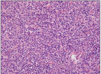
Organization type A+B2, Masaoka staging I) (Fig. 2).

DISCUSSION

The majority of thymomas are found in the anterior mediastinum. Only a few thymic masses arising out of the anterior mediastinum have been described in the literature, and these have been found in an ectopic thymus location such as the neck, pulmonary hilus, or posterior mediastinum [1,2]. A thymoma occurring in the middle mediastinum is ex- tremely rare. The thymus arises embryologically from the third pharyngeal pouch and branchail cleft on each side. The thymic masses from each side then move toward each other and migrate from the midline to the anterior mediastinum and their final position. Failure of the thymic gland to migrate during embryogenesis leads to ectopic thymic tissue. The in- cidence of ectopic thymic tissue is 3% to 5% in the retro- innomiate vein (i.e., a paratracheal site) [3].

Surgical resection has been used for both a firm diagnosis

and treatment in previously reported cases of a middle media-

stinum thymoma [4]. Endobronchial ultrasound-guided biopsy

may be useful, as in this case. Sakurai et al. [5] reported that

(2)

Su Ryeun Chung, et al

− 268 − Fig. 1. Preoperative heart contrast computed tomography scan shows round shaped mass in the right paratracheal area (arrow).

Fig. 2. Postoperative pathology slide shows the thymoma (H&E stain, ×200, Type A+B2).

18F-fluorodeoxyglucose positron emission tomography (PET) scan and 11C-acetate PET are useful for diagnosing a thymo- ma from the middle mediastinum as ectopic thymic tissue.

Castleman disease is an atypical lymphoproliferative dis- order, and masses are commonly located in the chest. Most patients are asymptomatic and lesions are accidentally found on chest X-ray as rounded mediastinal masses, often mistaken for a thymoma [6]. The most effective treatment for Castleman disease is surgical resection.

Because of malignant changes, patients with unicentric Castleman disease may need more postoperative im- munosuppressive therapy or radiation. Thus, it is important to pathologically confirm the diagnosis [7].

Thymomas rarely arise in the middle mediastinum, and thymoma is not considered in the differential diagnosis of middle mediastinum masses. But thymomas appear to have malignant potential, and the overall rate of thymoma re- currence is approximately 20%. So a thymoma should be considered in the differential diagnosis of a mediastinal mass [2,8]. A complete resection of a thymoma is useful for both treatment and diagnosis.

REFERENCES

1. Shimosato Y, Mukai K. Tumors of the thymus and related

lesion. In: Rosai J, editor. Atlas of tumor pathology: fascicle 21, third series. Washington: Armed Force Institute of Pathology; 1995. p. 33-247.

2. Kojima K, Yokoi K, Matsuguma H, et al. Middle media- stinal thymoma. J Thorac Cardiovasc Surg 2002;124:639-40.

3. Ashour M. Prevalence of ectopic thymic tissue in myasthe- nia gravis and its clinical significance. J Thorac Cardiovasc Surg 1995;109:632-5.

4. Nakamura H, Adachi Y, Fujioka S, Miwa K, Haruki T, Taniguchi Y. Thoracoscopic resection of middle mediastinal noninvasive thymoma: report of a case. Surg Today 2007;

37:787-9.

5. Sakurai H, Kaji M, Suemasu K. Thymoma of the middle me- diastinum: 11C-acetate positron emission tomography imag- ing. Ann Thorac Surg 2009;87:1271-4.

6. Keller AR, Hochholzer L, Castleman B. Hyaline-vascular and plasma-cell types of giant lymph node hyperplasia of the mediastinum and other locations. Cancer 1972;29:670- 83.

7. Jongsma TE, Verburg RJ, Geelhoed-Duijvestijn PH. Castle- man’s disease: A rare lymphoproliferative disorder. Eur J Intern Med 2007;18:87-9.

8. Monden Y, Nakahara K, Iioka S, et al. Recurrence of thy- moma: clinicopathological features, therapy, and prognosis.

Ann Thorac Surg 1985;39:165-9.

수치

Fig. 2. Postoperative pathology slide shows the thymoma (H&E  stain, ×200, Type A+B2).

참조

관련 문서

Sandberg D, Lavyne M (1999) Symptomatic spinal epidural lipomatosis after local epidural corticosteroid injections:

The influence of normalized middle school physical education and after school sports club on personality. Kim

▪ 실세계의 객체에서 불필요한 부분을 제거하여 필요한 부분만을 간결하고 이해하기 쉬운 클래스 로 만드는 작업. ▪

Likewise, the ripple effect of M&A on the value of the acquiring company -- which could not be determined using existing M&A theoretical

The close substitutes are R&D efforts, technologies, and goods that significantly constrain the exercise of market power with respect to the relevant

One might try to solve the problem by copying the first d elements of x to a temporary array, moving the remaining n − d elements left d places, and then copying the first d

(바) “액체 밀봉드럼 (Liquid seal drum)”이라 함은 플레어스택의 화염이 플레어시 스템으로 거꾸로 전파되는 것을 방지하거나 또는 플레어헤더에

[r]