Original Article
Urogenit Tract Infect 2017;12(2):82-88http://crossmark.crossref.org/dialog/?doi=10.14777/uti.2017.12.2.82&domain=pdf&date_stamp=2017-08-25
The Risk Factors of Recurrent Febrile Urinary Tract Infection
within 1 Year in Urinary Stone Patients with Acute Obstructive Pyelonephritis
Sin Woo Lee, Sol Yoon, Jungmo Do, Deok Ha Seo1, Chunwoo Lee1, Seong Uk Jeh, See Min Choi, Sung Chul Kam1, Jeong Seok Hwa, Ky Hyun Chung1, Jae Seog Hyun
Department of Urology, Gyeongsang National University Hospital, Jinju, 1Department of Urology, Gyeongsang National University Changwon Hospital, Changwon, Korea
Purpose: To identify and evaluate the risk factors for the development of recurrent febrile urinary tract infection (fUTI) among patients with previous urinary stone and acute obstructive pyelonephritis (OPN).
Materials and Methods: We retrospectively reviewed the medical records of 52 patients, who had urinary tract stones presented with OPN, between 2010 and 2015. Following their initial treatment, patients who were subsequently admitted with fUTI were included.
Results: The mean age of patients was 62.2±14.6 years, and the mean follow-up duration was 26.0±20.39 months. Escherichia coli was found to be the dominating organism (68.2%, 15/22) in the initial urine culture. Patients were divided into two groups: The recurrent fUTI group (n=23) and the non-recurrent fUTI group (n=29).
Between these two groups, significant differences were found with respect to diabetes history (recurrent group: 47.8% vs. non-recurrent group; 17.2%, p=0.018), stone location (kidney, 60.9% vs. ureter, 31.0%, p=0.031), and initially positive urine culture (60.9% vs. 27.6%, p=0.016). In a multivariate analysis, having an initially positive urine culture (95% confidence interval, 1.130-224.117;
p=0.040) was identified as being an independent risk factor for developing recurrent fUTI. In a multivariate analysis, the initial laboratory test finding of acute renal insufficiency (ARI, p=0.019) and presence of a kidney stone (p=0.022) were significant factors associated with a newly-diagnosed-positive urine culture diagnosis.
Conclusions: Having an initially positive urine culture was a significant risk factor for the development of recurrent fUTI in urinary stone patients with acute OPN.
In addition, repeated urine tests were also needed in patients with ARI or renal stones during the follow-up period.
Keywords: Urinary calculi; Urinary tract infections; Pyelonephritis
Copyright 2017, Korean Association of Urogenital Tract Infection and Inflammation. All rights reserved.
This is an open access article distributed under the terms of the Creative Commons Attribution Non-Commercial License (http://creativecommons.org/licenses/by-nc/4.0) which permits unrestricted non-commercial use, distribution, and reproduction in any medium, provided the original work is properly cited.
Received: 12 December, 2016 Revised: 26 April, 2017 Accepted: 27 May, 2017
Correspondence to: Jae Seog Hyun http://orcid.org/0000-0002-8820-4117
Department of Urology, Gyeongsang National University Hospital, Gyeongsang National University School of Medicine, 79 Gangnam-ro, Jinju 52727, Korea
Tel: +82-55-750-8195, Fax: +82-55-757-4503 E-mail: [email protected]
INTRODUCTION
Obstructive pyelonephritis (OPN) is a complicated urinary tract infection (UTI). It is an emergent urological condition
that can cause septicemia with increased risk of mortality [1,2]. About 10% of patients with septic shock due to UTI had an accompanying condition of urinary tract obstruction (UTO). Compared with UTI patients without obstruction,
patients with such obstruction showed longer hospital stays (12.8 days vs. 8.3 days, p=0.04) and a significantly higher mortality (27.0% vs. 11.0%, p=0.03) [3]. The causes of OPN are various and include ureteral stricture, urinary stone, and malignancy. However, the most common reason for obstruction in OPN is the presence of urinary stone. Rahman et al. [4] reported that about two-thirds of OPN patients was accompanied by the presence of urinary stone. Other studies have reported that patients with a history of previous ureteral stone accompanied by sepsis had a mortality rate as high as 19%, if appropriate ureteral decompression were not performed [5]. Furthermore, if decompressive procedure was delayed due to the so-called “weekend effect,” the mortality rate could be at risk of being elevated [6].
Therefore, appropriate stone management and infection control were crucial in OPN patients with a urinary stone.
However, many patients have experienced recurrence of UTI, even after having received treatment both for urinary stone and original UTI. Vahlensieck et al. [7] reported that 33% of patients showed recurrent UTI. Moreover, de Cogain et al. [8] also reported that about 32% of patients showed recurrent UTI during a 5-year follow-up period. We believe that it is important to manage and minimize stone activity and stone recurrence, as well as to reduce recurrent febrile UTI (fUTI). However, we have noticed that studies on the risk factors for recurrent fUTI following OPN treatment were heavily limited.
Hence, our goal in this study was to evaluate the risk factors for recurrent fUTI in acute OPN patients with urinary stones.
MATERIALS AND METHODS
We retrospectively reviewed the medical record of patients who had urinary stone with acute OPN between May 2010 and December 2015. This study was approved by the institutional review board of the Gyeongsang National University Hospital (IRB GNUH 2016-11-009). All patients visited the emergency room (ER) at a single center (Gyeongsang National University Hospital) and were admitted, via ER, to the department of either urology or nephrology for further evaluation and treatment.
The inclusion criteria for patients were having had both acute pyelonephritis (APN) and urolithiasis. The diagnosis of APN was based on clinical symptomology (ex. fever;
flank pain), laboratory testing (for leukocytosis; pyuria), and physical examination (costovertebral angle tenderness) [9]. All patients had a ureteral stone with UTO. Those patients who also had an accompanying renal stone were retained.
The exclusion criteria were having an alternative diagnosis or an alternative source of UTI or another cause of urinary obstruction (ex., ureteral compression by stricture or malignancy; bladder outlet obstruction). Patients who had minimal symptoms and not admitted were also excluded.
Lastly, patients whose records containing incomplete data or whose follow-up duration was short (<3 months) were excluded. The term ‘recurrent fUTI’ was defined as APN or recurrence of OPN with (either a remnant or recurred) stone, requiring re-admission to receive intravenous antibiotics. All patients were divided into two groups (a recurrent fUTI group and a non-recurrent group) within 1 year after discharge.
The initial imaging studies were abdominal contrast/
non-contrast computed tomography (CT) and kidney ureter bladder (KUB) X-ray. The term ‘obstruction’ was defined as having a presence of upper urinary tract dilation with hydronephrosis, as shown by CT imaging. After the treatment, follow-up imaging studies were performed with KUB (for radiopaque stones) or non-contrast abdominal CT (for radiolucent stones). Decompressive procedures, percutaneous nephrostomy (PCN) tube insertion and ureteral stent insertion, were performed. PCN tube insertion was done by an interventional radiologist. Ureteral stent insertion was done by a urologist on duty, with administration of local anesthesia. Patients with ureteral stent were maintained on a Foley catheter for maximal decompression for a minimum of 2-3 days. In addition, the term ‘pyuria’ was defined as having the presence of over 5 white blood cells per high-power field. The initial bladder urine culture was done for all patients in the ER, and an additional PCN urine culture was performed in patients who had PCN tube or ureteral stent. Before urinary stone treatments [such as ureteroscopy (URS), retrograde intrarenal surgery (RIRS), percutaneous nephrolithotomy (PCNL) or extracorporeal shock wave lithotripsy (ESWL)], all patients received a course of preoperative broad spectrum antibiotics for a minimum of 3 days. After urine culture was found to contain no bacterial growth or became culture-negative for infection, urinary stone treatments were started.
We identified demographic variables (age; gender),
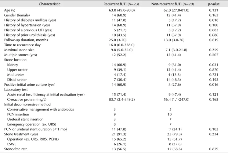
Table 1. Comparison of the characteristics between the two groups (recurrent fUTI vs. non-recurrent fUTI)
Characteristic Recurrent fUTI (n=23) Non-recurrent fUTI (n=29) p-value
Age (y) 63.0 (49.0-90.0) 62.0 (27.0-81.0) 0.131
Gender (female) 14 (60.9) 12 (41.4) 0.163
History of diabetes mellitus (yes) 11 (47.8) 5 (17.2) 0.018
History of hypertension (yes) 14 (60.9) 11 (37.9) 0.100
History of a previous UTI (yes) 5 (21.7) 5 (17.2) 0.683
History of prior urolithiasis (yes) 10 (43.5) 11 (37.9) 0.686
Follow-up duration, months 25.0 (3-70) 13.0 (3.0-76) 0.619
Time to recurrence day 16.0 (6.0-338.0)
Maximal stone size 9.8 (5.0-35.0) 7.1 (3.0-21.8) 0.259
Multiple stones (yes) 12 (52.2) 12 (41.4) 0.507
Stone location
Kidney 14 (60.9) 9 (31.0) 0.031
Upper ureter 9 (39.1) 12 (41.4) 0.870
Mid ureter 4 (17.4) 4 (13.8) 0.721
Distal ureter 7 (30.4) 14 (48.3) 0.193
Positive initial urine culture (yes) 14 (60.9) 8 (27.6) 0.016
Laboratory test
Acute renal insufficiency at initial evaluation (yes) 15 (71.4) 9 (47.4) 0.121
C-reactive protein (mg/L) 83.7 (2.4-349.2) 56.4 (1.1-247.0) 0.165
Initial decompressive method
Conservative management with antibiotics 3 5
PCN insertion 9 10
Ureteral stent insertion 3 7
Emergency operation (ex. URS) 8 7
PCN or ureteral stent duration (≥1 mo) 11 (47.8) 7 (24.1) 0.103
Stone treatment (yes) 21 (91.3) 23 (79.3) 0.234
Operation (ex. URS, RIRS, PCNL) 15 (65.2) 15 (51.7)
ESWL 6 (26.1) 8 (27.6)
Stone-free rate 13 (56.5) 17 (58.6) 0.879
Values are presented as median (range), number (%), or number only.
fUTI: febrile urinary tract infection, PCN: percutaneous nephrostomy, URS: ureterostomy, RIRS: retrograde intrarenal surgery, PCNL: percutaneous nephrolithotomy, ESWL: extracorporeal shockwave lithotripsy.
previous medical history [diabetes mellitus (DM);
hypertension; UTI; urolithiasis episode], and radiologic studies to determine stone size, number, location, and stone-free status. The term ‘stone-free status’ was defined as an absence of residual stone fragments on follow-up imaging studies. Pearson’s chi-square test and the Mann-Whitney U-test were used to correlate clinical variables. Univariate and multivariate Cox regression analyses were used to define the independent risk factors for recurrent fUTI and positive urine cultures which were newly diagnosed as being positive or which were newly comprised of different organisms than had originally been found in the initial urine culture.
RESULTS
The mean age of patients was 62.2±14.6 years, and the mean follow-up duration was 26.0±20.39 months. After
infection control and stone management, 23 patients presented recurrent fUTI during the 1-year follow-up period.
Patients were divided into two groups (the recurrent fUTI group [n=23] and the non-recurrent fUTI group [n=29]).
In terms of baseline characteristics, differences in age, gender, and previous treatment histories (UTI and uroli- thiasis) were not significant. However, the rate of DM history was significantly higher in the recurrent fUTI group (47.8%
vs. 17.2%, p=0.018) than in the non-recurrent group. In the recurrent fUTI group, the median time to recurrent fUTI was 16.0 (6.0-338.0) days, and the mean duration of hospitalization was 8.2±41 days. The rate of early fUTI recurrence within the first month was 43.5% (10/23). With regards to stone location, the recurrent fUTI group showed a higher rate of having an accompanying renal stone than that shown by the non-recurrent fUTI group (60.9% vs.
31.0%, p=0.031). The rate of initially positive voided or PCN urine culture was also significantly higher in the
Table 2. Organisms isolated in the urine culture Organisms isolated in the initial urine culture
(n=22)
Organisms isolated in recurrent UTI
(n=23) Escherichia coli (ESBL+E. coli) 15 (8/15) 11 (7/11)
Staphylococcus species 2 2
Klebsiella pneumoniae 1 3
Enterobacter species 1 1
Enterococcus species 1 3
Pseudomonas 1 2
Proteus species 1
Streptococcus species 1
UTI: urinary tract infection, ESBL+: extended-spectrum beta-lactamase positive.
Table 3. Risk factors for recurrent febrile urinary tract infection
Univariate analysis Multivariate analysis
OR 95% CI p-value OR 95% CI p-value
Age (continuous) 1.032 0.990-1.075 0.135 1.029 0.915-1.157 0.633
DM history 4.400 1.243-15.574 0.022 5.200 0.550-49.163 0.150
History of a previous urinary tract infection 1.333 0.335-5.311 0.683
Position of stone (kidney vs. ureter) 3.457 1.096-10.906 0.034 8.730 0.761-100.196 0.082 Initial urine culture (positive vs. negative) 4.083 1.270-13.131 0.018 15.917 1.130-224.117 0.040 Acute renal insufficiency (>1.2 mg/dl) 2.778 0.752-10.260 0.125 3.061 0.302-31.026 0.344 Ureteral catheter duration (≥1 mo, yes) 2.794 0.800-9.760 0.107 0.316 0.029-3.422 0.343
Treatment (operation vs. ESWL) 1.750 0.568-5.393 0.330
Ureteral catheter duration (≥1 mo, yes) 0.429 0.593-9.176 0.225
Stone free after treatment (yes) 1.090 0.360-3.297 0.879
OR: odds ratio, CI: confidence interval, DM: diabetes mellitus, ESWL: extracorporeal shockwave lithotripsy.
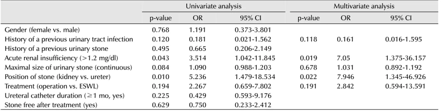
Table 4. Risk factors for positive urine cultures that were newly diagnosed or different organism with the initial urine culture
Univariate analysis Multivariate analysis
p-value OR 95% CI p-value OR 95% CI
Gender (female vs. male) 0.768 1.191 0.373-3.801
History of a previous urinary tract infection 0.120 0.181 0.021-1.562 0.118 0.161 0.016-1.595 History of a previous urinary stone 0.495 0.665 0.206-2.149
Acute renal insufficiency (>1.2 mg/dl) 0.043 3.514 1.042-11.845 0.019 7.05 1.375-36.157 Maximal size of urinary stone (continuous) 0.084 1.090 0.988-1.203 0.678 1.031 0.892-1.192 Position of stone (kidney vs. ureter) 0.010 5.236 1.479-18.534 0.022 7.946 1.345-46.926
Treatment (operation vs. ESWL) 0.194 2.267 0.659-7.802 0.191 2.842 0.594-13.591
Ureteral catheter duration (≥1 mo, yes) 0.225 0.429 0.593-9.176 Stone free after treatment (yes) 0.629 0.750 0.233-2.412 OR: odds ratio, CI: confidence interval, ESWL: extracorporeal shockwave lithotripsy.
recurrent fUTI group when compared with the non-recurrent group (60.9% vs. 27.6%, p=0.016). According to the initial laboratory testing, the number of patients with acute renal insufficiency (ARI) were comparatively higher in the recurrent fUTI group-but without statistical significance.
The most commonly used decompressive procedure was PCN tube insertion (19/52, 36.5%). After stone treatment,
the stone free-rate was likewise insignificant (p=0.879, Table 1).
E. coli was found to be the dominating organism in both the initial urine culture (68.2%, 15/22) and in that of recurrent fUTI culture (11/23). Of those in the recurrent fUTI group, 14 (60.9%) patients showed positive urine cultures that were newly diagnosed as being positive or different from those found in the initial urine culture.
Compared with the initial urine culture, the number of Klebsiella and Enterococcus species has somewhat increased (Table 2).
When we analyzed the suspected risk factors for recurrent fUTI, significant differences were found in DM history (yes, p=0.022), stone location (kidney, p=0.034), and initially- positive urine culture (positive, p=0.018) in a univariate analysis. In a multivariate analysis, an initially positive urine culture (positive, 95% confidence interval [CI], 1.130- 224.117; p=0.040) was identified as being an independent risk factor for the subsequent recurrence of fUTI (Table 3).
In both univariate and multivariate analyses, a finding of ARI on the initial laboratory testing (95% CI, 1.375-36.157;
p=0.019), as well as the presence of a kidney stone (95%
CI, 1.345-46.926; p=0.022), were factors significantly associated with a newly-diagnosed-positive urine culture diagnosis (Table 4).
DISCUSSION
In our study, 44.2% (23/52) of OPN patients with a urinary stone showed recurrent fUTI. Of them, 10 patients showed early fUTI recurrence within the first month. These rates were relatively high compared with previous studies.
Vahlensieck et al. [7] reported a rate of 33.0%; de Cogain et al. [8] reported a UTI recurrence rate of 32.0% during the follow-up period. However, as compared with these studies, our patients were generally older (62.2 vs. 56.0 and 54.7-55.6 years). There were also differences in the baseline characteristics between both studies (ex., patients’
previous history; urinary stone status) and the diagnosis of (and definition of what constituted) recurrent UTI.
Perhaps these differences contributed to the discrepant results.
The risk factors for developing recurrent fUTI were a history of previous DM treatment, combination of a renal stone, and an initially positive urine culture in univariate analysis. Kofteridis et al. [10] reported that an elderly APN patient with DM showed an increased risk of bacteremia, long hospitalization, and mortality. In another study, it was found that comorbidity factors, such as DM, could influence urinary stone activity and the recurrence of UTI after the treatment of infection and stone [11]. DM, as a comorbidity factor, may be associated with stone activity and UTI recurrence; however, multivariate analysis did not find DM to be an independent risk factor (p=0.150). Additionally, in diabetic patients, glucose control could have been an important factor. However, the lack of information regarding, for instance, HbA1c or fasting glucose level is one of the limitations of our study. Of the total number of patients, patients who had both ureteral stone and renal stone showed a comparatively higher rate of UTI recurrence and the greater likelihood of remnant stone. A large number of renal stone patients had a relatively large stone burden and were at elevated risk of developing infected stone (i.e., struvite stone and calcium carbonate apatite stone). These
may be associated with UTI recurrence.
According to our multivariate analysis, an initially positive urine culture was found to be the only independent risk factor for recurrent fUTI (p=0.040). A positive urine culture could be considered as being more significant UTI and may influenced the recurrence of fUTI, following the treatment and management of stones and infection. In a urine culture examination, we collected mid-stream urine (from all patients) and PCN urine (from PCN tube insertion patients). If there were bacterial species of more than 105 colony-forming units per milliliter between them in quantitative counts, we regarded this as being a ‘positive’
urine culture. However, the results from bladder urine and PCN urine of patients with complete obstruction could sometimes differ from one another. For instance, in our study, the pelvic urine cultures of 8 patients tested positive;
of those, 6 patients tested as having the same bacterial species in both their bladder and their pelvic urine.
However, the other 2 patients tested positive for PCN urine culture while negative for bladder urine culture. Eswara et al. [12] reported that-among readmitted patients who developed sepsis after having undergone treatment for a stone-there was only 64% correspondence between whether both their stone culture and their urine culture tested positive. Additionally, another previous study reported that stone culture results were better correlated with pelvic, rather than bladder, urine culture results [13].
When OPN patients presenting with fever visited the ER, antibiotic treatment could be started before testing the pelvic urine culture. However, antibiotic levels of intra-pelvic urine rise slowly. Therefore both tests of bladder urine culture and pelvic urine aspiration culture were needed at initial evaluation [14,15]. In our study, we conducted ureteral stent insertion under local anesthesia and did not routinely collect pelvic urine samples. This could be a limitation of our study. However, we always checked urine cultures after a decompressive procedure to determine whether patients would test negative for infection.
E. coli was, by far, the most common organism found in urine cultures. In our study, about 44% of patients had accompanying renal stones, excluding full staghorn stone patients. Therefore, urine cultures with urease-producing bacteria (the Klebsiella, Proteus species, for instance)-
which are common in infected stones-may have been small in number. Having an ARI or kidney stone were
found to be a risk factor for developing new fUTI (as indicated by the presence of new and different organisms in the most recent urine culture as compared with those which had originally been found in the initial urine culture).
In the case of ARI, we could not check for a previous history of chronic renal failure. However, patients who took hemodialysis were not included. The ARI found in patients upon initial laboratory testing may have been associated with complete ureteral obstruction or delayed urinary stone treatment. In another study, the rate of post-operative UTI was lower in stone-free patients than in patients with remnant stone (38% vs. 64%) [11]. However in our study, the stone-free rate was not different between the two groups. Recurrent UTI was higher in patients with renal stone; and having a renal stone was a significant factor for a new diagnosis of fUTI (Table 4) or for being at risk of suffering a recurrence of fUTI (Table 3). Patients with an accompanying renal stone had an elevated risk of having a remnant stone that had previously been exposed to infected urine; and this remnant stone may have been an influence on the onset of post-operative complications, such as recurrent fUTI.
Due to recurrent fUTI, 10 patients were re-admitted within the first month after discharge. Among them, 8 patients underwent URS with or without RIRS or PCNL, and the remaining 2 patients were treated with ESWL. Increased intra-renal pressure and urine extravasation during URS were associated with an elevated risk of post-operative fever [16]. Auge et al. [17] reported that the irrigation pressure of RIRS using an access sheath was significantly lower than for URS. An access sheath had a protective effect against pyelovenous and pyelolymphatic backflow. Similarly, as compared with URS, retroperitoneal laparoscopic ureterolithotomy showed a lower rate of post-operative fever (4.9% as compared with 13.9%, p=0.043). In another study, the rate of post-operative APN within the first month was 7.4% in patients who had undergone RIRS. Early antibiotic treatment before 1 week could decrease the chance of APN about 63.0% [18]. Therefore, appropriate antibiotic treatment and careful attention are needed when an OPN patient undergoes URS. There has been some concerns that ureteral stent insertion could increase the risk of exacerbating urinary infection by stone manipulation and that it would be less effective for draining urine as compared with a large-caliber PCN tube [19]. However,
previous studies have shown that either PCN or a ureteral stent could effectively relieve obstruction and infection associated with urinary stone [20,21]. Recently, Wang et al. reported that, as compared with the insertion of a PCN tube, URS together with a course of strong antibiotics could also be an effective and safe management protocol for fighting sepsis associated with obstructing ureteral stone [22]. Similar to this, the differences between the decompression method (ureteral stent insertion vs. PCN tube insertion) and stone treatment method (URS and RIRS vs. ESWL) were not found to be significant in our study (Table 1).
Our study has several limitations to consider. Our results were derived from retrospective data and the number of patients included was relatively small. Moreover, additional data regarding the status of infection (ex., stone culture, blood culture) or stone (ex., urinary calculi formation) were somewhat lacking. However, the presentation of OPN with urinary stone has been a relatively common disease; and proper management and careful monitoring remain important. We are focused more on the risk of recurrent fUTI associated with the presence of stones than on the treatment of stones. To prevent and respond to the recurrence of fUTI after stone treatment, a range of research is still needed; and one could find this study meaningful in this regard. Moreover, further studies, such as an investigation into the efficacy of low-dose antibiotic prophylaxis for high-risk patients or large scale studies of OPN patients with stone, may also be necessary.
CONCLUSIONS
We found that having an initially positive urine culture was a significant risk factor for the development of recurrent fUTI in urinary stone patients with acute OPN. Moreover, the median time to recurrence was only 16 days. Therefore, cautionary short term follow-up is necessary in these patients. Additionally, repeated urine tests were also needed in patients found to have ARI or renal stones during the follow-up period.
CONFLICT OF INTEREST
No potential conflict of interest relevant to this article was reported.
REFERENCES
1. Yamamoto Y, Fujita K, Nakazawa S, Hayashi T, Tanigawa G, Imamura R, et al. Clinical characteristics and risk factors for septic shock in patients receiving emergency drainage for acute pyelonephritis with upper urinary tract calculi. BMC Urol 2012;12:4.
2. Yoshimura K, Utsunomiya N, Ichioka K, Ueda N, Matsui Y, Terai A. Emergency drainage for urosepsis associated with upper urinary tract calculi. J Urol 2005;173:458-62.
3. Reyner K, Heffner AC, Karvetski CH. Urinary obstruction is an important complicating factor in patients with septic shock due to urinary infection. Am J Emerg Med 2016;34:694-6.
4. Rahman NU, Meng MV, Stoller ML. Infections and urinary stone disease. Curr Pharm Des 2003;9:975-81.
5. Borofsky MS, Walter D, Shah O, Goldfarb DS, Mues AC, Makarov DV. Surgical decompression is associated with decreased mortality in patients with sepsis and ureteral calculi.
J Urol 2013;189:946-51.
6. Blackwell RH, Barton GJ, Kothari AN, Zapf MA, Flanigan RC, Kuo PC, et al. Early intervention during acute stone admissions:
revealing "the weekend effect" in urological practice. J Urol 2016;196:124-30.
7. Vahlensieck W, Friess D, Fabry W, Waidelich R, Bschleipfer T.
Long-term results after acute therapy of obstructive pyelonephritis. Urol Int 2015;94:436-41.
8. de Cogain MR, Lieske JC, Vrtiska TJ, Tosh PK, Krambeck AE.
Secondarily infected nonstruvite urolithiasis: a prospective evaluation. Urology 2014;84:1295-300.
9. Jiang JT, Li WG, Zhu YP, Sun WL, Zhao W, Ruan Y, et al.
Comparison of the clinical efficacy and safety of retroperitoneal laparoscopic ureterolithotomy and ureteroscopic holmium laser lithotripsy in the treatment of obstructive upper ureteral calculi with concurrent urinary tract infections. Lasers Med Sci 2016;31:915-20.
10. Kofteridis DP, Papadimitraki E, Mantadakis E, Maraki S, Papadakis JA, Tzifa G, et al. Effect of diabetes mellitus on the clinical and microbiological features of hospitalized elderly patients with acute pyelonephritis. J Am Geriatr Soc 2009;
57:2125-8.
11. Iqbal MW, Youssef RF, Neisius A, Kuntz N, Hanna J, Ferrandino MN, et al. Contemporary management of struvite stones using
combined endourologic and medical treatment: predictors of unfavorable clinical outcome. J Endourol 2016;30:771-7.
12. Eswara JR, Shariftabrizi A, Sacco D. Positive stone culture is associated with a higher rate of sepsis after endourological procedures. Urolithiasis 2013;41:411-4.
13. Mariappan P, Smith G, Bariol SV, Moussa SA, Tolley DA. Stone and pelvic urine culture and sensitivity are better than bladder urine as predictors of urosepsis following percutaneous nephrolithotomy: a prospective clinical study. J Urol 2005;
173:1610-4.
14. Browne RF, Zwirewich C, Torreggiani WC. Imaging of urinary tract infection in the adult. Eur Radiol 2004;14 Suppl 3:
E168-83.
15. Ng CK, Yip SK, Sim LS, Tan BH, Wong MY, Tan BS, et al.
Outcome of percutaneous nephrostomy for the management of pyonephrosis. Asian J Surg 2002;25:215-9.
16. D'Addessi A, Bassi P. Ureterorenoscopy: avoiding and managing the complications. Urol Int 2011;87:251-9.
17. Auge BK, Pietrow PK, Lallas CD, Raj GV, Santa-Cruz RW, Preminger GM. Ureteral access sheath provides protection against elevated renal pressures during routine flexible ureteroscopic stone manipulation. J Endourol 2004;18:33-6.
18. Alezra E, Lasselin J, Forzini T, Francois T, Viart L, Saint F.
Prognostic factors for severe infection after flexible ureteroscopy: Clinical interest of urine culture the day before surgery?. Prog Urol 2016;26:65-71.
19. Marien T, Miller NL. Treatment of the infected stone. Urol Clin North Am 2015;42:459-72.
20. Mokhmalji H, Braun PM, Martinez Portillo FJ, Siegsmund M, Alken P, Kohrmann KU. Percutaneous nephrostomy versus ureteral stents for diversion of hydronephrosis caused by stones: a prospective, randomized clinical trial. J Urol 2001;165:1088-92.
21. Pearle MS, Pierce HL, Miller GL, Summa JA, Mutz JM, Petty BA, et al. Optimal method of urgent decompression of the collecting system for obstruction and infection due to ureteral calculi. J Urol 1998;160:1260-4.
22. Wang CJ, Hsu CS, Chen HW, Chang CH, Tsai PC. Percutaneous nephrostomy versus ureteroscopic management of sepsis associated with ureteral stone impaction: a randomized controlled trial. Urolithiasis 2016;44:415-9.