• 검색 결과가 없습니다.

Blood Flow and Skin Temperature Increases by Monochromatic Infrared Energy Irradiation J K S P T

N/A
N/A
Protected

Academic year: 2021

Share "Blood Flow and Skin Temperature Increases by Monochromatic Infrared Energy Irradiation J K S P T"

Copied!
6
0
0

로드 중.... (전체 텍스트 보기)

전체 글

(1)

Blood Flow and Skin Temperature Increases by Monochromatic Infrared Energy Irradiation

Jae-Hyoung Lee, PT, PhD

1

, Gi Won Kim, PT, PhD

2

1

Department of Physical Therapy, Electrotherapy Research Laboratory for Tissue Growth & Repair, Wonkwang Health Science University,

2

Department of Physical Therapy, Suwon Women’s College

Purpose: The purpose of this study was to determine the effect of monochromatic infrared energy (MIRE) on the blood flow of the superficial radial artery and local skin temperature in healthy subjects.

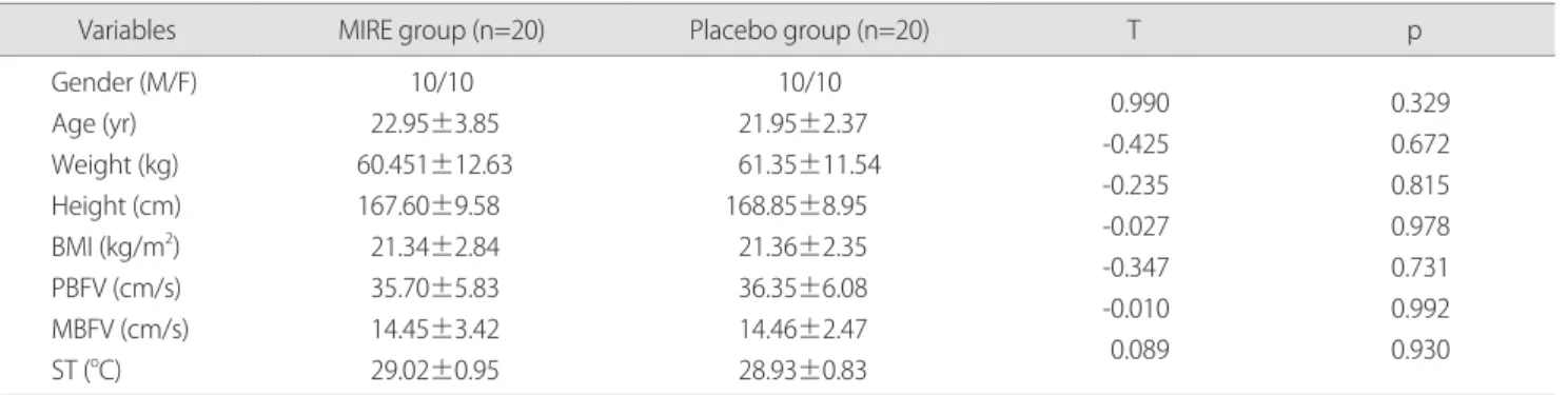
Methods: Forty healthy volunteers were recruited and randomly assigned to MIRE group (n=20) and placebo group (n=20).

The MIRE group received a 890 nm MIRE irradiation on the forearm using two therapy pads for 30 minutes. The therapy pad was composed of an array of 60 diodes. MIRE unit was set at bar 8, that corresponds to a diode power of 10 mW and a power density of 63 mW/cm

2

. The placebo group received sham MIRE. Peak blood flow velocity (PBFV), mean blood flow velocity (MBFV), and skin temperature (ST) were measured pre- and post-MIRE irradiation.

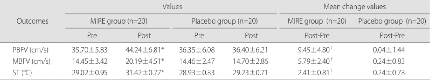
Results: There was a significant difference in PBFV (p<0.001), MBFV (p<0.001), and ST (p<0.001) between the pre- and post- treated values in the MIRE group. In contrast, no significant difference was found between the pre- and post-treated values in the placebo group. There was significant difference in mean change values from baseline of PBFV (p<0.001), MBFV (p<0.001), and ST (p<0.001) between the MIRE group and the placebo group. There was a significant increase in PBFV (p<0.001), MBFV (p<0.001), and ST (p<0.001) following MIRE irradiation.

Conclusion: The arterial blood flow and local skin temperature of the forearm in the healthy subjects were significantly increased following MIRE irradiation.

Keywords: Monochromatic infrared energy, Arterial blood flow, Skin temperature

I. Introduction

Monochromatic infrared energy (MIRE) therapy is a treatment that uses a near-infrared light with a single wavelength or a very small range of wavelengths. According to the U.S. Food and Drug Administration (FDA), this kind of infrared lamp is a device intended for medical purposes that emits energy at infrared frequencies ranging from 700 nm to 50,000 nm to provide topical healing. The Anodyne Therapy System

that uses 890 nm of near infrared light energy has been FDA approved since 1994 and it has been used for increasing circulation and reducing pain.

1

Recently, MIRE has been used as a treatment modality for several indications, including peripheral diabetic neuropathy, pain management, and wound healing.

According to the manufacturer, the mechanism of action of a 890 nm of MIRE is primarily to increase microcirculation and local skin temperature of the target area.

1

The increase in skin microcirculation produced by light energy is believed to be due to the activation of nitric oxide (NO) synthesis in the irradiated area.

2,3

Nitric oxide synthase (NOS) absorbs light energy, resulting to its activation. NO is an endothelial- derived relaxing factor (EDRF) and a powerful vasodilator and it is produced from the amino acid L-arginine by the Received March 21, 2012 Revised June 4, 2012

Accepted June 7, 2012

Corresponding author Jae-Hyoung Lee, [email protected]

Copyright © 2012 by The Korean Society of Physical Therapy

This is an Open Access article distributed under the terms of the Creative Commons Attribution Non-Commercial License (http://creativecommons.org/licenses/by-nc/3.0/) which permits unrestricted non-commercial use, distribution, and reproduction in any medium, provided the original work is properly cited.

The J ournal of K orean S ociety of P hysical T herapy Original Article

(2)

Original Article

enzymatic action of NOS in many cells. Its production by vascular endothelial cells and hemoglobin in red blood cells are particularly important in the regulation of blood flow. NO activates guanylate cyclases causing the dephosphorylation of guanosine-5’-triphosphate to cyclic guanosine monophosphate (cGMP). The increase in intracellular cGMP inhibits Ca

++

entry into the cell causing the relaxation of contractile proteins in vascular smooth muscle resulting to vasodilation. In addition, NO activates K

+

channels, which leads to hyperpolarization and relaxation of vascular smooth muscle.

4

Meanwhile, Minson et al.

5

proposed two independent mechanisms contributing to the increase in skin circulation during local heating. These are the fast- responding vasodilator system which is mediated by the axon reflexes and a more slowly responding vasodilator system that relies on local production of NO.

According to previous studies, MIRE has been shown to improve diabetic foot ulcers

6,7

and recalcitrant dermal lesions, including venous ulcers, diabetic ulcers, and a wound related to scleroderma.

8

Powell et al.

9

reported that MIRE can improve activities of daily living and reduce the number of falls and the fear of falling in the elderly population with diabetic peripheral neuropathy (DPN). Some authors have suggested that MIRE improve sensation and pain in the foot of patients with DPN. Harkless et al.

10

and Leonard et al.

11

have reported that MIRE improve sensation and pain on the feet of elderly patients with DPN. The study of DeLellis et al.

12

and Powell et al.

13

reported a significant improvement in foot sensation with DPN following MIRE.

However, some research studies reported that MIRE application did not provide improvement in peripheral sensation and pain. The study of Nawfar and Yacob

14

, Lavery et al.

15

, and Clifft et al.

16

revealed that MIRE was not effective in increasing sensation in subjects with DPN. Franzen- Korzendorfer et al.

17

did not demonstrate any effects of MIRE on sensation and pain in adults with DPN.

The rate and quality of wound healing following the use of MIRE may be related to local increases in NO concentration.

Increases in NO have been demonstrated to correlate with vasodilatory and anabolic responses.

3

Given the conflicting and controversial findings of many authors regarding the

effectiveness of MIRE in wound healing and pain relief. This study was conducted for the purpose of determining the effect of MIRE on the blood flow of the superficial artery using Doppler ultrasound, with concomitant local skin temperature in healthy subjects.

II. Materials and Methods

1. Subjects

Forty-five healthy volunteers were recruited by convenient sampling. Five subjects were excluded because they had a history of smoking for many years. Subjects were excluded if they met the following criteria: had history of diabetic mellitus, neuropathy, peripheral vascular disease, had skin lesions over forearm, under medication causing vasodilation, had active malignancy, and were pregnant. The experimental procedure was explained to the subjects, and a written informed consent was obtained before enrollment. This study was performed in accordance to the principles and ethical standards on human trial. Forty subjects of whom 20 were men and 20 were women, mean age was 22.45±3.19 years (range 19~35), were included. No dropout was reported after inclusion and randomization. The subjects were randomly assigned to two groups by drawing 1 of 40 envelopes labeled A (MIRE group, n=20) and B (Placebo group, n=20). Subjects were unaware of the code for active or sham MIRE until the data analysis was completed. The subjects were assessed by an independent examiner. Demographic data regarding age, gender, body weight, height and body mass index (BMI) of the participants were collected. The study was performed in a quiet room, in which the temperature was kept at 24

±1

o

C. The subjects were asked to avoid having meals and coffee, vigorous exercise 2 hours prior to the experiment.

The subjects were lying in a comfortable supine position for 30 minutes prior to the trial.

2. Intervention

In the MIRE group, the subjects received MIRE (Model 480

Anodyne Therapy Professional System; Anodyne Therapy

LLC., Tampa, FL, USA). Anodyne Therapy System is a device

that delivers MIRE through gallium aluminum arsenide diodes,

(3)

with a wavelength of 890 nm, that are mounted in flexible therapy pads (3 cm×7.5 cm; 22.5 cm

2

). The therapy pad has an array of 60 diodes. Each diode has a diameter of 0.45 cm (0.16 cm

2

), and each has a power of 13 mW and a power density of 82 mW/cm

2

. The two therapy pads were placed on the volar surface of the proximal and distal portion of the right forearm in direct contact with the skin. The dosage of the MIRE unit was set at bar 8, that corresponds to a diode power of 10 mW and a power density of 63 mW/cm

2

with 30 minutes delivering a total energy density of 113 J/cm

2

. For the placebo group, the procedures were the same except that the sham Anodyne Therapy System was used by turning the switch off and disconnect the output circuit.

3. Outcome measures 1) Blood flow velocity (BFV)

An ultrasound system (MyLabOne, Esaote, Genova, Italy) was used to measure the blood flow velocity. The radial artery at the right wrist was insonated with a 5.6 MHz pulse wave using a vascular and small parts linear array ultrasound transducer (SL3323, Esaote, Genova, Italy). The transducer was directed to the skin superficial to the radial artery with an angle of approximately 45 degrees in a supine position.

Aquasonic

100 (Parker Laboratories, Inc., Fairfield, NJ, USA) was used as acoustic coupling medium. Pressure to the tissue was carefully avoided. Peak blood flow velocity (PBFV) and mean blood flow velocity (MBFV) were obtained and electronically stored on the computer’s hard disc.

Doppler ultrasound measurement was performed before and immediately after the MIRE irradiation. All measurements were repeated three times each session.

2) Skin temperature (ST)

The ST was measured using an electronic thermometer (Tri-R Instrument Inc., Rockville Centre, New York, USA). The thermistor was sensitive to temperature changes of ±0.2

o

C.

The skin thermistor probe which was placed 4 cm proximal to the distal wrist crease on the anterior aspect of the right forearm recorded the ST. Temperature measurements were performed before and immediately after the MIRE irradiation.

4. Statistical analysis

The data were presented as mean and standard deviation.

The value of the changes was obtained by subtracting the pre-treated value from post-treated value. Paired t-test was used to determine the significant difference in mean value between the pre- and post-treatment of BFV and ST within each group. Independent t-test was used to compare the significant difference in mean value of demographic data and the baseline characteristics between the two groups.

Independent t-test was used to compare the significant difference in mean changes of BFV and ST between the two groups. The level of statistical significance was set at a two- tailed p-value of 0.05. Statistical analysis was performed on a personal computer by using SPSS ver. 10.0 (SPSS Inc., Chicago, IL, USA).

Table 1. Demographic data and baseline characteristics in both groups

Variables MIRE group (n=20) Placebo group (n=20) T p

Gender (M/F) Age (yr) Weight (kg) Height (cm) BMI (kg/m

2

) PBFV (cm/s) MBFV (cm/s) ST (

o

C)

10/10 22.95±3.85 60.451±12.63 167.60±9.58

21.34±2.84 35.70±5.83 14.45±3.42 29.02±0.95

10/10 21.95±2.37 61.35±11.54 168.85±8.95

21.36±2.35 36.35±6.08 14.46±2.47 28.93±0.83

0.990 -0.425 -0.235 -0.027 -0.347 -0.010 0.089

0.329 0.672 0.815 0.978 0.731 0.992 0.930 Values are number or mean±standard deviation.

M: male, F: female, BMI: body mass index, PBFV: peak blood flow velocity, MBFV: mean blood flow velocity, ST: skin temperature.

(4)

III. Results

1. Demographic data and baseline characteristics Forty healthy subjects aging 19 to 35 years old participated in the study. Their body weight ranged from 42 to 88 kg, height from 155 to 190 cm with a BMI of 16.41 to 28.58 kg/m

2

. No significant difference was seen in the demographic data and baseline characteristics of participants between the two groups (Table 1).

2. Blood flow velocity

Table 2 shows the values and mean change values of PBFV and MBFV in the radial artery in the MIRE and placebo group.

There was significant difference in PBFV (t=-7.794, p<0.001) and MBFV (t=-10.519, p<0.001) between pre- and post- treated values in the MIRE group. In contrast, no significant difference was found between pre- and post-treated values in the placebo group. There was a significant difference in mean change values from baseline of PBFV (t=-8.404, p<0.001) and MBFV (t=-9.790, p<0.001) between the MIRE group and the placebo group. There was a significant increase in blood flow in the radial artery following MIRE irradiation.

3. Skin temperature

Table 2 shows the values and mean change values of ST of the forearm in the MIRE and placebo group. There was a significant difference in ST (t=-13.330, p<0.001) between pre- and post-treated values in the MIRE group. There was no significant difference in ST in the placebo group. There was a significant difference in mean change values from

baseline of ST (t=-8.601, p<0.001) between the MIRE group and the placebo group. There was significant increase in ST following MIRE irradiation.

4. Other outcome

In MIRE group, the subjects reported slightly warm (1/20, 5%), warm (11/20, 55%), slightly hot (4/20, 20%) and tolerable hot (4/20, 20%) sensation underneath the therapy pads during MIRE irradiation. No adverse effects were observed during the irradiation.

IV. Discussion

The present study demonstrated that MIRE can increase the blood flow of the superficial radial artery as measured by Doppler ultrasound among healthy subjects. PBFV was increased from baseline by 23.9% in the MIRE group and by 0.4% in the placebo group. MBFV was increased from baseline by 39.7% in the MIRE group and by 1.7% in the placebo group. Effect size (d) of PBFV and MBFV were 1.36 (95% CI, 0.65~2.02) and 1.59 (95% CI, 0.88, 2.31), respectively. The improvement of PBFV and MBFV were important benefit relative to a control with relative difference (RD) of 23.51 % and 38.05%. This was the first study to examine the effects of MIRE on superficial arterial blood flow using a Doppler ultrasound among healthy subjects.

Two previous studies have demonstrated that the Anodyne Therapy System has the potential to increase blood flow.

Burke

1

reported that a 45-minute MIRE treatment increases blood flow by 400% over baseline. Without MIRE, blood flow

Table 2. The values and mean change values of PBFV, MBFV and ST in the MIRE and placebo group Outcomes

Values Mean change values

MIRE group (n=20) Placebo group (n=20) MIRE group (n=20) Placebo group (n=20)

Pre Post Pre Post Post-Pre Post-Pre

PBFV (cm/s) MBFV (cm/s) ST (

o

C)

35.70±5.83 14.45±3.42 29.02±0.95

44.24±6.81*

20.19±4.51*

31.42±0.77*

36.35±6.08 14.46±2.47 28.93±0.83

36.40±6.21 14.70±2.86 29.23±0.71

9.45±4.80

5.79±2.40

2.41±0.81

0.04±1.44 0.24±0.83 0.24±0.78 Values are number or mean±standard deviation.

PBFV: peak blood flow velocity, MBFV: mean blood flow velocity, ST: skin temperature.

*Significantly differences between pre and post treatment (p<0.001).

Significantly differences between groups (p<0.001).

(5)

increases only by 40% in 2 diabetic feet using a scanning laser Doppler. However, Burke’s research is a case study and it did not provide dosage for MIRE application, and therefore it was difficult to compare its results to this present study. Our results, however, were similar to a study conducted recently by Mak and Cheing

18

wherein a 30-minute MIRE treatment yielded a 30% increase in the microcirculation of the skin in the foot of healthy subjects. They used MIRE with a diode power of 12 mW and a power density of 60 mW/cm

2

. The results have demonstrated an increased in the blood flow following a 890 nm of MIRE treatment applied for 30 minutes.

Our study used a MIRE dosage with a diode power of 10 mW and power density of 63 mW/cm

2

for 30 minutes.

To compare the flood flow, our study measured the superficial blood flow of the radial artery at the wrist area using a Doppler ultrasound while Mak and Cheing

18

measured the superficial skin blood flow at the foot using a laser Doppler flowmetry (LDF). Eicke et al.

19

reported that radial artery blood flow assessment using a Doppler ultrasound and laser Doppler perfusion monitor at the fingertips were correlated significantly (r=0.616, p=0.004), and thus, both methods are suitable to monitor blood flow changes, and validate the use of Doppler ultrasound as an alternative approach to LDF in healthy subjects.

In this study, the ST at the application site of the diodes was elevated using an electronic thermometer equipped with a skin thermistor probe. Concomitantly, all subjects have reported a mild warm to tolerable hot sensation. ST was increased from baseline by 8.3% in the MIRE group and by 0.8% in the placebo group. The ST was 7.5% greater in the MIRE group relative to the placebo group, although important benefits could not be ascertained yet. The concomitant increase in ST may provide an explanation for the observed findings, as it has been well recognized that a variation in tissue temperature will cause a corresponding increase in arterial blood flow. The study of Noble et al.

20

has shown that MIRE, similar to Anodyne Therapy System, could increase ST in the healthy human forearm. They observed that ST increased significantly following a 45-minute MIRE irradiation using 4.0 J/cm

2

. In contrast, Mak and Cheing

18

observed a mild elevation in ST at the foot after MIRE application, but no

significant difference was found among the MIRE, warm pack and sham MIRE groups. This difference may be due to the differing measurement sites used in ST assessment. In their study, they measured the ST at the third metatarsal head just distal area to the therapeutic pads, while this study measured the ST at the area directly under the therapeutic pad.

The dilation of skin blood vessels is mediated primarily by EDRFs and sympathetic nerve. EDRFs such as NO and prostacyclin, which are produced predominantly by endothelial cells can increase microcirculation due their vasodilatory effects. The increase in blood flow as a result of NO release into the smooth muscles can cause shearing forces on the arterial endothelial cells that leads to a secondary increase in arterial circulation.

3,21

In addition, local heating evokes an increase in skin blood flow by sympathetic axon-reflex.

22

These effects are clinically significant in wound management given that an increase in blood flow may accelerate wound healing process and reduce pain by augmenting the delivery of oxygen and nutrients to the site of injury.

23

The present study demonstrated that the PBFV, the MBFV, and the ST were significantly greater in the MIRE group than in the placebo group. The improvement of PBFV and MBFV were important benefits relative to a control group. The results suggest that MIRE irradiation can increase blood flow in the superficial radial artery with a concomitant increase in the local skin temperature of healthy subjects. The MIRE irradiation, therefore, can be used to improve blood flow.

This study was performed on healthy subjects. Further clinical research is therefore needed to confirm the results of this study especially among patients with various types of wounds and who are experiencing pain.

Author Contributions

Research design: Lee JH, Kim GW Acquisition of data: Lee JH, Kim GW

Analysis and interpretation of data: Lee JH, Kim GW Drafting of the manuscript: Lee JH

Research supervision: Lee JH

(6)

Acknowledgements

1. This work was supported partly by the Academic Research Fund of Wonkwang Health Science University (2012).

2. We wish to thank all the volunteers who participated in this study, and Apsun Inc. (Korea) for supplying Anodyne Therapy device.

References

1. Burke TJ. 5 Questions--and answers--about MIRE treatment. Adv Skin Wound Care. 2003;16(7):369-71.

2. Samoilova KA, Zhevago NA, Menshutina MA et al. Role of nitric oxide in the visible light-induced rapid increase of human skin microcirculation at the local and systemic level: I. diabetic patients.

Photomed Laser Surg. 2008;26(5):433-42.

3. Samoilova KA, Zhevago NA, Petrishchev NN et al. Role of nitric oxide in the visible light-induced rapid increase of human skin microcirculation at the local and systemic levels: II. healthy volunteers. Photomed Laser Surg. 2008;26(5):443-9.

4. Jin RC, Loscalzo J. Vascular nitric oxide: formation and function. J Blood Med. 2010;2010(1):147-62.

5. Minson CT, Berry LT, Joyner MJ. Nitric oxide and neurally mediated regulation of skin blood flow during local heating. J Appl Physiol.

2001;91(4):1619-26.

6. Nather A, Sim YE, Chew LLj et al. Anodyne therapy for recalcitrant diabetic foot ulcers: a report of four cases. J Orthop Surg (Hong Kong). 2007;15(3):361-4.

7. Hunter S, Langemo D, Hanson D et al. The use of monochromatic infrared energy in wound management. Adv Skin Wound Care.

2007;20(5):265-6.

8. Horwitz LR, Burke TJ, Carnegie D. Augmentation of wound healing using monochromatic infrared energy. Exploration of a new technology for wound management. Adv Wound Care.

1999;12(1):35-40.

9. Powell MW, Carnegie DH, Burke TJ. Reversal of diabetic peripheral neuropathy with phototherapy (MIRE) decreases falls and the fear of falling and improves activities of daily living in seniors. Age Ageing. 2006;35(1):11-6.

10. Harkless LB, DeLellis S, Carnegie DH et al. Improved foot sensitivity and pain reduction in patients with peripheral neuropathy after treatment with monochromatic infrared photo energy--MIRE. J Diabetes Complications. 2006;20(2):81-7.

11. Leonard DR, Farooqi MH, Myers S. Restoration of sensation, reduced pain, and improved balance in subjects with diabetic peripheral neuropathy: a double-blind, randomized, placebo- controlled study with monochromatic near-infrared treatment.

Diabetes Care. 2004;27(1):168-72.

12. DeLellis SL, Carnegie DH, Burke TJ. Improved sensitivity in patients with peripheral neuropathy: effects of monochromatic infrared photo energy. J Am Podiatr Med Assoc. 2005;95(2):143-7.

13. Powell MW, Carnegie DE, Burke TJ. Reversal of diabetic peripheral neuropathy and new wound incidence: the role of MIRE. Adv Skin Wound Care. 2004;17(6):295-300.

14. Nawfar SA, Yacob NB. Effects of monochromatic infrared energy therapy on diabetic feet with peripheral sensory neuropathy: a randomised controlled trial. Singapore Med J. 2011;52(9):669-72.

15. Lavery LA, Murdoch DP, Williams J et al. Does anodyne light therapy improve peripheral neuropathy in diabetes? A double-blind, sham-controlled, randomized trial to evaluate monochromatic infrared photoenergy. Diabetes Care. 2008;31(2):

316-21.

16. Clifft JK, Kasser RJ, Newton TS et al. The effect of monochromatic infrared energy on sensation in patients with diabetic peripheral neuropathy: a double-blind, placebo-controlled study. Diabetes Care. 2005;28(12):2896-900.

17. Franzen-Korzendorfer H, Blackinton M, Rone-Adams S et al.

The effect of monochromatic infrared energy on transcutaneous oxygen measurements and protective sensation: results of a controlled, double-blind, randomized clinical study. Ostomy Wound Manage. 2008 ;54(6):16-31.

18. Mak MC, Cheing GL. Immediate effects of monochromatic infrared energy on microcirculation in healthy subjects. Photomed Laser Surg. 2012;30(4):193-9.

19. Eicke BM, Milke K, Schlereth T et al. Comparison of continuous wave Doppler ultrasound of the radial artery and laser Doppler flowmetry of the fingertips with sympathetic stimulation. J Neurol.

2004;251(8):958-62.

20. Noble JG, Lowe AS, Baxter GD. Monochromatic infrared irradiation (890 nm): effect of a multisource array upon conduction in the human median nerve. J Clin Laser Med Surg. 2001;19(6):291-5.

21. Buerk DG, Barbee KA, Jaron D. Nitric oxide signaling in the microcirculation. Crit Rev Biomed Eng. 2011;39(5):397-433.

22. Nicotra A, Asahina M, Mathias CJ. Skin vasodilator response to local heating in human chronic spinal cord injury. Eur J Neurol. 2004

;11(12):835-7.

23. Hancock CM, Riegger-Krugh C. Modulation of pain in

osteoarthritis: the role of nitric oxide. Clin J Pain. 2008;24(4):353-

65.

수치

Table 1. Demographic data and baseline characteristics in both groups
Table 2 shows the values and mean change values of PBFV  and MBFV in the radial artery in the MIRE and placebo group

참조

관련 문서

The purpose of this study was to examine the effect of shadowing using English movies on listening and speaking skills, learning interest, and attitudes

The purpose of this study is to investigate the effect of the “problem solving and programming” area of ​​information subject on perception and

The purpose of this study is to clarify the effect of identification affirmation according to K-Pop star image on the desire of middle school students to participate

The purpose of this research was to identify the mediating effect of unconditional self-acceptance and the moderated mediating effect of emotional

The purpose of this study is to examine the effects of flow experience through the dance sports media, a dance sports image, and identification on

The purpose of this study was to examine the effects of 8 weeks of Zumba dance exercise for obese middle-aged women on blood lipid index and vascular aging.. The subjects of

The purpose and necessity of this study was to investigate the effect of the 8-week GX complex exercise program on the stress index, blood lipid and APG

The purpose of this study was to analyse effect of primer solution on degree of conversion two-step self-etch adhesive with a Fourier transform infrared