KJTCVS
The Korean Journal of Thoracic and Cardiovascular SurgeryClinical Research Clinical Value of Intraoperative Flow Measurements of
Brachiocephalic Arteriovenous Fistulas for Hemodialysis
Jonggeun Lee, M.D. 1 , Seogjae Lee, M.D., Ph.D. 1 , Jee Won Chang, M.D., Ph.D. 1 , Su Wan Kim, M.D., Ph.D. 1 , Jung-Kook Song, M.D., Ph.D. 2
1
Department of Thoracic and Cardiovascular Surgery, Jeju National University Hospital, Jeju National University School of Medicine;
2Department of Preventive Medicine, Jeju National University School of Medicine, Jeju, Korea
ARTICLE INFO
Received August 26, 2019 Revised December 14, 2019 Accepted December 24, 2019 Corresponding author Jee Won Chang Tel 82-64-717-1704 Fax 82-64-717-1131 E-mail jwchang@jejunu.ac.kr ORCID
https://orcid.org/0000-0001-9331-9405
Background: The aim of this study was to analyze the clinical outcomes of autogenous brachiocephalic arteriovenous fistulas and to investigate the factors associated with 1-year patency after initiation of hemodialysis.
Methods: We retrospectively reviewed the medical records of 41 patients who under- went surgery to create an autogenous brachiocephalic arteriovenous fistula between January 2015 and December 2017, received hemodialysis at the same hospital for longer than 1 year, and were monitored for their vascular access status. Intraoperative flow was measured using transit-time ultrasonography.
Results: The 1-year primary and secondary patency rates were 61% (n=25) and 87.8%
(n=36), respectively. The functional group (subjects who required no intervention to main- tain patency within the first year after hemodialysis initiation) displayed a significantly higher median intraoperative flow rate (450 mL/min) than the non-functional group (sub- jects who required intervention at least once regardless of 1-year patency) (275 mL/min) (p=0.038). Based on a receiver operating characteristic curve analysis, all patients were ad- ditionally subdivided into a high-flow group (>240 mL/min) and a low-flow group (≤240 mL/min). The high-flow group included a significantly greater number of functional bra- chiocephalic arteriovenous fistulas than the low-flow group (74.2% vs. 20%, respectively;
p=0.007).
Conclusion: Transit-time flow, as measured with intraoperative transit-time ultrasonog- raphy, was associated with patency without the need for intervention at 1 year after initi- ation of hemodialysis.
Keywords: Renal dialysis, Surgical arteriovenous shunt, Chronic kidney failure
Copyright © The Korean Society for Thoracic and Cardiovascular Surgery. 2020. All right reserved.
This is an Open Access article distributed under the terms of the Creative Commons Attribution Non-Commercial License (http://creativecommons.org/licenses/
Introduction
End-stage renal disease (ESRD) is a major public health problem, and preserving vascular access for maintenance hemodialysis is a lifelong challenge for ESRD patients. Au- togenous arteriovenous fistulas (AVFs) are the preferred method of vascular access for hemodialysis maintenance in ESRD patients due to their relatively long patency, low rate of infectious or thrombotic complications, and dura- bility despite repeated needling [1,2]. However, maturation failure is one of the most serious limitations to patency.
According to a previous report, 17% of autogenous accesses failed within the first 3 months [3]. The aim of the present
study was to analyze the clinical outcomes of autogenous brachiocephalic AVFs in order to investigate the factors as- sociated with patency without the need for intervention.
Methods
Between January 2015 and December 2017, 3 surgeons operated on 81 patients to create autogenous antecubital brachiocephalic AVFs for hemodialysis access. Among these patients, 41 continued hemodialysis at the same hos- pital for at least 1 year. Patients were followed up with re- garding their vascular access status. We retrospectively re- viewed the medical records of these 41 patients.
https://doi.org/10.5090/kjtcs.2020.53.3.121 pISSN: 2233-601X eISSN: 2093-6516
Korean J Thorac Cardiovasc Surg. 2020;53(3):121-126
https://doi.org/10.5090/kjtcs.2020.53.3.121
KJTCVS
Preoperative assessment
First, an appropriate site for AVF creation was selected.
If the diameter of the forearm veins was less than 2 mm under compression, and if the veins were stiff on palpation or showed signs of thrombophlebitis following recent in- travenous injection or blood sampling, no forearm AVF was created and a brachiocephalic AVF was chosen instead.
Physical examinations and routine laboratory tests were conducted preoperatively. Physical examinations for bra- chiocephalic AVFs involved measuring the diameter of the antecubital and upper arm cephalic veins while compress- ing the proximal upper arm with a stretchable rubber phle- botomy tourniquet, palpation of the upper arm cephalic vein under compression to examine elasticity, and Allen’s test. If the upper arm cephalic vein had a gross tourni- quet-derived diameter larger than 3 mm and was elastic upon palpation without any fibrotic segment, we per- formed brachiocephalic AVF. Venography was performed selectively in patients with a history of hemodialysis cathe- ter insertion into the ipsilateral subclavian or internal jug- ular vein. Informed consent was obtained before surgery.
Operative techniques
Brachiocephalic AVF creation was performed under lo- cal anesthesia or brachial plexus block. The final decision regarding creation of the AVF site was made based on the vascular conditions identified during surgery. Before anas- tomosis, the diameter of the upper arm cephalic vein was measured intraoperatively by inserting a 3-mm metal- tipped vascular dilator without resistance. The vein was gently dilated using saline injection without excessive pres- sure in order to prevent intimal damage. End-to-side anas- tomosis was performed with a polypropylene 7-0 (Ethicon Inc., Somerville, NJ, USA) continuous running suture. In- traoperative transit-time flow (TTF) was measured with a handheld 3-mm TTF probe (HT313; Transonic Systems Inc., Ithaca, NY, USA) by encircling the cephalic vein ap- proximately 5–10 mm proximal to the anastomosis site at 5 minutes post-anastomosis. Measurements were repeated until the flow stabilized. The wound was closed layer by layer after bleeding was controlled.
Surveillance of arteriovenous fistula
At a renal replacement therapy unit AVF surveillance was conducted to monitor for pain during hemodialysis, prolonged bleeding after hemodialysis, and abnormal
physical examination findings. Patients were also moni- tored for findings such as high venous pressure or subopti- mal blood flow in order to identify imminent AVF failure.
The criteria for intervention included a high-pitched or systolic bruit on auscultation, inability to attain an optimal dialysis flow rate of 250 mL/min, an increase in dialysis venous pressure above 150 mm Hg, or a rise of 30 to 50 mm Hg above the baseline value. Any symptoms related to AVF such as pain, limb swelling, or hand ischemia were also considered to indicate AVF failure. Primary patency was defined as the duration between AVF surgery and the first intervention to restore or maintain patency, including percutaneous angioplasty, surgical revision, or thrombec- tomy. Secondary patency was defined as the duration be- tween AVF surgery and permanent AVF failure.
Statistical analysis
We divided the patients into a functional and a non- functional group in order to analyze the clinical outcomes.
The functional group was defined by a patent AVF at 1 year after initiation of hemodialysis without the need for intervention. The non-functional group was defined by an AVF requiring intervention regardless of patency. Based on the receiver operating characteristic (ROC) curve analysis conducted to obtain the cutoff value of intraoperative TTF, the included patients were also divided into a high-flow and a low-flow group. Between-group comparisons were made using the Fisher exact test for categorical variables and the t-test (or the Wilcoxon rank-sum test for variables that did not show a normal distribution) for continuous variables. All statistical analyses were performed using IBM SPSS ver. 24.0 (IBM Corp., Armonk, NY, USA) and R ver. 3.4.4 (R Project for Statistical Computing, Vienna, Austria; (https://www.r-project.org/). Within R, the pROC [4] and Epi [5] packages were used for a ROC curve analy- sis. All p-values of less than 0.05 were considered to indi- cate statistical significance.
This study was approved by the Institutional Review Board of Jeju National University Hospital (IRB approval no., JEJUNUH 2019-08-008).
Results
The study included 24 (58.5%) men and 17 women with
a mean age of 64.5 years (range, 39–88 years). The under-
lying diseases of ESRD were diabetes (n=27, 65.9%), hyper-
tension (n=15), glomerulonephritis (n=1), and polycystic
kidney disease (n=1). No definite cause was reported in 1
Jonggeun Lee, et al. Intraoperative Flow of Arteriovenous Fistula KJTCVS
case. We defined hemodialysis initiation as 3 consecutive successful hemodialysis treatments with brachiocephalic AVF without any symptoms or signs of AVF malfunction.
Under this definition, the average duration from AVF cre- ation to the initiation of hemodialysis was 94.2 days. The 1-year primary and secondary patency rates were 61%
(n=25) and 87.8% (n=36), respectively.
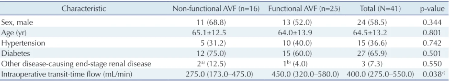
The characteristics of the functional and non-functional groups are presented in Table 1. No statistically significant differences were found with regard to age, sex, or underly- ing diseases between the 2 groups. The median values of intraoperative TTF (mL/min) were 275.0, 450.0, and 400.0 in the non-functional AVF group, the functional group, and the total study population, respectively, with a statisti- cally significant difference between the functional and non-functional groups (p=0.038). A box plot was used to illustrate the interquartile range, the range containing the central 95% of observations, and the maximum and mini- mum values (Fig. 1).
The non-functional group included 8 patients with AVFs that were patent at 1 year after single percutaneous angio-
plasty, 3 patients with AVFs that were patent at 1 year with percutaneous angioplasty or thrombolysis performed more than twice, 3 patients for whom attempts to initiate hemo- dialysis failed in spite of intervention, and 2 patients with AVFs that showed early failure within 3 months after he- modialysis initiation. Based on the radiological findings, the factors requiring intervention were maturation failure (n=6), juxta-anastomotic stenosis (n=4), non-anastomotic multifocal venous stenosis (n=4), thrombophlebitis (n=1), and central venous stenosis (n=1). Of the 5 cases that lacked patency at 1 year after initiation, 3 involved matura- tion failure, 1 involved multifocal venous stenosis, and 1 involved thrombophlebitis. All of the cases of juxta-anas- tomotic stenosis recovered patency after a single interven- tion.
The ROC curve of the intraoperative TTF revealed an optimal cut-off of 240 mL/min with a relatively high sensi- tivity (92.0%) and a moderate specificity (50.0%) (Fig. 2).
The area under the ROC curve was 0.695, indicating that this test was relatively poor at discriminating between those with and those without functional AVF. When the Table 1. Clinical demographics of functional and non-functional AVFs at 1 year after initiation of maintenance hemodialysis
Characteristic Non-functional AVF (n=16) Functional AVF (n=25) Total (N=41) p-value
Sex, male 11 (68.8) 13 (52.0) 24 (58.5) 0.344
Age (yr) 65.1±12.5 64.0±13.9 64.5±13.2 0.801
Hypertension 5 (31.2) 10 (40.0) 15 (36.6) 0.742
Diabetes 12 (75.0) 15 (60.0) 27 (65.9) 0.501
Other disease-causing end-stage renal disease 2
a)(12.5) 1
b)(4.0) 3 (7.3) 0.550
Intraoperative transit-time flow (mL/min) 275.0 (173.0–475.0) 450.0 (320.0–580.0) 400.0 (275.0–550.0) 0.038
c)Values are presented as number (%), mean±standard deviation, or median (range).
AVF, arteriovenous fistula.
a)
Glomerulonephritis and unknown kidney disease.
b)Polycystic kidney disease.
c)The Wilcoxon rank-sum test was performed on these data.
1,000
800
600
400
200
AVF status
Non-functional AVF Functional AVF
Intraoperative TTF (mL/min)
0
Fig. 1. A box plot of intraoperative TTF by AVF status regarding 1-year patency (functional or non-functional). AVF, arteriovenous fistula; TTF, transit-time flow.
1.0
0.8
0.6
0.4
0.2
1 Specificity
1.0
Sensitivity
0.0 0.2 0.4 0.6 0.8
Sensitivity: 92.0%
Specificity: 50.0%
PV+: 20.0%
PV : 25.8%
Model: patency 1 year transit-time flow Optimal cutoff value: 240 mL/min AUC: 0.695 (0.513 0.877), p<0.001
Fig. 2. Receiver operating characteristic curve. PV+, positive pre-
dictive value; PV-, negative predictive value; AUC, area under the
curve.
https://doi.org/10.5090/kjtcs.2020.53.3.121
KJTCVS
patients were divided into high-flow (TTF >240 mL/min) and low-flow (TTF ≤240 mL/min) groups based on the re- sults of ROC analysis, the high-flow group displayed a sig- nificantly larger proportion of functional AVFs than the low-flow group (74.2% and 20%, respectively; p=0.007) (Table 2). The high-flow group included 8 cases of non- functional AVFs, 4 of which were due to maturation fail- ure, 3 of which were attributed to juxta-anastomotic and non-anastomotic venous stenosis, and 1 of which resulted from thrombophlebitis. Patency was not restored in 2 of these cases, one of which involved maturation failure and the other thrombophlebitis.
Discussion
Brachiocephalic AVFs represent a traditional option for upper-arm autogenous vascular access that has been re- ported to have a higher patency rate than forearm autoge- nous arteriovenous access [6]. Bae et al. [7] also reported that brachiocephalic AVF was associated with a shorter time to first needling and fewer interventions before matu- ration than radiocephalic AVF. However, brachiocephalic AVFs have their own limitations, such as an association with cephalic arch stenosis, which has a reported incidence of as high as 77% in patients with brachiocephalic AVF [8].
Because the cephalic arch may be the sole outflow of bra- chiocephalic AVF, these AVFs can lead to subsequent thrombotic or stenotic failure that requires surgical inter- ventions, such as central transposition [9]. The present study showed that cases of venous stenosis, including jux- ta-anastomotic and non-anastomotic multifocal venous stenoses, required intervention. However, the exact cause could not be identified because of limited clinical informa- tion regarding factors such as vein diameter or preopera- tive radiological evaluation of the vein. The factors related to the maturation and patency of brachiocephalic AVFs should be further investigated.
In a systematic review of 34 studies, the primary patency rate for autogenous AVFs was found to be 72% at 6 months and 51% at 18 months, whereas the secondary patency rate was 86% at 6 months and 77% at 18 months [3]. Our results revealed 1-year primary and secondary patency rates of 61% and 87.8%, respectively, which are similar to those found in the aforementioned review. However, Lin et al. [10]
found that comprehensive care that included patient edu- cation and access flow surveillance followed by early inter- vention improved the 1-year secondary patency rate to 94%
in the high-flow group (intraoperative TTF >200 mL/min) and 80% in the low-flow group for radiocephalic AVFs. At our institution, nephrologists and nurses in the renal re- placement therapy unit are the health professionals pri- marily in charge of AVF care. A multi-disciplinary ap- proach is needed to improve AVF outcomes in the future.
Parameters related to the patency of various types of au- togenous AVFs, including older age, distal location, or small venous diameter, were negative prognostic factors for AVF maturation. Ahn et al. [11] reported that previous ip- silateral central venous catheterization was an independent risk factor for maturation failure, and increased body mass index or obesity-related decrease in intraoperative flow was associated with decreased AVF patency [12]. Intraoperative TTF was a predictor of patency, especially for radiocephal- ic AVFs. In 1998, Johnson et al. [13] reported for the first time that intraoperative TTF was correlated with the AVF outcome and that radiocephalic AVF flow less than 170 mL/min predicted failure in up to 56% of AVFs within 90 days. Another report by Won et al. [14] found that a radio- cephalic AVF flow less than 160 mL/min was associated with a high early failure rate, and intraoperative low flow was determined to be an independent risk factor. A report by Berman et al. [15] was the first study to establish the minimal flow rates needed for maturation and subsequent patency of brachiocephalic AVF. According to the report of Berman et al. [15], a cutoff of 308 mL/min was needed for Table 2. Clinical characteristics based on intraoperative TTF
Characteristic Low-flow TTF
(≤240 mL/min) (n=10)
High-flow TTF
(>240 mL/min) (n=31) Total (N=41) p-value
Sex, male 7 (70.0) 17 (54.8) 24 (58.5) 0.480
Age (yr) 64.7±12.9 64.4±13.5 64.5±13.2 0.949
Hypertension 4 (40.0) 11 (35.5) 15 (36.6) 1.000
Diabetes 8 (80.0) 19 (61.3) 27 (65.9) 0.447
Other disease-causing end-stage renal disease 0 3
a)(9.7) 3 (7.3) 0.564
Functional arteriovenous fistula 2 (20.0) 23 (74.2) 25 (61.0) 0.007
Values are presented as number (%) or mean±standard deviation.
TTF, transit-time flow.
a)