자기공명영상 소견을 통한 척추 성형술 후 골 시멘트 누출의 예측
원광대학교 의과대학 정형외과학교실 김창수․김태균․채수욱․이 영
Prediction of Bone Cement Leakage in Patients Receiving Vertebroplasty by MRI Finding
Chang Su Kim, Tae Kyun Kim, Soo Uk Chae, Young Yi
Department of Orthopedic Surgery, School of Medicine, Wonkwang University Hospital, Iksan, Korea
Objectives: To identify MRI predictors of bone cement leakage, we compared pre operative MRI and postoperative CT.
Materials and Methods: Between October 2004 to March 2009, percuataneous vertebroplasties were performed in 58 patients from 167 patients of spine compression fracture. Among them, 37 patients took MRI before vertebroplasty and CT after operation to figure bone cement leakage. In 37 patients, 45 vertebras (T9; 1, T10; 3, T11; 5, T12; 8, L1; 11, L2; 9, L3; 4, L4; 3, L5; 1) preoperative MRI was taken to measure the presence of cortical disruption of the vertebral body and vacuum or cystic portion, severity of body compression, bone cement amount and bone cement amount/ severity of body compression ratio.
Results: In postoperative CT scan of 42 vertebrae with bone cement, leakage was detected in 17 vertebrae (37.7%). However, no patients displayed any neurological symptoms or required surgery.
Endplate cortical disruption was related to an increase risk of intervertebral bone cement leakage (P<0.05). Bone cement leakage tended to occur less frequently when there is a vacuum or fluid collection (P<0.05). No other factors showed significant relation with cement leakage.
Conclusions: Vertebroplasty group in magnetic resonance imaging of cortical damage to the vertebral endplates and fluid collection or vacuum changes when that can be useful to predict the leakage of bone cement after vertebroplasty group findings suggest.
Key Words: Osteoporotic vertebral compression fracture, Vertebroplasty, Bone cement leakage, MRI
Received: June 10, 2012 Revised: June 22, 2012 Accepted: August 14, 2012
Corresponding Author: Tae Kyun Kim, Department of Orthopaedic Surgery, School of Medicine, Wonkwang University, 344-2, Shinyong-dong, Iksan 570-711, Korea
Tel: +82-63-859-1360, Fax: +82-63-852-9329 E-mail: [email protected]
* 본 논문은 2012년 원광대학교 병원의 연구비 지원을 받아 이루어 졌음.
평균 수명의 증가로 골다공증이 있는 노인 환자에
게서의 척추 압박골절이 증가하고 있다.
1,2척추 압박
골절은 골다공증의 가장 흔한 합병증의 하나이며 일
상생활 정도의 활동에 의해서도 발생할 수 있다. 척
추 압박골절의 치료에 있어서 기존의 전통적인 방법
으로는 척추 고정술과 같은 수술적 요법과 장기간의
침상안정 등의 보존적 방법이 있다.
3그러나 최근에
는 polymethylmethacrylate (PMMA)를 이용한 경피적
척추성형술이 도입되어 널리 시술되고 있다. 경피적 척추성형술은 척추체 내에 골시멘트, 알코올 등의 각종 물질을 주입시키는 시술로 1984년 Deramond 등이 처음으로 통증을 일으키는 척추체의 혈관종에 골시멘트를 주입하여 통증 완화를 보고한 이래, 골 다공증으로 인한 압박골절, 척추체를 침범하는 악성 종양등에 대해서도 뛰어난 통증 완화를 보고하였 다.
4,5특히 척추 성형술은 보존적 치료의 효과가 없 는 골절에 대해 시도하는 치료로 비교적 안전하며 통증 경감, 척추 압박 변형 진행 억제 또는 회복, 폐 기능 감소 방지 및 조기 보행을 가능하게 한다.
6-10하지만 골시멘트의 인접조직으로 누출에 의한 부작 용이 발생할 수 있어 시술에 주의를 요하며 시멘트 의 누출은 저자에 따라 약 11~81%정도로 흔히 발 생한다고 보고되고 있다.
11-13또한 척추체 정맥 및 신경관으로의 골시멘트 누출에 의한 폐색전증이나 신경손상과 같은 심각한 합병증을 초래할 수 있음이 보고된 바 있다.
14-16이에 저자들은 시술 전 시행한 척추 자기 공명 영상을 통해 골시멘트 누출에 영향 을 줄 수 있는 위험인자를 분석해 보고자 하였다.
대상 및 방법
2004년 10월부터 2009년 3월까지 골다공증성 척 추 압박 골절로 내원한 167명의 환자 중 58명의 환 자에 대해 척추 성형술을 시행하였으며 그 중 37명 대해 척추 성형술 시행 전 척추 자기공명 영상촬영 을 시행하였으며 척추 성형술 시행 후 컴퓨터 단층 촬영을 시행하여 골 시멘트 누출 유무를 확인 하였 다. 이들의 평균 연령은 68.5세(58~75)였으며 남자 가 6명, 여자가 31명이였다. DEXA (Dual Energy X-ray Absorptiometry)를 이용한 골밀도 검사(BMD) 에서 T-score가 전례에서 -3.0 이하였으며 총 37명 의 환자, 척추 45개(T9; 1, T10; 3, 11번 흉추: 5건, 12 번 흉추: 8건, 1번 요추: 11건, 2번 요추: 9건, 3번 요 추: 4건, 4번 요추: 3건, 5번 요추: 1건)에 대하여 척 추 성형술을 시행하였으며 시술 전 시행한 척추 자 기공명 영상을 통해 척추체 종판 피질골 손상 여부 와 척추내 진공변화(vacuum change) 또는 액체 집적 유무, 척추 압박 골절률, 골 시멘트 주입량, 골 시멘
트 주입량 대 척추 압박 골절률의 상대적 비율을 측 정하였다. 척추 압박 골절률은 근접부위 정상 척추 체를 기준으로 골절된 척추체의 전면과 후면 그리고 척추체 중간의 가장 압박골절이 심한 부위 3지점의 높이 감소율의 평균값으로 정의하였다.
경피적 척추 성형술은 한명의 술자에 의해 시행되 었으며, 모든 시술은 수술장에서 시행하였다. 시술 은 fentanyl과 midazolam 전처치 후 생체 모니터링하 에 시술을 진행하였으며 복와위에서 C형 방사선 투 시기하에 시술대상 척추체를 확인하고 시술 부위에 국소 마취를 하였다. 골생검 바늘을 양쪽 척추경을 통하여 척추체 앞쪽 1/3지점까지 삽입하였으며 각 척추체의 시술은 양쪽 척추경을 통하여 하는 것을 원칙으로 하였으나 한쪽만 시행해도 충분하다고 판 단되었던 3예에서는 한쪽만 시행하였다. 골시멘트 주입 전 조영제를 이용한 척추체 조영을 시행하여 정맥내의 직접 주입과 누출여부를 확인하였으며, polymethylmethacrylate (PMMA, CMW3 bone cement, Depuy international Ltd, blackpool, England) 15cc에 소 량의 바륨-조영제(barium sulfate)를 첨가하고 PMMA monomer용액 5cc를 혼합하여 이를 20cc 주사기에 넣은 후, 다시 공기가 들어가지 않게 유의하며 1cc주 사기에 1cc씩 담아 준비하였다. 골 시멘트의 유동성 이 치약 정도의 유동성을 보일 때 손의 압력을 이용 하여 주입하였으며 C형 방사선 투시기를 이용하여 측면과 전후상으로 반복적으로 관찰하며 주입하였 다. 골 시멘트 주입 중 척추체외로 골 시멘트 누출이 발견되면 즉시 주입을 멈추고 척수강 또는 신경공으 로의 누출 및 정맥을 통한 색전 유무를 확인하였다.
척수강 또는 신경공으로의 누출 및 정맥을 통한 색
전이 없을 때는 골 시멘트 주입을 멈추었다가 다시
누출 유무를 확인하면서 골 시멘트 누출이 더 심해
지지 않는 한도 내에서 척추체에 충분히 골 시멘트
가 퍼질 때까지 주입하였다. 이 후 밖에 남아 있는
골 시멘트가 중합 반응에 의한 발열 후 경화되는 것
을 확인하고 주사침을 제거하였다. 술 후 모든 예에
서 수술 당일 전산화 단층 촬영을 시행하여 골 시멘
트 누출을 확인하였으며 시술 전 시행한 척추 자기
공명 영상을 통해 확인된 각각의 소견과 골 시멘트
유출과의 관련성을 분석하고자 SPSS version 12를
Table 1. Site of cement leakage
Site of cement leakage Number Percentage (%) Anterior external vertebral
venous plexus
8 77
Disc space 4 23.6
Basivertebral vein 3 17.6
Anterior internal vertebral venous plexus
2 11.8
Table 2. Bone cement amount and bone cement amount/
severity of body compression ratio Bone cement
amount Case Severity of body
compression ratio
~5cc 5 58%
5~6cc 9 64%
6cc~ 3 55%
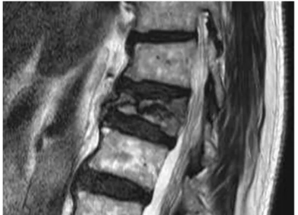
Fig. 1. A 76 year male who had undergone verte- broplasty of L1 body due to acute compression fracture T1 weighted sagittal image show cortical defect at vertebreal upper and lower endplate.
이용한 dependent T test를 이용하였고, 유의 수준은
P value 0.05 미만으로 하였다.결 과
척추 성형술 후 시행한 컴퓨터 단층촬영에서 골 시멘트를 주입하였던 45개의 척추체중 17개(37.7%) 의 척추체에서 골 시멘트 누출이 관찰되었다. 그러 나 골 시멘트 누출로 인한 신경증상을 보이거나 수 술이 필요한 누출은 없었다. 누출이 가장 흔하게 일 어나는 곳은 전방 척추관외 정맥총(anterior external vertebral venous plexus)을 통한 누출로 8예(47%)였으 며 그 외 누출 부위로 추간판(disc space) 4예(23.6%), 기저 추체 정맥(basivertebral vein) 3예(17.6%), 전방 척추관내 정맥총(anterior internal vertebral venous plexus) 2예 였다(11.8%)(Table 1). 주입된 골 시멘트 의 양은 평균 5.1cc (2~9.5)였으며 골 시멘트 누출이 있었던 환자군에서의 평균 주입량은 5.3cc (4~9.5) 였고, 골 시멘트 누출이 없었던 환자군에서의 평균 주입량은 4.9cc (2~8)였다(Table 2). 골 시멘트 누출 이 있었던 환자군에서 높은 결과를 보였으나 통계학 적인 의미는 없었다(P>0.05). 골절과 동반된 척추체 종판 피질골 손상은 15예에서 관찰되었고 그 중 4예
에서 골절된 피질골을 통한 시멘트 누출이 관찰되었 다. 척추체 종판의 피질골 손상은 추간강 시멘트 누 출과 유의한 상관 관계를 보였고(P<0.05)(Fig. 1). 척 추내 골절과 같이 형성된 액체집적 또는 진공변화가 있을 때 유의하게 골 시멘트의 유출이 적었다 (P<0.05)(Fig. 2). 그 외 척추 자기 공명 영상을 통해 측정한 척추 압박 골절률 및 골 시멘트 주입량 대 척추 압박 골절률의 상대 비율에서는 골 시멘트 누 출과 유의한 상관관계를 보이지 않았다(P>0.05)
고 찰
경피적 척추 성형술은 Deramond 등에 의해 1984 년 처음 소개되었으며, 이후 골다공증을 동반한 척 추체 압박골절에서 동통의 감소나 조기 거동을 위해 사용되고 있고 점차 사용 빈도가 증가하고 있다. 척 추 성형술을 시행한 후 통증은 시술 직후부터 감소 하며 대부분 24시간 내에는 호전된다. 동통 감소의 기전은 정확히 알려졌지 않지만, 파괴된 추체의 안 정화나 발열 및 화학적 독성에 의한 감각신경 말단 의 파괴에 의한 것으로 생각되고 있다.
17,18많은 저자 들에 의해 골다공증에 의한 압박골절에 대한 경피적 척추성형술의 통증 완화 효과가 보고되어졌으나 시 술에 따른 합병증 또한 많이 발생한다고 보고되고
있다.
19,20척추성형술 후 합병증은 대부분이 골 시멘
Fig. 2. (A) T2 weighted sagittal image shows dark signal intensity line rimmed cystic area (white arrow) in vertebral body. (B) At T1 weighted image, cystic lesion and surround bone marrow is low signal intensity. (C) After bone cement injection, Fluoroscope image shows bone cement in vertebral body without leakage.
트 누출과 연관이 있으며 심각한 부작용으로는 화농 성 척추염, 골 시멘트 국소적 누출에 의한 척수 및 신경근 손상과 하지 마비,
21-23정맥 혈관을 따라 누 출된 골 시멘트 조각에 의한 폐동맥 색전, 난원공을 통한 뇌동맥 색전, 심낭염 등도 보고되었다.
24-27또한 Lin
28은 척추성형술 시 골 시멘트의 추간판 유출이 있었던 환자들에서 추후 인접 척추체 골절이 증가한 다고 보고하였다. 본 저자들은 이러한 골시멘트의 유출 가능성을 미리 예측할 수 있는 방사선학적 소 견이 있다면 이를 통해 시술 중 또는 시술 후 발생 할 수 있는 합병증에 대해 대비하고 골다공증에 의 한 압박 골절의 수술 결정에 있어서 도움을 줄 수 있을 것으로 생각 하여 연구를 시작하였다. 골 시멘 트의 누출을 예방하는 방법으로는 골시멘트의 불투 명도를 높이고, 생검침 주입시 척추경의 내측벽이 파손되지 않도록 주의하며 골 시멘트의 조영이 추체 후방 1/4지점에 이르면 즉시 주입을 정지하고 주입 압력을 단절적으로 하는 것이 보고되었으며,
29Peh 등은 중심부에 골절이 위치한 경우 생검바늘의 끝을 외측으로 위치시키면 추간판으로의 유출을 줄일 수 있다고 하였다.
30또한 Ryu 등
31은 약 2cc내외의 골시 멘트 주입만으로도 척추체 강도를 유지할 수 있어 소량의 주입도 골 시멘트 누출을 예방할 수 있는 하 나의 방법이라고 보고하였으나, 본 연구에서는 골 시멘트의 주입양이 골 시멘트 누출군에서 보다 많 은 것으로 나왔으나 통계학적인 의미는 없었다.
MiRovsky 등
32은 척추 종판의 피질골 골절이 있을
때 골 시멘트 누출이 증가됨을 보고하였으며 Koh 등
33은 종판의 피질골 골절이 있을 때 골 시멘트 누 출이 유의하게 증가되며 반대로 진공 변화 또는 낭 성 변화가 있을 때 골 시멘트 누출이 유의하게 감소 된다고 보고하였다. 저자들의 연구에서도 척추성형 술 전 시행한 자기 공명 영상에서 척추 종판의 피질 골 손상이 있을 경우 골 시멘트 누출을 유의하게 증 가시키는 결과를 보였으며 척추내 골절과 같이 형성 된 진공 변화 또는 액체 집적이 있을 때 유의하게 골 시멘트 누출이 감소되는 소견을 보여 위의 저자 들과 일치하는 결과를 얻을 수 있었다. 본 연구에서 는 연구대상이 적었다는 점과 척추 종판 피질골의 손상 여부만을 대상으로 분석한 제한점이 있어 추후 피질골의 손상 정도를 포함한 연구를 통해 보완이 필요할 것으로 사료되며 골 시멘트 누출을 예측하여 예방할 수 있는 방법에 대해서도 지속적인 연구가 필요할 것으로 사료된다.
결 론
척추 성형술 후 골 시멘트의 누출을 미리 예측할 수 있는 소견으로는 척추체 종판 피질골의 손상 및 액체 집적 또는 진공변화 등을 들 수 있다. 이러한 소견을 진단할 수 있는 자기공명영상 검사가 척추 성형술 전 골 시멘트의 누출을 미리 예측하는 데 유 용하다.
A B C
참 고 문 헌