산화스트레스에 대한 EGCG (Epigallocatechin Gallate)의 INS-1 세포 보호효과와 기전
메리놀 병원1, 백인제 임상의학연구소 분자치료연구실2, 인제대학교 의과대학 내과학교실3
김미경
1,2․정혜숙
2․윤창신
2․권민정
2,3․고경수
2,3․이병두
2,3․박정현
2.3The Protective Effect of EGCG on INS-1 Cell in the Oxidative Stress and Mechanism
Mi Kyung Kim1,2, Hye Sook Jung2, Chang Shin Yoon2, Min Jeong Kwon2,3, Kyung Soo Koh2,3, Byung Doo Rhee2,3, Jeong Hyun Park2,3
Maryknoll General Hospital1, Molecular Therapy Lab., Paik Memorial Institute for Clinical Research, Inje University2, Department of Internal Medicine, College of Medicine, Inje University3
Abstract
Background: Oxidative stress is important in both diabetic complications and the development and the progression of type 2 diabetes via the effects on the pancreatic β-cells. EGCG (epigallocatechin galleate), a major constituent of green tea, has been known to have beneficial effects on various diseases through the mechanisms of antioxidant and cell signaling modulation. But, very small numbers of studies were published about the direct effects of EGCG on the pancreatic β cell lines. We performed this study to see the protective effect of EGCG on pancreatic β cell line under H2O2 and the mechanisms of this phenomenon.
Methods: We used INS-1 cells and hydrogen peroxide as an oxidative stressor. Their viabilities were verified by MTT assay and FACS. The activity of glutathione peroxidase was assessed by total glutathione quantification kit. Western blot and semi-quantitative RT-PCR for the catalase, SOD (superoxide dismutase), PI3K and Akt were performed. Functional status of INS-1 cells was tested by GSIS (glucose stimulated insulin secretion).
Results: The biological effects of EGCG were different according to its concentrations. 10 μM EGCG effectively protected hydrogen peroxide induced damage in INS-1 cells. The expression and the activity of SOD, catalase and the glutathione peroxidase were significantly increased by EGCG. EGCG significantly increased PI3K and Akt activity and its effect was inhibited partially by wortmannin. GSIS was well preserved by EGCG.
Conclusion: EGCG in low concentration effectively protected INS-1 cells from the oxidative stress through the activation of both antioxidant systems and anti-apoptosis signaling. Further studies will be necessary for the more detailed mechanisms and the clinical implications. (KOREAN DIABETES J 32:121~130, 2008) Key Words: Antiapoptosis, Antioxidant, EGCG, INS cell, Oxidative stress
접수일자: 2008년 3월 6일, 통과일자: 2008년 4월 16일, 책임저자: 박정현, 인제대학교 의과대학 내과학교실
* 본 연구는 2006년 대한당뇨병학회 연구비(제13회 바이엘 연구비)지원으로 이루어졌음.
서 론
최근 몇 년 전부터 제1형 당뇨병에서 뿐만 아니라 제2형 당뇨병에서도 베타세포의 역할이 많이 강조되고 있다1-4). 성 인이 된 후에도 베타세포는 조금씩은 증식할 수 있는 것으 로 알려져 있는데 제2형 당뇨병의 경우에는 세포자연사 (apoptosis)의 정도가 그것을 훨씬 상회할 정도로 커져 있어 전체적으로 베타세포의 양이 점점 줄어들고 이로 인해 고혈 당의 정도가 악화된다고 알려져 왔다.
여러 가지 자극들이 베타세포의 자연사(apoptosis)를 촉 진하는 것으로 알려져 있는데, 그 중에서 대표적인 것이 고 혈당에 의한 당독성(glucotoxicity), 지방산에 의한 지질독성 (lipotoxicity), 활성 산소기(reactive oxygen species, ROS) 들의 증가에 의한 산화스트레스 등이다5). 이 중 산화스트레 스는 당뇨병의 합병증을 일으키는데 중심적인 역할을 하는 것으로 잘 알려져 있었으나 최근에는 당독성, 지질독성 등 의 여러 가지 자극들의 마지막 단계로서 직간접적으로 베타 세포 자체의 세포자연사(apoptosis)를 촉진시키는 데에도 중 요한 역할을 하는 것으로 보고되어 있다. 베타세포는 산화 스트레스에 매우 취약한 세포로 알려져 있는데, 그 이유의 하나로 베타세포에는 항산화 작용에 관여하는 효소가 매우 적은 것으로 보고되어 있다5-9).
녹차에서 추출된 여러 가지 물질들 중에서 epigallocatechin gallate (EGCG)는 가장 강력한 생물학적 작용을 가진 것으 로 알려졌고, 그 작용이 여러 가지 질환들에서 증명되어 왔 다10). 대표적인 질환으로서 여러 종류의 악성 암12-19), 심혈 관질환11), 중추 신경계 내의 여러 가지 퇴행성(degenerative)
질환들20-23)에서의 연구 결과들이 보고되어 있다. 당뇨병에
있어서는 일본인들에서 녹차를 많이 섭취하는 사람일수록 당뇨병의 발생이 적었다는 최근 보고가 있었고24), 몇 가지 동물 모델들에서 인슐린감수성을 증가시키고 혈당을 감소시 키는 효과가 보고되어 있다25-28). 한편으로 암조직에서는 EGCG가 암세포의 세포자연사를 촉진한다고 알려져 있는
반면13-18), 신경변성질환이나 피부질환에서는 오히려 해로운
외부 자극들에 의한 세포자연사를 방지하는 것으로 보고되
어 있다20-23). 이러한 상반된 현상은 EGCG가 세포 종류와
그 농도에 따라 정반대의 생물학적인 효과를 나타내기 때문 인 것으로 생각되고 있다.
EGCG는 항산화제로 잘 알려진 비타민 C보다 항산화 효과 가 10배나 높다고 알려져 있다11,17,30-32)
. 최근에는 앞서의 여러 가지 생물학적 활성효과가 고전적으로 생각했던 항산화제로서 의 효과뿐만 아니라 EGCG와 그것의 여러 가지 대사물들이
세포 내에서 신호 전달 체계에 관여하고 그러한 것들을 변화 시킴으로써 다양한 효과를 보인다고 알려져 있다21,32-37).
저자들은 베타세포자연사(apoptosis)의 중요한 원인 중 하나인 산화스트레스로부터 EGCG가 INS-1 세포를 보호 할 수 있는지를 알아보고, 그 기전에 대해서 조사하고자 하 였다.
방 법
1. 세포배양
INS-1 세포(passage 30~40)는 10% 우태아 혈청, 11 mM 포도당, 24 mM NaHCO3, 10 mM HEPES, 50 μM 2-mercaptoethanol이 포함된 RPMI 1640 (Sigma, MO) 배 지에 37℃, 5% CO2 배양기 안에서 배양하였다.
2. 반응 조건
RPMI 1640배지에 EGCG (Sigma, MO) 10 μM을 포함 하는 다양한 농도를 넣고 24시간 pre-incubation을 한 다음 과산화수소수(Sigma, MO) 80 μM를 5시간 반응시켰다.
PI3K 저해제인 wortmannin (Cell signaling, MA) 사용 시 에는 H2O2처리 한 시간 전에 반응시켰다.
3. MTT법
세포의 생존율을 알아보기 위해서 MTT (Amresco, OH) 법을 사용하였다. 모든 반응이 끝나면 배지를 버리고 우태 아 혈청이 포함되지 않은 배지 90 μL에 5 mg/mL MTT 10 μL를 넣고 2시간 반응시켰다. 다시 배지를 버린 뒤 DMSO 100 μL를 넣고 쉐이커에서 1~2분간 섞은 후 570 nm에서 흡광도를 측정하였다.
4. FACSort
Apoptosis marker인 Annexin V (BD bioscience, CA)와 Propidium Iodide (BD bioscience, CA)를 염색한 후 flow cytometry를 이용하여 확인을 하였다. 반응이 끝난 세포를 Trypsin-EDTA를 사용하여 분리한 후 1,500 rpm으로 5분 간 원심 분리하여 상층액은 버렸다. Binding solution (140 mM NaCl, 10 mM HEPES pH 7.4, 2.5 mM CaCl2)을 넣 고 섞어준 다음 1500 rpm으로 5분간 원심 분리하여 상층액 을 버리고 Annexin V 3 μL와 PI 10 μL를 15분간 반응시킨 후 FACS buffer (1% FBS, 0.1% NaN3) 300 μL를 넣고 flow cytometry에 흘려 보낸 뒤 분석하였다.
5. Glutathione Peroxidase 활성 측정
모든 반응이 끝나면 mammalian tissue lysis buffer (Sigma, MO) 100 μL를 넣고 세포를 융해시키고 12,000 rpm, 10분간 4℃에서 원심 분리하여 상층액만을 깨끗한 e-tube에 옮기고 사용하기 전까지 70℃에 보관하였다. 활성 측정은 Glutathione peroxidase assay kit (Cayman, MI)를 사용하여 측정하였다.
6. Reverse-Transcription Polymerase Chain Reaction
Trizol reagent (Invitrogen, CA)를 사용하여 RNA를 분리 하고 nano-drop을 이용하여 RNA를 정량하였다. Accupower RT/PCR premix (Bioneer, Daejeon, Korea)에서 제작한 각 각의 primer (Table 1)들을 사용하여 RT-PCR을 시행하였 다. Reverse transcription과정은 42℃에서 1시간, PCR 과정 은 94℃에서 2분간 예비 변성을 하고 94℃에서 30초, 48℃
에서 30초, 68℃에서 30초를 30사이클을 돌린 후 마지막 확 장은 7분간 한 다음 4℃에서 보관하였다. 만들어진 PCR 생 성물은 1.5% 아가로즈 젤에 전기영동 하여 SL-20 DNA Image Visualizer에 비추어 GelDoc densitometry로 관찰하 였다.
7. Western Blotting
모든 반응이 끝나면 단백질을 분리하기 위해서 mammalian tissue lysis buffer (Sigma, MO) 100 μL를 넣어 세포를 융 해시키고 12,000 rpm, 10분간 4℃에서 원심 분리하여 상층 액 만을 깨끗한 e-tube에 옮기고 사용하기 전까지 -70℃에 보관하였다. Western blot 실험을 시작하기 전 BCATM protein assay kit (PIERCE, IL)로 단백질을 정량한 다음 15% SDS-PAGE에 한 well당 30 μg을 로딩하여 전기영동
을 하고 20V에서 PVDF membrane에 옮긴 후 5% SKIM MILK로 blocking하였다. 1차 antibody phospho-PI3K p85 (Tyr458) (1:1000), total Akt (1:1000), phospho-Akt (Ser473) (1:1000), Caspase 3 (Cell signaling, MA) (1:1000), Mn -SOD (Upstate, NY) (1:2000), Catalase (Abcam, Cambridge, UK) (1:2000)를 4℃에서 overnight 반응하였다. 2차 antibody Rabbit (Biorad, CA) (1:3000)을 실온에서 1시간 반응한 뒤 AP-conjugated development kit (Biorad, CA)로 발색하 였다.
8. 인슐린분비능
모든 반응이 끝난 뒤 PBS로 한 번 세포를 씻어주고 5 mM 포도당, 2% 우태아 혈청이 포함된 RPMI 1640배지를 넣고 5시간 동안 반응을 시켰다. 그 뒤 5 mM 포도당이 포 함된 Krebs-Ringer Buffer (119 mM NaCl, 4.75 mM KCl, 2.54 mM CaCl2, 1.2 mM MgSO4, 1.2 mM KH2PO4, 5 mM NaHCO3, 20 mM HEPES, pH 7.4), 25 mM 포도당이 포함된 Krebs-Ringer Buffer를 각각 넣고 1시간 다시 배양 한 후 배지를 e-tube에 옮긴 다음 4℃에서 12,000 rpm으로 10분간 원심 분리하여 상층액만을 분리하였다. Rat/mouse Insulin ELISA kit (Linco Research, MO)를 사용하여 인슐 린 단백질 양을 측정하였다.
결 과
1. 산화스트레스하에서 EGCG의 INS-1 세포 생존 감소 방지 효과와 농도에 따른 차이
앞서의 논문에서 저자들은 INS-1 세포에서 50% 정도의 세포 죽음을 야기하는 적정의 H2O2 농도와 처리시간을 각각 80 μM, 5시간을 산화스트레스의 조건으로 정하였다38). EGCG 농도를 0, 5, 10, 50,100 μM로 하였을 때 EGCG 10 μM에서
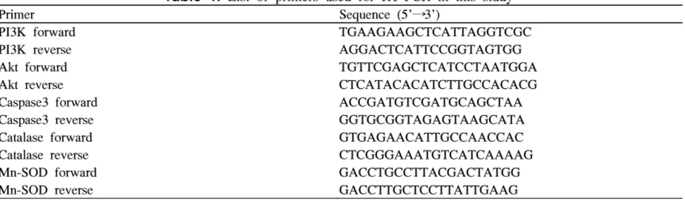
Table 1. List of primers used for RT-PCR in this study
Primer Sequence (5'→3')
PI3K forward TGAAGAAGCTCATTAGGTCGC
PI3K reverse AGGACTCATTCCGGTAGTGG
Akt forward TGTTCGAGCTCATCCTAATGGA
Akt reverse CTCATACACATCTTGCCACACG
Caspase3 forward ACCGATGTCGATGCAGCTAA
Caspase3 reverse GGTGCGGTAGAGTAAGCATA
Catalase forward GTGAGAACATTGCCAACCAC
Catalase reverse CTCGGGAAATGTCATCAAAAG
Mn-SOD forward GACCTGCCTTACGACTATGG
Mn-SOD reverse GACCTTGCTCCTTATTGAAG
A
B
Fig. 1. The cell viability assessed by MTT assay (A) and FACS (B). Cell viability against H2O2 (80 μM) induced toxicity in INS-1 cells was different on the concentration of EGCG. In the histogram, the results obtained from four independent experiments are reported as means ± S.D. (A). FACS analysis after double staining with annexin V/propidium iodide (FL1:AnnexinV, FL2:PI). Dot plots from a representative FACS experiment are shown (B).
A
B
C
D
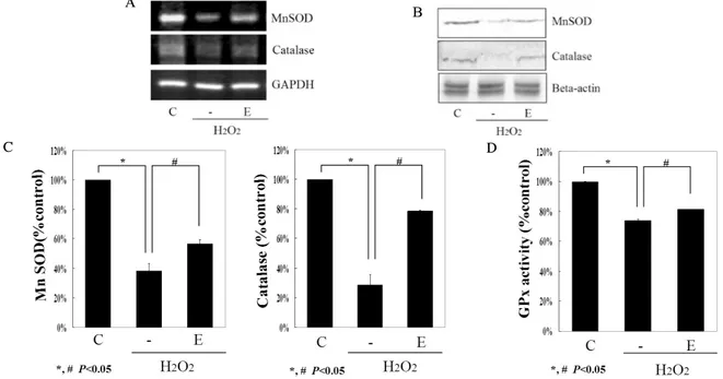
Fig. 2. EGCG changes antioxidant enzymes expression. Total RNA was isolated from INS-1 cells incubated for 24 h without (control; C) or with hydrogen peroxide (H2O2) and in the presence of EGCG (E). (A) Expression of MnSOD, catalase, and endogenous control GAPDH was evaluated by RT-PCR. A representative experiment of three is shown. (B) MnSOD and Catalase expression evaluated by western blot analysis. A representative experiment of three is shown. (C) Densitometric analyses of western blot are reported as means ± S.D. of the three different experiments. (D) GPx activity was evaluated by Glutathione peroxidase assay kit.
H2O2만 처리한 군과 비교하여 20% 정도의 세포 생존도의 증가가 관찰되었다(P < 0.05) (Fig. 1A). 그러나 50 μM 이 상의 농도에서는 H2O2만 처리한 군보다 오히려 세포 생존 율이 감소하였으며, 이는 농도가 증가됨에 따라 더 감소하 였다. FACS 결과에서도 EGCG 10 μM가 INS-1 세포에서 비슷한 세포 보호 효과가 있음을 확인하였다(Fig. 1B).
2. EGCG에 의한 항산화 체계의 활성화
항산화 효소의 mRNA와 단백질 변화를 보기 위해 RT PCR과 western blot을 시행하였다. MnSOD의 mRNA 농도 는 EGCG를 넣어준 실험군에서 통계적으로 의미 있게 증가 되었고, western blot에서도 그 단백질의 양이 유의하게 증 가됨을 관찰할 수 있었다(P < 0.05)(Fig. 2A-C). Catalase의 경우에도 RT-PCR과 western blot에서 H2O2만 처리한 군보 다 EGCG 실험군에서 의미 있게 증가되었다(P < 0.05) (Fig. 2A-C). H2O2만 처리한 군에서는 glutathione 효소 활 성도가 대조군에 비해 30% 정도 감소하였고 EGCG를 넣어 준 군에서는 의미있는 증가를 보였다(P < 0.05) (Fig. 2D).
3. EGCG에 의한 Anti-apoptosis System의 활성화
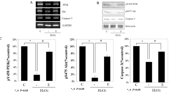
EGCG 처리군에서 PI3K mRNA가 유의하게 증가되었으 며, western blot에서도 EGCG가 활성화된 PI3K activity를 현저하게 증가시켰다(P < 0.05) (Fig. 3). 산화스트레스 하 에서 EGCG는 Akt mRNA를 통계적으로 의미 있게 증가시 켰으며, pS473 Akt 단백질도 EGCG군에서 유의하게 증가 되었다(P < 0.05) (Fig. 3). GSK3-β mRNA와 단백질의 양 은 H2O2만 처리한 군과 비교해서 EGCG 처리한 군에서 특 별한 차이를 보이지 않았다(data not shown). Caspase 3 mRNA은 H2O2만 처리한 군과 큰 차이가 없었으나 western blot에서는 caspase 3 단백질의 양이 EGCG군에서 통계적 으로 유의하게 덜 감소되었다(P < 0.05) (Fig. 3).
4. PI3K Inhibitor 전처리 후, EGCG의 세포 보호 효 과의 부분적 감소
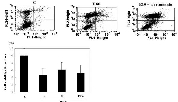
PI3K inhibitor인 wortmannin을 각각의 군에 처리 후 앞 의 실험대로 과산화수소와 EGCG를 처리하여 살아 있는 세 포의 양을 FACS로 측정하였다. 살아있는 세포의 수는 H2O2만 처리한 군에서는 40%로 감소되었고 EGCG 처리군
A
B
C
Fig. 3. EGCG modulated cell signalings related to the apoptosis. Total RNA was isolated from INS-1 cells incubated for 24 h without (control; C) or with hydrogen peroxide (H2O2) and in the presence of EGCG (E). (A) Expression of PI3K, Akt, total caspase 3, and endogenous control beta-actin was evaluated by RT-PCR. A representative experiment of three is shown. (B) Phosphorylation of PI3K and Akt and total caspase 3 were evaluated by western blot analysis. A representative experiment of three is shown. (C) Densitometric analyses of western blot are reported as means ± S.D. of the three different experiments
에서는 증가되었다. EGCG 처리한 군에서 wortmannin과 함께 처리 시 살아 있는 세포의 수가 wortmannin을 처리하 지 않은 군보다 더 감소하여, 그것의 % 감소분은 50%에 달 하였다(Fig. 4).
% inhibition
of protection = 100 - % protection with wortmannin
% protection with EGCG (without wortmannin) 5. EGCG의 포도당 자극에 의한 인슐린분비 증가 효과
포도당 농도를 증가시켜 인슐린분비의 변화를 측정하였 다. 대조군에서는 포도당 농도 5 mM에 비해 25 mM에서 인슐린분비의 증가가 2배 정도 증가되었다. 5 mM과 25 mM 각각의 포도당 농도에서, H2O2만 처리한 군에서는 인 슐린분비가 현저하게 감소되었고 포도당 농도를 증가시킬 때 인슐린분비의 증가는 대조군에 비해 현저히 낮았다.
EGCG 처리군에서는 각각의 농도에서의 인슐린분비능뿐만 아니라, 5 mM에서 25 mM 포도당 농도에서의 인슐린분비 능의 증가도 H2O2만 처리한 군보다 의미 있게 증가됨을 관 찰할 수 있었다(P < 0.05) (Fig. 5).
고 찰
본 연구에서는 녹차의 활성 성분 중 가장 강력한 것의 하 A
B
Fig. 4. The cell viability assessed by FACS after treatment of PI3K inhibitor. Cell viability against H2O2 (80 μM) induced toxicity in INS-1 cells was increased with 10 μM of EGCG (E) and partially decreased with PI3K inhibiotor (E+W).
FACS analysis after double staining with annexin V/propidium iodide(FL1:AnnexinV, FL2:PI). Dot plots from a representative FACS experiment are shown (A). In the histogram, the results obtained from three independent experiments are reported as means ± S.D. (B).
Fig. 5. Insulin secretion from INS-1cells in response to glucose (5 and 25 mM) concentration after a 24-h incubation with control medium (control), medium containing H2O2
with EGCG (E) and without EGCG. Data are means ± S.D. of three separate experiments.
나인 EGCG가 췌장 베타세포주를 산화스트레스 하에서 직 접적으로 보호하는 효과가 있는지를 살펴보고자 하였다. 앞 서의 여러 가지 연구들에서, EGCG가 암세포에서는 세포의 사멸을 촉진시키는 데 반하여12-19) 여러 가지 다른 퇴행성질 환에서는 세포를 보호하는 효과가 있었다11,20-23). 베타세포 에 대해서는 4일간 당뇨병 모델 쥐에 EGCG를 복강 내 주사 하여 췌장 소도의 양이 줄어들었다는 보고39)와 RINm5F 세 포에서 EGCG가 cytokine에 의한 췌장 베타세포 손상을 줄 였다40)는 서로 상반된 보고가 있었다. 본 연구에서 EGCG 가 INS-1세포의 생존능을 증가시키는 효과는 농도에 따라 차이가 있었는데, 다른 연구에서도 EGCG가 농도에 따라 세포자연사(apoptosis)에 대하여 이중적인 결과를 보인다고 보고해 왔다. 어떤 암세포에서는 배양 시간이 24시간 이상 일 경우에는 40 μM 이상의 농도에서, 48시간 배양 시에는 20 μM에서도 세포자연사가 의미 있게 증가하는 결과를 보 고하였다15). 대부분 세포자연사를 유도하는 EGCG의 농도 는 100 μM이나 200 μM 이상으로 고농도의 EGCG를 사용
하였다12,13,18). 그에 반해 다른 질환 모델, 즉 상피 세포나 뇌
세포의 퇴행성질환을 예방하는 연구에서는 대체로 낮은 농 도, 즉 50 μM 이하에서 세포의 생존을 증가시켜서 그 질환 에 도움이 되는 것으로 알려져 있다. 췌장 베타세포주를 이 용한 본 연구에서도 세포의 생존이 증가되는 EGCG의 농도 는 10 μM로 대체적으로 낮았고, 이러한 결과는 EGCG 농 도에 따른 세포 생존에 대한 다른 연구에서와 비슷한 결과 를 보이고 있다17). 그리고 일정 농도 이상의 고농도에서는 오히려 세포자연사를 더 촉진시키는 결과가 관찰되었다. 이 러한 실험 조건들에서의 결과가 앞으로 임상적으로 이용 되기 위해서는 더 많은 연구가 계속되어야 할 것으로 생각 된다.
어떤 기전으로 EGCG가 베타세포를 산화스트레스에 의 한 자연사(apoptosis)로부터 보호할 수 있는가에 대해 살펴 본 바로는, 전통적으로 잘 알려진 항산화 작용뿐만 아니라, 베타세포의 생존에 중요한 역할을 하는 세포 내 신호전달 체계에도 영향을 미친다는 사실을 본 연구를 통해 확인하였 다. 췌장의 베타세포는 다른 세포들과 비교해서 유난히 항 산화 효소의 양이 적고 그 효과가 약한 것으로 알려져 있는 데5-9), 본 연구에서도 EGCG는 항산화 체계를 효과적으로 증가시킨다는 사실을 몇 가지 결과를 통해서 볼 수 있었다.
이러한 결과는 항산화제로 잘 알려진 녹차에서 추출한 물질 이 췌장 베타세포주에서도 그러한 효과를 나타내는 것을 입 증한 것으로 볼 수 있다.
그러나 다른 세포주에서는 단지 단순한 항산화제로만 작
용하는 것이 아니라 EGCG가 PI3K/Akt 체계를 변화시켜 세포의 세포자연사를 억제한다는 것이 몇 가지 연구들에서 알려져 왔다20-23). G93A라는 운동신경세포에서 H2O2로 산 화스트레스를 주었을 때, EGCG가 생존 신호가 되는 PI3K/Akt를 증가시키고20), 사멸의 신호인 GSK-3-β를 감소 시켜 세포자연사(apoptosis)를 방지하였다고 보고된 바 있 고, 다른 종류의 N18D3 세포에서도 EGCG가 비슷한 세포 전달 체계를 변화시켜서 신경세포를 보호할 수 있다고 발표 하였다21). 그 밖에도 EGCG는 세포 생존에 관계하는 MAP kinase 신호 전달에도 관여하는데, 이 경우에는 세포의 농도 에 따라 다른 효과를 보인다고 알려져 왔다. 즉 nanomole이 나 micromole 농도에서는 MAP kinase를 활성화시켜 세포 자연사를 방지하지만, 높은 농도에서는 이와는 반대로 MAP kinase를 억제시키고 JNK를 증가시켜 결국은 세포의 생존 율을 감소시킨다는 보고가 있다32,34). 이렇듯, EGCG의 세포 전달 체계에 대한 효과는 세포의 종류나 모델이 되는 병이 나 자극에 따라 다른 효과를 보일 수 있을 것으로 생각되어 왔다. 본 연구에서는 췌장 베타세포에 산화스트레스를 주었 을 때 EGCG가 PI3K/Akt 신호 전달 체계에 어떤 영향을 미치는지 살펴 보았다. 결과에서 보는 바와 같이 EGCG가 caspase-3를 더 적게 변화시키고, PI3K activity와 Akt의 인 산화를 증가시켜 췌장 베타세포에서도 단순히 ROS를 없애 거나 항산화 효소를 증가시키는 작용 이외에도 이러한 세포 전달 체계에 영향을 미쳐서 세포 생존을 증가시키는 역할을 하고 있다는 것을 알 수 있었다. PI3K 억제제인 wortmannin 을 처리하였을 때 EGCG의 세포 보호 효과가 절반 정도 감 소되는 본 연구 결과로 미루어 볼 때, 췌장 베타세포인 INS-1 세포에서는 산화스트레스에 대한 EGCG의 긍정적인 효과는 항산화 작용과 세포 생존에 관여하는 중요한 세포 전달 체계를 모두 공유하여 일어나는 것으로 생각될 수 있다.
본 연구에서는 EGCG가 INS-1 세포에서 산화스트레스에 서도 인슐린분비 작용을 회복시킬 수 있음을 보였다. 다른 당뇨병 모델 동물 실험에서 녹차는 혈청 인슐린농도를 증가 시켜서 혈당을 조절하기도 했지만, 간에서 당 생성을 감소 시키고 말초 조직에서 인슐린의 작용을 증가시키는 효과들 도 보고되어 왔다26-28). 어떤 연구에서는 인슐린분비는 증가 시키지 않고 오히려 말초 조직에서의 인슐린저항성을 개선 시키거나 잘 알려져 있지 않은 단백질을 발현시켜서 당 대 사를 개선시킬 가능성에 대해서도 보고했었다25).
결론적으로 여러 가지 역학 연구들에서 당뇨병 발병 감 소의 효과가 있는 것으로 알려진 녹차 추출물 EGCG는, 항
산화 효과와 항세포자연사(apoptosis) 작용을 통하여 췌장 베타세포를 산화스트레스로부터 직접적으로 보호할 수 있을 것으로 생각된다. 향후 임상에서 실제로 활용되기 위해서는 좀 더 많은 연구가 필요할 것이다.
요 약
연구배경: 산화스트레스는 만성 당뇨병 합병증의 발생에 도 중요할 뿐 아니라 췌장 베타세포에 대한 직접적이거나 간접적인 영향으로 제2형 당뇨병의 발생과 진행에도 중요한 역할을 하는 것으로 밝혀지고 있다. 녹차의 주요 추출물 중 의 하나인 EGCG (epigallocatechin galleate)는 각종 암 치 료를 비롯하여, 심혈관계질환 및 뇌변성질환에 좋은 영향을 미친다고 알려져 왔으며 임상 연구에서도 당뇨병의 발생을 감소시키는 것으로 알려져 왔다. 그 기전은 전통적으로 잘 알려진 항산화제로서의 역할 뿐 아니라 세포 전달 체계의 변화를 통해서 이루어지는 것으로 보고되고 있다. 그러나 췌장 베타세포에서 EGCG가 직접적으로 미치는 영향에 대 해서는 많이 알려져 있지 않다. 본 연구는 과산화수소를 이 용한 산화스트레스하에서 EGCG가 INS-1 세포를 보호할 수 있는지를 살펴보고 그 기전을 보고자 하였다.
방법: INS-1 세포는 RPMI 1640배지에서 배양하였고, 산화스트레스는 과산화수소수를 이용한 모델을 사용하였다.
세포의 생존능은 MTT assay와 annexin V와 porpium iodide (PI)를 사용한 FACS로 확인하였다. glutathione peroxidase 활성도는 total glutathione quantification kit로 측정하였고 catalase, SOD (superoxide dismutase), PI3K와 Akt의 mRNA와 활성도는 Western blot과 semi-quantitative RT-PCR를 이용하여 측정되었다. PI3K 억제제인 wortmannin 을 전처리한 후 세포의 생존 정도를 FACS로 측정하였다.
INS-1 세포의 기능적인 측면은 포도당 자극에 의한 인슐린 분비(GSIS)로 평가하였다
결과: 산화스트레스하에서의 EGCG의 효과는 농도에 따 라 차이가 나서, 10 μM의 EGCG에서는 세포 생존도의 증 가가 관찰되었으나, 50 μM 이상의 농도에서는 세포 생존율 이 감소하였으며, 이는 농도가 증가됨에 따라 더 감소하였 다. SOD, catalase와 glutathione peroxidase의 mRNA 발현 과 단백질의 양은 EGCG 처리에 의해 유의하게 증가되었 다. EGCG는 PI3K와 Akt 활성도를 증가시켰고, PI3K 억제 제에 의해 EGCG의 효과는 부분적으로 감소되었다. 포도당 자극에 의한 인슐린분비는 EGCG에 의해 잘 보존되었다.
결론: EGCG는 낮은 농도에서 INS-1 세포를 산화스트레스
로부터 효과적으로 보호할 수 있었고, 항산화효과와 세포자 연사(apoptosis)방지에 관여하는 세포 신호 전달을 기전으로 하고 있었다. 더 명확한 기전과 임상적 적용을 위해서는 더 많은 연구가 있어야 할 것이다.
참 고 문 헌
1. Christopher JR: Type 2 Diabetes-a Matter of B cell Life and Death? Science 307:380-84, 2005
2. Donath MY, Ehses JA, Maedler K, Shumann DM, Ellingsgaard H, Eppler E, Reinecke M: Mechanisms of beta cell death in type 2 diabetes. Diabetes 54(Suppl 2):S108-13, 2005
3. Butler AE, Janson J, Bonner-Weir S, Ritzel R, Rizza RA, Butler PC: Beta-cell deficit and increased beta-cell apoptosis in humans with type 2 diabetes.
Diabetes 52:102-10, 2003
4. Kahn SE: The relative contributions of insulin resistance and β-cell dysfunction to the pathophysiology of Type 2 diabetes. Diabetologia 46:3-19, 2003
5. Robertson RP, Harmon J, Tran PO, Poitout V: Beta cell glucose toxicity, lipotoxicity, and chronic oxidative stress in type 2 diabetes. Diabetes 53 (Suppl.
1):S119-24, 2004
6. Sakuraba H, Mizukami H, Yagihashi N, Wada R, Hanyu C, Yagihashi S: Reduced beta-cell mass and expression of oxidative stress-related DNA damage in the islet of Japanese type II diabetic patients.
Diabetologia 45:85-96, 2002
7. Green K, Brand MD, Murphy MP: Prevention of mitochondrial oxidative damage as a therapeutic strategy in diabetes. Diabetes 53 (Suppl 1):S110-8, 2004
8. Evans JL, Goldfine ID, Maddux BA, Grodsky GM:
Are oxidative stress-activated signaling pathways mediators of insulin resistance and beta-cell dysfunction? Diabetes 52:1-8, 2003
9. Kajimoto Y, Kaneto H: Role of oxidative stress in pancreatic β-cell dysfunction. Ann N Y Acad Sci 1011:168-76, 2004
10. Beecher GR, Warden BA, Merken H: Analysis of tea polyphenols. Proc Soc Exp Biol Med 220:267-70,
1999
11. Knekt P, Kumpulainen J, Jarvinen R, Rissanen H, Heliovaara M, Reunanen A, Hakulinen T, Aromaa A:
Flavonoid intake and risk of chronic diseases. Am J Clin Nutr 76:560-8, 2002
12. Masuda M, Suzui M, Weinstein IB: Effects of epigallocatechin-3-gallate on growth, epidermal growth factor receptor signaling pathways, gene expression, and chemosensitivity in human head and neck squamous cell carcinoma cell lines. Clin Cancer Res 7:4220-9, 2001
13. Liang YC, Lin-shiau SY, Chen CF, Lin JK: Suppression of extracellular signals and cell proliferation through EGF receptor binding by (-)-epigallocatechin gallate in human A431 epidermoid carcinoma cells. J Cell Biochem 67:55-65, 1997
14. Sachinidis A, Seul C, Seewald S, Ahn H, Ko Y, Vetter H: Green tea compounds inhibit tyrosine phosphorylation of PDGF β-receptor and transformation of A172 human glioblastoma. FEBS Lett 471:51-5, 2000
15. Shimizu M, Deguchi A, Lim JT, Moriwaki H, Kopelovich L, Weinstein IB: (-)-Epigallocatechin gallate and polyphenon E inhibit growth and activation of the epidermal growth factor receptor and human epidermal growth factor receptor-2 signaling pathways in human colon cancer cells. Clin Cancer Res 11:2735-46, 2005 16. Yang GY, Liao J, Li C, Chung J, Yurkow EJ, Ho CT, Yang CS: Effect of black and green tea polyphenols on c-jun phosphorylation and H2O2 production in transformed and non-transformed human bronchial cell lines: possible mechanisms of cell growth inhibition and apoptosis induction. Carcinogenesis 21:2035-9, 2000
17. Peng G, Wargovich MJ, Dixon DA: Anti-proliferative effects of green tea polyphenol EGCG on Ha-Ras- induced transformation of intestinal epithelial cells.
Cancer Lett 238:260-70, 2006
18. Hou Z, Sang S, You H, Lee MJ, Hong J, Chin KV, Yang CS: Mechanism of action of (-)-epigallocatechin -3-gallate: auto-oxidation-dependent inactivation of epidermal growth factor receptor and direct effects on
growth inhibition in human esophageal cancer KYSE 150 cells. Cancer Res 65:8049-56, 2005
19. Albrecht DS, Clubbs EA, Ferruzzi M, Bomser JA:
Epigallocatechin-3-gallate (EGCG) inhibits PC-3 prostate cancer cell proliferation via MEK- independent ERK1/2 activation. Chem Biol Interact 171:89-95, 2008
20. Koh SH, Kwon H, Kim KS, Kim J, Kim MH, Yu HJ, Kim M, Lee KW, Do BR, Jung HK, Yang KW, Appel SH, Kim SH: Epigallocatechin gallate prevents oxidative-stress-induced death of mutant Cu/Zn- superoxide dismutase (G93A) motoneuron cells by alteration of cell survival and death signals.
Toxicology 202:213-25, 2004
21. Koh SH, Kim SH, Kwon H, Kim JG, Kim JH, Yang KH, Kim J, Kim SU, Yu HJ, Do BR, Kim KS, Jung HK: Phosphatidylinositol-3 kinase/Akt and GSK-3 mediated cytoprotective effect of epigallocatechin gallate on oxidative stress-injured neuronal- differentiated N18D3 cells. Neurotoxicology 25:793-802, 2004 22. Katiyar SK, Afaq F, Azizuddin K, Mukhtar H:
Inhibition of UVB-induced oxidative stress-mediated phosphorylation of mitogen-activated protein kinase signaling pathways in cultured human epidermal keratinocytes by green tea polyphenol (-) -epigallocatechin-3-gallate. Toxicol Appl Pharmacol 176:110-7, 2001
23. Levites Y, Amit T, Youdim MB, Mandel S:
Involvement of protein kinase C activation and cell survival/cell cycle genes in green tea polyphenol (-)-epigallocatechin 3-gallate neuroprotective action. J Biol Chem 277:30574-80, 2002
24. Iso H, Date C, Wakai K, Fukui M, Tamakoshi A JACC Study Group: The relationship between green tea and total caffeine intake and risk for self-reported type 2 diabetes among Japanese adults. Ann Intern Med. 144:554-62, 2006
25. Tsuneki H, Ishizaka M, Terasawa M, Wu JB, Sasaoko T, Kimura I: Effect of Green tea on blood glucose levels and serum proteomic patterns in diabetic (db/db) mice and on glucose metabolism in healthy humans. BMC Pharmacol 4:18, 2004
26. Sabu MC, Smitha K, Kuttan R: Anti-diabetic diabetic of green tea polyphenols and their role in reducing oxidative stress in experimental diabetes. J.
Ethanopharmacol. 83:109-16, 2002
27. Waltner-Law ME, Wang XL, Law BK, Hall RK, Nawano M: Epigallocatechin gallate, a constituent of Green tea represses hepatic glucose production. J Biol Chem 277:34933-40, 2002
28. Wu LY, Juan CC, Ho LT, Hsu YP, Hwang LS: Effect of green tea supplementation on insulin sensitivity in Sprague-Dawley rats. J Agric Food Chem 52:643-8, 2004
29. Song Y, Manson JE, Buring JE, Sesso HD, Liu S:
Associations of dietary flavonoids with risk of type 2 diabetes, and markers of insulin resistance and systemic inflammation in women: a prospective study and cross-sectional analysis. J Am Coll Nutr 24:
376-84, 2005
30. Doronicheva N, Yasui H, Sakurai H: Chemical structure-dependent differential effects of flavonoids on the catalase activity as evaluated by a chemiluminescent method. Biol Pharm Bull 30:213-7, 2007
31. Scalbert A, Morand C, Manach C, Remesy C: Absorption and metabolism of polyphenols in the gut and impact on health, Biomed. Pharmacother 56:276 -82, 2002 32. Williams RJ, Spencer JP, Rice-Evans C: Flavonoids:
antioxidants or signalling molecules? Free Radic Biol Med. 36:838-49, 2004
33. Agullo G, Gamet-Payrastre L, Manenti S, Viala C, Remesy C, Chap H, Payrastre B: Relationship between flavonoid structure and inhibition of phosphatidylinositol 3-kinase: a comparison with tyrosine kinase and protein kinase C inhibition. Biochem Pharmacol 53:
1649-57, 1997
34. Balasubramanian R, Efimova T, Eckert RL: Green tea polyphenol stimulates a Ras, MEKK1, MEK3, and p38 cascade to increase activator protein 1 factor -dependent involucrin gene expression in normal human keratinocytes. J Biol Chem 277:1828-36, 2002 35. Agullo G, Gamet-Payrastre L, Manenti SC, Remesy
C, Chap H, Payrastre B: Relationship between flavonoid structure and inhibition of phosphatidylinositol 3-kinase: a comparison with tyrosine kinase and protein kinase C inhibition. Biochem Pharmacol 53:1649-57, 1997
36. YaWen C, ChunFa H, KehSung T, RongSen Y, ChengChieh Y, ChingYao Y, ShoeiYn L, ShingHwa L: The role of phosphoinositide 3-Kinase/Akt signaling in low-dose mercury-induced mouse pancreatic β-cell dysfunction in vitro and in vivo. Diabetes 55:1614-24, 2006
37. Lu M, Bi CS, Gong XG, Chen HM, Sheng XH, Deng TL, Xu KD: Anti-proliferative effects of recombinant iron superoxide dismutase on HepG2 cells via a redox-dependent PI3k/Akt pathway. Appl Microbiol Biotechnol. 76:193-201, 2007
38. 권민정, 정혜숙, 김미경, 강성훈, 서광욱, 송재광, 윤태 연, 전민경, 하태환, 윤창신, 김미경, 이우제, 노정현, 권 수경, 김동준, 고경수, 이병두, 임경호, 이순희, 박정현:
INS-1 세포에서 항산화 효과를 통한 Quercetin의 세포 보호 효과. 당뇨병 31:383-90, 2007
39. Yun SY, Kim SP, Song DK: Effects of (-)-epigallocatechin -3-gallate on pancreatic beta-cell damage in streptozotocin -induced diabetic rats. Eur J Pharmacol. 541:115-21, 2006
40. Han MK: Epigallocatechin gallate, a constituent of green tea, suppresses cytokine-induced pancreatic beta-cell damage. Exp Mol Med. 35:136-9, 2003