Imaging Science in Dentistry 2015; 45: 117-22 http://dx.doi.org/10.5624/isd.2015.45.2.117
Introduction
With edentulous patients, it is difficult to secure suitable positioning when taking panoramic radiographs and also it is hard to assess the positioning by means of inspecting anatomical structures such as the occlusal plane, ante- ro-posterior position of the anterior teeth, and symmetry in their panoramic radiographs after taken. Several stud-
ies have examined the difficulties of taking panoramic radiographs of edentulous patients. Without distinguish- ing between dentulous and edentulous images, Dhillon et al.1 noted that most panoramic radiographs had at least one error. Glass et al.2 reported that 89% of panoramic radiographs of edentulous patients had at least one error, which included the following positions: chin too high, too far forward, tongue not raised, chin too low, head tilted, head turned, and too far back. Batenburg et al.3 showed that panoramic radiographs without positioning reproduc- ibility were not reliable for diagnosis and evaluation of the edentulous mandible. Accuracy and reproducibility of edentulous patients’ panoramic radiographs are import- ant for dental diagnosis and treatment planning because
A new bite block for panoramic radiographs of anterior edentulous patients: A technical report
Jong-Woong Park1, Khanthaly Symkhampha2, Kyung-Hoe Huh1, Won-Jin Yi1, Min-Suk Heo1,*, Sam-Sun Lee1, Soon-Chul Choi1
1Department of Oral and Maxillofacial Radiology and Dental Research Institute, School of Dentistry, Seoul National University, Seoul, Korea
2Division of Oral and Maxillofacial Radiology, Department of Basic Science, Faculty of Dentistry, University of Health Sciences, Vientiane, Laos
AbstrAct
Purpose: Panoramic radiographs taken using conventional chin-support devices have often presented problems with positioning accuracy and reproducibility. The aim of this report was to propose a new bite block for panoramic radiographs of anterior edentulous patients that better addresses these two issues.
Materials and Methods: A new panoramic radiography bite block similar to the bite block for dentulous patients was developed to enable proper positioning stability for edentulous patients. The new bite block was designed and implemented in light of previous studies. The height of the new bite block was 18 mm and to compensate for the horizontal edentulous space, its horizontal width was 7 mm. The panoramic radiographs using the new bite block were compared with those using the conventional chin-support device.
results: Panoramic radiographs taken with the new bite block showed better stability and bilateral symmetry than those taken with the conventional chin-support device. Patients also showed less movement and more stable positioning during panoramic radiography with the new bite block.
conclusion: Conventional errors in panoramic radiographs of edentulous patients could be caused by unreliability of the chin-support device. The newly proposed bite block for panoramic radiographs of edentulous patients showed better reliability. Further study is required to evaluate the image quality and reproducibility of images with the new bite block.(Imaging Sci Dent 2015; 45: 117-22)
Key words: Panoramic, Radiography; Bite Block; Reproducibility of Results
Copyright ⓒ 2015 by Korean Academy of Oral and Maxillofacial Radiology
This is an Open Access article distributed under the terms of the Creative Commons Attribution Non-Commercial License(http://creativecommons.org/licenses/by-nc/3.0) which permits unrestricted non-commercial use, distribution, and reproduction in any medium, provided the original work is properly cited.
Imaging Science in Dentistry·pISSN 2233-7822 eISSN 2233-7830
*This study was supported by grant no. 04-2012-0058 from the SNUDH Research Fund.
Received February 2, 2015; Revised March 3, 2015; Accepted March 15, 2015
*Correspondence to : Prof. Min-Suk Heo
Department of Oral and Maxillofacial Radiology, School of Dentistry, Seoul Nation- al University, 101 Daehak-ro, Jongno-gu, Seoul 110-749, Korea
Tel) 82-2-2072-3016, Fax) 82-2-744-3919, E-mail) [email protected]
*표 선 두께
=
1/0.3ptpanoramic radiographs have been widely used in cases of the screening of cysts, foreign bodies, and neoplasms,4-6 finding bone resorption and osteopenia of the jaws,7-9 and installing implants.10
Conventional bite blocks and chin-support devices, such as the ones included with the Orthopantomograph® OP100 (Im- aging Instrumentarium, Tuusula, Finland) (Figs. 1A and B,
respectively), are widely available for positioning anterior edentulous patients during panoramic radiography.11 The chin-support device is more commonly used than the con- ventional bite block because the height of the bite block is 12 mm, which seems to provide an insufficient vertical dimen- sion for the edentulous area.
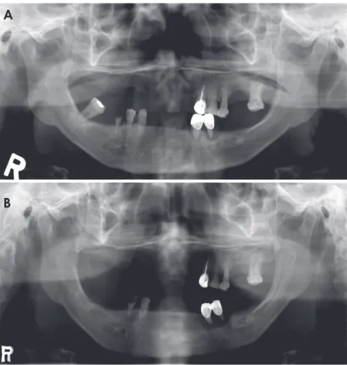
However, using the chin-support device for panoramic radiography has also presented some problems. Pano- ramic radiographs using the chin-support device have frequently shown insufficient inter-maxillary space and lack of reproducibility (Fig. 2). According to the study of Park et al.,12 the reproducibility of panoramic radiographs of edentulous patients using a conventional chin-support device was worse than that of dentulous patients. There- fore, a new bite block for edentulous patients is required to improve the accuracy and reproducibility of panoramic radiographs of edentulous patients by improving the accu- racy and reproducibility of positioning patients for taking panoramic radiographs.
The aim of this report is to propose a new bite block for panoramic radiographs of edentulous patients that could
A B
Fig. 1. A. The conventional bite block for anterior edentulous pa- tients. B. The chin-support device for anterior edentulous patients.
Fig. 2. A and B. Two panoramic radio- graphs acquired from the same anterior edentulous patient using a conventional chin-support device show lack of repro- ducibility in the inter-maxillary vertical dimension.
A
B
enable a greater degree of accuracy and reproducibility in patient positioning, and reduce the weaknesses of pan- oramic radiographs taken with a chin-support device.
Materials and Methods
A new bite block for edentulous patients was designed to bring reproducibility when taking panoramic radiographs to a similar level as that of a bite block for dentulous pa- tients. The new bite block for edentulous patients involved modifications in the bite portion that could compensate for the missing anterior teeth and resorbed anterior alveolar bone tissue of anterior edentulous patients. The dimen- sions of the missing and resorbed tissues were determined by referring to previous studies.
In detail, the new bite block (Fig. 3A) was designed to position anterior edentulous patients in the same manner as
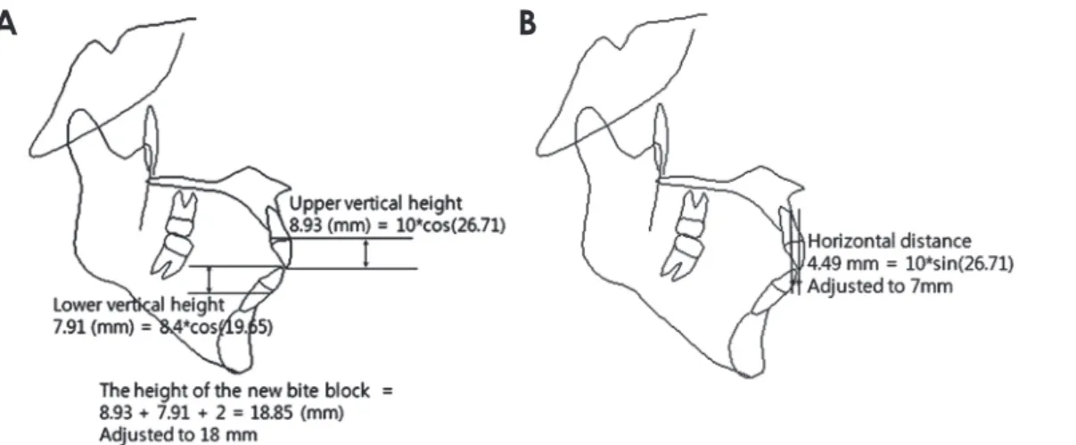
anterior dentulous patients who could bite into the notch- es of a conventional standard bite block (Fig. 3B) while undergoing panoramic radiography. The new bite block needed to compensate for the area of missing anterior teeth and resorbed alveolar bone. To compensate for those areas, statistical analysis of measured tooth lengths, facio-lingual inclinations, and resorbed anterior alveolar bone dimen- sions were investigated in the literature. According to Vol- chansky and Cleaton-Jones,13 the mean sizes of teeth were 10.0mm among the maxillary central incisors and 8.4mm among the mandibular central incisors in those aged 20 to 40 years. Furthermore, the mean facio-lingual inclination of the maxillary central incisors was 33.50˚, and that of the mandibular central incisors was 26.44˚.14 The mean angle between the occlusal plane and Frankfort horizontal plane was 6.79˚.15 Thus, the angle between the maxillary central incisor and vertical plane was 26.71˚, and the angle between the mandibular central incisor and vertical plane was 19.65˚. Also, the extent of anterior alveolar bone re- sorption was 5.90mm in non-denture wearers and 6.69mm in denture wearers in the maxillary central incisor area, and 7.07 mm in non-denture wearers and 13.03mm in denture wearers in the mandibular central incisor area.16
Using these values, the vertical dimension and the an- tero-posterior horizontal distance (horizontal distance) of the new bite block was determined. For the vertical dimension, the vertical vector of the anterior tooth por- tion was calculated to be 16.85mm, and the thickness of the bite position (notch) of the conventional standard bite block for dentulous patients of the Orthopantomograph® OP100 (2mm) was taken into consideration. Thus, the vertical height of the new bite block was chosen to be 18.85mm, and adjusted to 18mm by trial and error (Fig.
A B
Fig. 3. A. The new bite block for anterior edentulous patients. B.
The conventional standard bite block for anterior dentulous pa- tients.
Fig. 4. A. The calculation to determine the height of the new bite block. B. The horizontal distance calculation to determine the antero-pos- terior dimension of the new bite block.
A B
4). For the horizontal distance, which is the difference be- tween the lengths from the panoramic radiograph sensor to the standard block’s notch and to the biting position of the new bite block, the horizontal vector of the anterior tooth portion was calculated to be 4.49mm, and the hori- zontal vector of the resorbed bone portion was considered to be in the range from 2.65mm to 3.00mm. The hori-
zontal distance for the new bite block was chosen to be 5mm (4.49mm), and was adjusted to 7mm by trial and error (Fig. 4).
Panoramic radiographs of anterior edentulous patients were taken using the new bite block. Then, the panoramic radiographs were compared with the radiographs taken previously using conventional chin-support device. Then
Fig. 5. Panoramic radiographs ac quired from anterior edentulous patients using the conventional chin-support device.
A. The radiograph shows insufficient inter-maxillary vertical height. B. The radiograph shows asymmetry although the patient’s jaw is not asymmetrical.
C. The panoramic radiographs acquired from an anterior edentulous patient us- ing the conventional chin-support device show horizontal magnification.
A
B
C
the reproducibility and technical errors of the radiographs using the conventional chin-support device were evaluated and compared to the radiographs using the new bite block.
results
Some radiographs taken with the chin-support device have shown insufficient inter-maxillary space (Fig. 5A), and others have lacked reproducibility of anatomical structural dimensions, especially in the horizontal direc- tion (Fig. 5B). The chin-support device might not provide appropriate reproducible and accurate positioning in tak- ing panoramic radiographs.
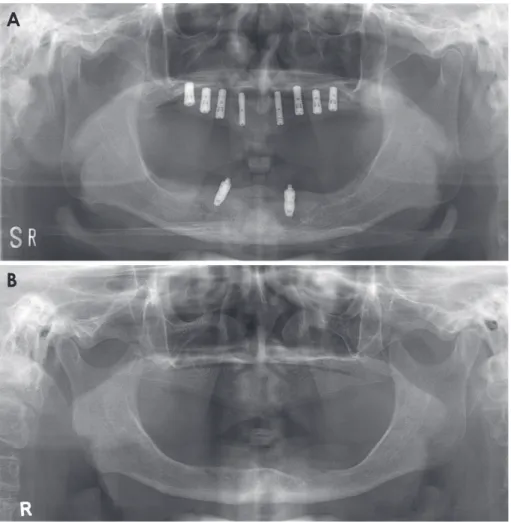
Panoramic radiographs taken with the new bite block showed better stability in inter-maxillary vertical space, antero-posterior position, and bilateral symmetry than radiographs taken with the conventional chin-support de- vice (Fig. 6). In addition, according to the radiographers’
statements, the patients using the new bite block showed less movement and more stable positioning while under- going panoramic radiography.
discussion
The panoramic radiographs of edentulous patients taken with the chin-support device showed some errors. These problems could have originated from the unreliability of the chin-support device in positioning patients while undergoing panoramic radiography. In other words, the chin-support device would provide inappropriate reproduc- ibility and inaccurate positioning support. This finding was in accordance with those of Park et al.,12 justifying the at- tempt to design a new bite block for panoramic radiographs of edentulous patients. Therefore, this new bite block was designed and implemented, and a patent was obtained on the bite block in the Republic of Korea (Registration No.
1299456).
The new bite block produced better inter-maxillary space, antero-posterior position, and bilateral symmetry in panoramic radiographs than the conventional chin-support device. These results might have originated from stable and accurate positioning of both of the jaws in taking panoram- ic radiographs with the new bite block.
Fig. 6. A and B. The panoramic radio- graphs taken using the new bite block showed a more suitable vertical height and antero-posterior position, and better bilateral symmetry than the panoramic radiographs taken using the chin-sup- port device.
A
B
However, ongoing evaluation of the new bite block should be carried out, including assessment of image qual- ity and reproducibility. It will take time to accumulate pan- oramic radiographs taken with the new bite block in order to perform these assessments.
references
1. Dhillon M, Raju SM, Verma S, Tomar D, Mohan RS, Lakhan- pal M, et al. Positioning errors and quality assessment in pan- oramic radiography. Imaging Sci Dent 2012; 42: 207-12.
2. Glass BJ, Seals RR Jr, Williams EO. Common errors in pan- oramic radiography of edentulous patients. J Prosthodont 1994; 3: 68-73.
3. Batenburg RH, Stellingsma K, Raghoebar GM, Vissink A.
Bone height measurements on panoramic radiographs: the ef- fect of shape and position of edentulous mandibles. Oral Surg Oral Med Oral Pathol Oral Radiol Endod 1997; 84: 430-5.
4. Kogon S, Bohay R, Stephens R. A survey of the radiographic practices of general dentists for edentulous patients. Oral Surg Oral Med Oral Pathol Oral Radiol Endod 1995; 80: 365-8.
5. Spyropoulos ND, Patsakas AJ, Angelopoulos AP. Findings from radiographs of the jaws of edentulous patients. Oral Surg Oral Med Oral Pathol 1981; 52: 455-9.
6. Swenson HM, Hudson JR. Roentgenographic examination of edentulous patients. J Prosthet Dent 1967; 18: 304-7.
7. Ortman LF, Hausmann E, Dunford RG. Skeletal osteopenia and residual ridge resorption. J Prosthet Dent 1989; 61: 321-5.
8. Soikkonen K, Ainamo A, Xie Q. Height of the residual ridge
and radiographic appearance of bony structure in the jaws of clinically edentulous elderly people. J Oral Rehabil 1996; 23:
470-5.
9. Wical KE, Swoope CC. Studies of residual ridge resorption.
I. Use of panoramic radiographs for evaluation and classifica- tion of mandibular resorption. J Prosthet Dent 1974; 32: 7-12.
10. Kaffe I, Ardekian L, Gelerenter I, Taicher S. Location of the mandibular foramen in panoramic radiographs. Oral Surg Oral Med Oral Pathol 1994; 78: 662-9.
11. Scarfe WC, Eraso FE, Farman AG. Characteristics of the Or- thopantomograph OP 100. Dentomaxillofac Radiol 1998; 27:
51-7.
12. Park JW, Huh KH, Yi WJ, Heo MS, Lee SS, Choi SC. Com- parison of the reproducibility of panoramic radiographs be- tween dentulous and edentulous patients. Imaging Sci Dent 2014; 44: 95-102.
13. Volchansky A, Cleaton-Jones P. Clinical crown height (length) - a review of published measurements. J Clin Periodontol 2001;
28: 1085-90.
14. Tong H, Kwon D, Shi J, Sakai N, Enciso R, Sameshima GT.
Mesiodistal angulation and faciolingual inclination of each whole tooth in 3-dimensional space in patients with near-nor- mal occlusion. Am J Orthod Dentofacial Orthop 2012; 141:
604-17.
15. Sato M, Motoyoshi M, Hirabayashi M, Hosoi K, Mitsui N, Shimizu N. Inclination of the occlusal plane is associated with the direction of the masticatory movement path. Eur J Orthod 2007; 29: 21-5.
16. Canger EM, Celenk P. Radiographic evaluation of alveolar ridge heights of dentate and edentulous patients. Gerodontolo- gy 2012; 29: 17-23.