Anti-proliferative and Antioxidant Activities of 1- methoxy-3-methyl-8-hydroxy-anthraquinone, a Hydroxyanthraquinoid Extrolite Produced by Amycolatopsis thermoflava strain SFMA-103
C. Ganesh Kumar
1*, Poornima Mongolla
1,4, Cheemalamarri Chandrasekhar
1,4, Yedla Poornachandra
1, Bandi Siva
2, K. Suresh Babu
2, and Kallaganti Venkata Siva Ramakrishna
31
Medicinal Chemistry and Biotechnology Division,
2Natural Products Chemistry Division,
3Nuclear Magnetic Resonance Centre, CSIR-Indian Institute of Chemical Technology, Uppal Road, Hyderabad 500007, India
4
Department of Biotechnology, Acharya Nagarjuna University, Nagarjunanagar, Guntur 522510, India
Received: May 4, 2017 / Accepted: June 26, 2017
Introduction
Soil comprises of a myriad of ecosystems harbouring diverse microorganisms, including bacteria, actinobacte- ria, fungi, etc. The continuous search for novel microbes from different soils around the world has resulted in the discovery of numerous bioactive compounds exhibiting a
diverse array of biological activities for the treatment of various diseases. Bioprospecting is a term recently coined to refer to the search for novel products or micro- organisms of economic importance from the world’s biota. The class actinobacteria accounts for a significant proportion of soil microflora and are widely distributed in both natural and man-made environments. Actino- bacteria are considered as a rich repertoire of diverse bioactive secondary metabolites (extrolites) and have gained increased attention since many of the clinically important antibiotics are produced by some rare genera Actinobacteria are prolific producers of a large number of natural products with diverse biological activi- ties. In the present study, an actinobacterium isolated from sunflower rhizosphere soil sample collected from Medak, Andhra Pradesh, South India was identified as Amycolatopsis thermoflava strain SFMA-103. A pigmented secondary metabolite in culture broth was extracted by using methanol and it was further puri- fied by silica gel column chromatography with methanol-chloroform solvent system. Structural elucidation studies based on UV-visible, 1D and 2D-NMR, FT-IR, and mass spectroscopic analyses confirmed the struc- ture as 1-methoxy-3-methyl-8-hydroxy-anthraquinone. It showed significant in vitro anticancer activity against lung cancer and lymphoblastic leukemia cells with IC
50values of 10.3 and 16.98 µM, respectively. In addition, 1-methoxy-3-methyl-8-hydroxy-anthraquinone showed good free radical scavenging activity by DPPH method with an EC
50of 18.2 µg/ml. It also showed other promising superoxide radical scavenging, nitric oxide radical scavenging and inhibition of lipid peroxidation activities. This is a first report of anti- proliferative and antioxidant activities of 1-methoxy-3-methyl-8-hydroxy-anthraquinone isolated from A.
thermoflava strain SFMA-103 which may find potential application in biotechnological and pharmaceutical fields.
Keywords: Anthraquinone, Amycolatopsis thermoflava, extrolite, antitumor, antioxidant
*Corresponding author
Tel: +91-40-27193105; Fax: +91-40-27193189 E-mail: [email protected]
© 2017, The Korean Society for Microbiology and Biotechnology
of actinobacteria, including vancomycin by Amycolatop- sis orientalis, rifamycin B from Amycolatopsis mediter- ranei, gentamicin C produced by Micromonopsora purpurea, erythromycin A by Saccharopolyspora eryth- raea and teicoplanin by Actinoplanes teichomyceticus [1].
Out of >8000 bioactive products recorded in the ABL database, 45.6% bioactives are produced by the prolific bioactive producers catering to the genus Streptomyces alone and 16% bioactives are produced by strains belonging to the rare genera of actinobacteria [2]. As the search for novel bioactive compounds continues, it has become apparent that the rate of discovery of new com- pounds from terrestrial streptomycetes has decreased to a large extent, whereas the rate of re-isolation of known compounds has increased [3]. Considering this fact, the emphatic search in the recent years has been reoriented towards actinobacteria from normal habitats and/or to discover new strains/species from diverse niches. The rationale behind such a strategy is to increase the proba- bility of identifying novel chemical entities by screening actinobacteria which were less focused in the previous natural product screening programs, since the isolation frequency of rare actinobacteria is much lower than that of the streptomycete strains isolated by conventional methods [2].
Bioactive extrolites such as anthraquinoid derivatives have a basic structure of 9,10-anthracenedione, i.e., a tricyclic aromatic compound with ketone groups present on the central ring in position C-9 and C-10. The func- tional groups, particularly hydroxyl, attached at specific positions have resulted in many anthraquinoid deriva- tives with diverse pharmacological applications [4].
Reports on anthraquinone-based extrolites from actino- bacteria are very limited which include actinorhodin, a benzoisochromanequinone polyketide antibiotic produced by Streptomyces coelicolor A3(2) [5]; blanchaquinone from Streptomyces strain MST-77755 [6]; tetracenomycin D from Streptomyces corchorusii AUBN(1)/7 [7];
lupinacidins A and B from endophytic Micromonospora sp. [8]; four anthraquinone derivatives such as 2- ethyl-1,8-dihydroxy-3-methyl-anthraquinone, 2-ethyl- 1-hydroxy-8-methoxy-3-methyl anthraquinone, 3,8- dihydroxy-1-propylanthraquinone and propylanthraqui- none-2-carboxylic ester from Micromonospora rhodorangea [9]; 2,3-dihydroxy-9,10-anthraquinone from Streptomyces galbus ERINLG-127 [10] and 2-hydroxy-9,10-anthraqui-
none from Streptomyces olivochromogenes ERINLG-261 [11]. In the present study, purification, structural eluci- dation and evaluation of cytotoxic and antioxidant activ- ities of 1-methoxy-3-methyl-8-hydroxy-anthraquinone from Amycolatopsis thermoflava strain SFMA-103 were carried out.
Materials and Methods
Microorganism and growth conditions
The actinobacterium strain SFMA-103 was previously isolated in our laboratory from the rhizosphere soil sam- ple from sunflower (Helianthus annuus L.) fields col- lected from Medak, Telangana, South India employing soil dilution technique on glycerol-asparagine-salts agar medium. The strain was maintained on yeast extract- malt extract-dextrose (YMD) agar medium at 4 ℃ for fur- ther study.
Morphological, cultural and molecular characterization Cultural and morphological characteristics of the strain was studied according to standard procedures on International Streptomyces Project (ISP) media such as tryptone-yeast extract agar (ISP-1), YMD agar (ISP-2), oat meal agar (ISP-3), starch-inorganic salts agar (ISP- 4), glycerol-asparagine-salts agar (ISP-5), peptone-yeast extract iron agar (ISP-6), tyrosine agar (ISP-7) and non- ISP media like nutrient agar and Czapek-Dox agar media and incubated at 35 ℃ for 7 days [12]. The micro- morphology of the strain cultured on ISP medium 2 at 40 ℃ for 5 days was examined under a light microscope [13]. Morphological properties such as colony character- istics, type of aerial hyphae, and growth of vegetative hyphae, fragmentation and colour pattern and spore for- mation were observed [14].
Extraction of genomic DNA of the strain was per-
formed according to the method described by Rainey and
coworkers [15]. The 16S rRNA gene was amplified using
forward primer (5'-AGA GTT TGA TCM TGG CTC AG-
3') and the reverse primer (5'-AAG GAG GTG WTC CAR
CC-3') on a PCR using an initial denaturation step at
98 ℃ for 3 min, followed by 35 cycles of denaturation at
94 ℃ for 1 min, primer annealing at 54℃ for 1 min, and
primer extension at 72 ℃ for 2 min. The final cycle was
achieved by a 5 min extension step at 72 ℃ and the PCR
product was analyzed in 1% agarose gel. The amplified
1.5 kb PCR product was eluted from the agarose gel using the GenElute
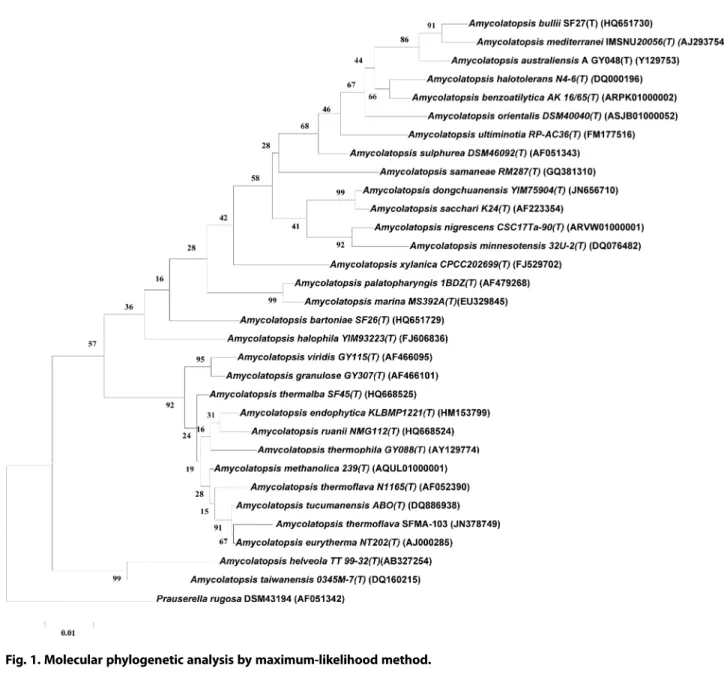
TMgel extraction kit (Sigma-Aldrich, USA) and was sequenced on a ABI 3730Xl DNA ana- lyzer (Applied BioSystems, USA). The phylogenetic posi- tion of the isolate (SFMA-103) was determined by performing a nucleotide sequence database search using the BLASTN program of NCBI database. The evolution- ary history was inferred by using the Maximum Likeli- hood method based on the Tamura-Nei model [16]. The tree with the highest log likelihood (-5843.1440) is shown. The percentage of trees in which the associated taxa clustered together is shown next to the branches.
Initial tree(s) for the heuristic search were obtained automatically by applying Neighbor-Joining and BioNJ algorithms to a matrix of pairwise distances estimated using the Maximum Composite Likelihood (MCL) approach, and then selecting the topology with superior log likelihood value. The tree is drawn to scale, with branch lengths measured in the number of substitutions per site. The analysis involved 32 nucleotide sequences.
Codon positions included were 1
st+ 2
nd+ 3
rd+ non-cod- ing. All positions with less than 95% site coverage were eliminated. That is, fewer than 5% alignment gaps, missing data, and ambiguous bases were allowed at any position. There were a total of 1370 positions in the final dataset. Evolutionary analyses were conducted using MEGA6 software [17].
Fermentation, isolation and purification of the extrolite Strain SFMA-103 was cultured in glycerol-asparag- ine-salts medium (pH 7.0) and incubated at 40 ℃ with agitation at 150 rpm in a New Brunswick Innova 43R shaker (Eppendorf North America, Hauppauge, USA) for 5 days. The fermented medium was later subjected to centrifugation (Sorvall RC 5C Plus, Kendro Lab Prod- ucts, USA) at 8200 ×g to remove the cell biomass result- ing in the cell-free supernatant. The extrolites present in the cell-free supernatant were extracted by absorption onto Diaion HP-20 (3%) resin. The resin was washed with water and then extracted with methanol to obtain the crude extract fractions. The fractions were analyzed by thin-layer chromatography (TLC) on silica gel 60 plates (F
254). Plates were developed in a methanol-chlo- roform (10:90, v/v) solvent mixture and visualized under UV light at 254 nm which revealed the presence of a sin- gle spot. Further, the crude extract fractions containing
the extrolite were pooled, concentrated under reduced pressure on a rotary vacuum evaporator (Rotavapor R- 205, Büchi, Switzerland) and further profiled by silica gel (60 −120 mesh) column (3 × 60 cm) chromatography.
The extrolite was eluted with a linear gradient of metha- nol-chloroform solvent system (0.5:95, v/v). The same solvent mixture was continued till the extrolite was com- pletely eluted and after drying resulted in golden yellow wax.
Structural characterization of the extrolite
The UV spectrum was measured by dissolving the purified extrolite in spectroscopic acetonitrile and recorded at 30 ℃ on a UV-visible double-beam spectro- photometer (Lambda 25, Perkin-Elmer, USA). 1D and 2D-NMR spectra were recorded on a Bruker Avance 300 and 600 MHz NMR spectrometers (Bruker, Switzerland) in DMSO-d
6at room temperature, and chemical shifts were represented in δ values expressed in ppm with tetramethylsilane as the internal standard. The Fourier transform infrared (FT-IR) spectrum was recorded using the Thermo-Nicolet Nexus 670 FT-IR spectrophotometer (ThermoFisher Scientific Inc., USA) at a resolution of 4 cm
-1in the wavenumber region of 400 −4,000 cm
-1. ESI- MS spectrum was recorded on a QSTAR XL Hybrid ESI-Q TOF mass spectrometer (Applied Biosystems Inc., USA).
Antioxidant assays
Antioxidant activity of purified extrolite was assessed on the basis of the free radical scavenging effect of the stable 1, 1-diphenyl-2-picrylhydrazyl (DPPH) with some modifications of a previously described method [18]. The diluted working solution of the purified extrolite was prepared in methanol. One millilitre of DPPH (0.002%
prepared in methanol) was mixed with 1 ml of purified
extrolite at different concentrations ranging from 10, 20,
40, 60, 80 and 100 μg/ml. The mixtures were shaken vig-
orously and left to stand in dark for 30 min. Absorbance
of the reaction mixtures were measured at 517 nm on a
UV-visible spectrophotometer (Lambda 25, Perkin-
Elmer, USA). Butyl hydroxytoluene (BHT) and α-
tocopherol were run in parallel as positive controls. The
radical scavenging activity was measured as a decrease
in the absorbance of DPPH. Lower absorbance of the
reaction mixture indicated higher free radical scaveng-
ing activity. DPPH radical scavenging activity was cal-
culated using the formula [19]: DPPH radical scavenging activity (%) = [(Absorbance of control − Absorbance of test sample) / (Absorbance of control)] × 100. The absor- bance of DPPH was plotted against the antioxidant con- centrations as standard curve to calculate the radical scavenging activity. Radical scavenging potential was expressed as EC
50value, which represents the concen- tration of purified extrolite at which 50% of the DPPH radicals scavenged. All tests were performed in tripli- cate and the values are represented as mean ± SD.
The superoxide radical scavenging activity of purified extrolite was performed according to the protocol described by Liu and coworkers [20]. The superoxide radicals were generated by phenazine methosulfate - nicotinamide adenine dinucleotide (PMS/NADH) sys- tem, which reduced nitroblue tetrazolium (NBT) to form a purple coloured formazan. Butyl hydroxytoluene (BHT) and α-tocopherol were run in parallel as positive controls. The scavenging activity of superoxide radical (%) was calculated from the plotted absorbance data for the dose-response curves and the IC
50values (in μM) were expressed as the mean of three independent exper- iments and the values are represented as mean ± SD.
The inhibition of lipid peroxidation was assayed by measuring the lipid peroxide decomposition product malondialdehyde (MDA), based on reaction with thio- barbituric acid using egg yolk as oxydizable substrate [21]. Butyl hydroxytoluene (BHT) and α-tocopherol were run in parallel as positive controls. The inhibition of lipid peroxidation was calculated from the plotted absor- bance data for the dose-response curves and the IC
50values (in μM) were expressed as the mean of three independent experiments and the values are repre- sented as mean ± SD. The nitric oxide (NO) scavenging assay was performed using the modified method of Marcocci et al. [22], which is based on release of nitric oxide from sodium nitroprusside and quantified by the Griess reaction. Varying concentrations of pure extrolite were solubilized in methanol and to these solutions, sodium nitroprusside (400 μl) and Griess reagent (220 μl) were added, followed by incubation at 25 ℃ for 1 h and then distilled water (2 ml) was added to each of these reaction mixtures. The absorbance was recorded at 546 nm and the IC
50values were calculated from dose- response curve. All tests were performed as three inde- pendent experiments and the results were expressed as
mean ± SD.
MTT cytotoxicity assay
The cytotoxicity of pure extrolite was studied using the 3-[4,5-dimethylthiazol-2-yl]-2,5-diphenyltetrazolium bromide (MTT) viability assay [23] which was deter- mined on the basis of measurement of in vitro growth inhibition of tumor cell lines in 96 well plates by cell- mediated reduction of tetrazolium salt to water insoluble formazan crystals using doxorubicin as a standard. The cytotoxicity was assessed against a panel of different human tumor cell lines: A549 derived from human alve- olar adenocarcinoma epithelial cells (ATCC No. CCL- 185), MDA-MB-231 derived from human breast adeno- carcinoma cells (ATCC No. HTB-26), DU145 derived from human prostate carcinoma (ATCC No. HTB-81), HepG2 derived from human hepatocellular carcinoma (ATCC No. HB-8065), COLO 205 derived from human colon carcinoma (ATCC No. CCL-222), MOLT-4 derived from human lymphoblastic leukemia cells (ATCC No.
CRL-1552) and HEK293 derived from human normal embryonic kidney cells (ATCC No. CRL-1573). Briefly, 1 × 10
6cells/well were seeded in 100 μl of DMEM and/or RPMI supplemented with 10% FBS in each well of 96- well microtitre plates and incubated for 24 h at 37 ℃ in a humidified 5% CO
2incubator (Model 2406, Shellab CO
2incubator, USA). The purified extrolite, diluted to the desired concentrations in culture medium, was added to the wells with respective vehicle control. After 48 h of incubation, the cells were subjected to MTT assay (5 mg/
ml MTT) and the plates were further incubated for 2 h.
The supernatant from each well was carefully removed, formazan crystals were dissolved in 100 μl of DMSO and absorbance was recorded at a wavelength of 540 nm on a multimode microplate reader (Infinite
®M200, Tecan, Switzerland). The IC
50values (50% inhibitory concentra- tion) were calculated from the plotted absorbance data for the dose-response curves. IC
50values (in μM) were expressed as the average of three independent experi- ments and the results were expressed as mean ± SD.
Results and Discussion
Taxonomy of strain SFMA-103
Morphological and cultural characteristics of strain
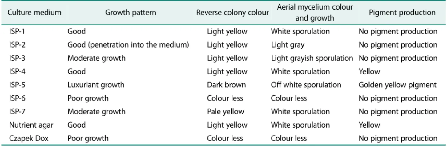
SFMA-103 are recorded in Table 1. The strain exhibited
luxuriant growth on ISP-5 and good growth on ISP-1, ISP-2, ISP-4 and nutrient agar. The growth was moder- ate on ISP-3 and ISP-7, while poor growth was observed on ISP-6 and Czapek Dox medium. A prominent golden yellow pigment production was observed on ISP-5 medium. Micromorphology of the strain grown on ISP medium 2 showed light grey aerial mycelium with light yellow colored substrate mycelium that fragmented into rod-shaped elements, which is the characteristic feature of Amycolatopsis sp. [13]. The phylogenetic position of the strain was determined by amplifying the 16S rRNA region and sequence of the strain was examined by BLAST analysis. The results revealed that the strain belongs to the genus Amycolatopsis, the suborder Pseud- onocardineae of the family Pseudonocardiaceae. Based on the phylogenetic tree (Fig. 1), the 16S rRNA genome sequence of the strain showed similarity with different Amycolatopsis strains. Based on the sequence homology, the strain SFMA-103 showed identity with both Amycol- atopsis eurytherma strain NT202 [24] and A. thermo- flava N1165 [13]. However, it is reported that A.
eurytherma strain NT202 showed no pigment produc- tion, while A. thermoflava N1165 showed soluble yellow pigment production which corroborates with the obser- vations made in the present study. Based on these com- parisons, the strain SFMA-103 was identified as A.
thermoflava and the 16S rRNA sequence is submitted with Genbank accession number JN378749. The strain SFMA-103 is maintained in the in-house culture collec- tion of the laboratory and designated as A. thermoflava strain ICTA-103.
Isolation, purification and identification of the bioactive extrolite
The fermentation of strain SFMA-103 was carried out in glycerol-asparagine-salts medium which produced the extrolite after 5 days of incubation. The culture filtrate revealed the presence of a single major spot on the TLC plate developed in a solvent mixture of methanol-chloro- form (10:90, v/v). The extrolite was extracted in metha- nol to obtain a crude extract (1.2 g/l) and purified by silica gel column chromatography in a solvent system of methanol-chloroform (0.5:95, v/v). The extrolite on the TLC plate was UV-active when visualized under UV light at a wavelength of 254 nm and was developed by spraying with anisaldehyde reagent followed by heating at 100 ℃ for 2−3 min which appeared as a blue coloured spot.
The pure extrolite was obtained as orange yellow coloured wax (30 mg/l) and the molecular formula of C
16H
12O
4was deduced based on EI-MS data, which showed a molecular ion peak at m/z 268.99 [M + H]
+[calcd. For C
16H
12O
4, 269, M + H]
+. UV (MeOH) scan spectrum of the purified extrolite showed absorbance peaks at 206, 287 and 420 nm. FT-IR spectrum of the purified extrolite showed a strong hydroxyl group absorption band at 3400 cm
-1, together with one car- bonyl group absorption band at 1558 cm
-1, suggesting the presence of hydroxyl and quinone groups.
1H NMR (300 MHz, DMSO-d
6) showed an ABX coupling system for aromatic signals at δ (ppm) 11.48 (1H, s, OH-1), 7.74 (1H, t, J = 8.4 and 6.7, H-6), 7.49 (1H, d, J = 8.4, H-7), 7.36 (1H, s, H-4), 7.32 (1H, d, J = 6.7, H-5), 7.13 (1H, s,
Table 1. Cultural characteristics of strain SFMA-103 on different International Streptomyces Project (ISP) and non-ISP media.
Culture medium Growth pattern Reverse colony colour Aerial mycelium colour
and growth Pigment production
ISP-1 Good Light yellow White sporulation No pigment production
ISP-2 Good (penetration into the medium) Light yellow Light gray No pigment production ISP-3 Moderate growth Light yellow Light grayish sporulation No pigment production
ISP-4 Good Light yellow White sporulation Yellow
ISP-5 Luxuriant growth Dark brown Off white sporulation Golden yellow pigment
ISP-6 Poor growth Colour less Colour less No pigment production
ISP-7 Moderate growth Pale yellow White sporulation No pigment production
Nutrient agar Good Light yellow White sporulation Yellow
Czapek Dox Poor growth Colour less Colour less No pigment production
H-2), 2.40 (3H, s, H
3-3) and 3.69 (3H, s, OMe). The
13C NMR (75 MHz, DMSO-d
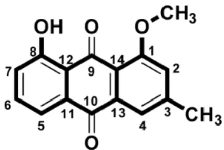
6) showed the presence of two ketals carbons with chemical shifts, δ (ppm): 184.9 (C-9) and 182.4 (C-10), twelve aromatic carbons with chemical shifts, δ (ppm): 160.89 (C-8), 156.9 (C-1), 139.0 (C-13), 137.2 (C-6), 132.5 (C-11), 128.0 (C-14), 123.0 (C-7), 122.34 (C-4), 118.8 (C-3), 118.5 (C-5), 115.5 (C-2), 113.7 (C-12), methoxy group at δ 55.9 ppm and one aromatic methyl group at δ 21.1 ppm, which is characteristic of an anthraquinone chromophore. The key HMBC and COSY correlations for the pure extrolite is shown in Fig. 2.
Based on the 1D and 2D NMR, FT-IR and mass spectral
data (Figs. S1 −S10), the chemical structure (Fig. 3) of the pure extrolite was determined as 1-methoxy-3- methyl-8-hydroxy-anthraquinone. Literature search Fig. 1. Molecular phylogenetic analysis by maximum-likelihood method.
Fig. 2. Key HMBC and COSY correlations of 1-methoxy-3-
methyl-8-hydroxy-anthraquinone.
revealed that there are no reports on this anthraqui- none from any microbial source.
Effect of 1-methoxy-3-methyl-8-hydroxy-anthraquinone on antioxidant activity
Considering the pharmacological importance of plant- derived anthraquinones [25], the pure extrolite from A.
thermoflava was assayed for antioxidant and anti-tumor activities. 1-methoxy-3-methyl-8-hydroxy-anthraquinone scavenged DPPH free radicals, superoxide anions, nitric oxide radicals and lipid peroxyl radicals in a dose-depen- dent manner as compared to the positive controls, BHT and α-tocopherol (Table 2). DPPH is an oxidizing radical
that forms a stable free radical which is reduced and sta- bilized by antioxidants. 1-methoxy-3-methyl-8-hydroxy- anthraquinone exhibited a significant DPPH radical scavenging activity (EC
50value of 18.2 ± 0.41 μg/ml) as compared to that of BHT, whereas α-tocopherol (positive control) exhibited a higher DPPH radical scavenging activity (EC
50value of 10.6 ± 0.32 μg/ml). In this context, 1-methoxy-3-methyl-8-hydroxy-anthraquinone reduced the free radicals to the corresponding hydrazine when it reacted with the released hydrogen ions. Further, the 1- methoxy-3-methyl-8-hydroxy-anthraquinone showed promising superoxide free radical scavenging activity (IC
50value of 11.9 ± 0.39 μg/ml) comparable to that of BHT and α-tocopherol (positive controls). Superoxide anions exert deleterious effects on biological system by forming singlet oxygen and hydroxyl radicals upon decomposition. The nitric oxide (NO) radical scavenging activity exhibited by 1-methoxy-3-methyl-8-hydroxy- anthraquinone was significant (IC
50value of 72.8 ± 0.25 μg/ml) as compared to the positive controls. 1-methoxy- 3-methyl-8-hydroxy-anthraquinone stabilized the lipid peroxidation product, malondialdehyde (MDA) and exerted significant inhibitory effect on lipid peroxidation with EC
50value of 31.4 ± 0.18 μg/ml as compared to that of BHT and α-tocopherol (positive controls). In biological systems, MDA acts as a mutagen and exerts its deleteri- ous effects by reacting with DNA bases to form DNA- DNA interstrand crosslinks or DNA-protein crosslinks [26, 27]. These results suggest that the antioxidant mechanism for 1-methoxy-3-methyl-8-hydroxy-anthra- quinone possibly depends on scavenging DPPH free rad- icals, superoxide anions, nitric oxide radicals and lipid peroxyl radicals.
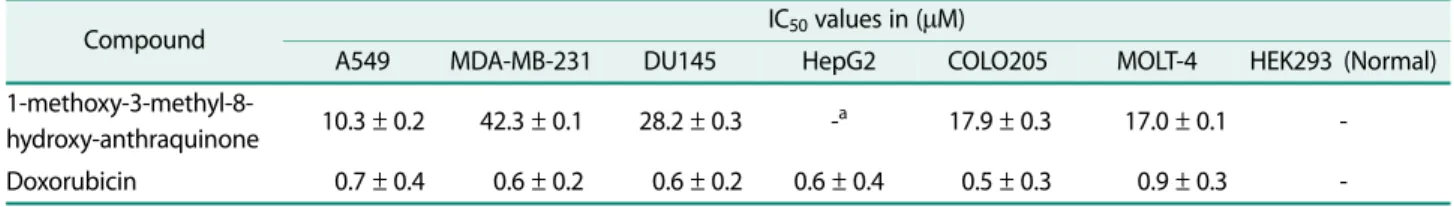
Effect of 1-methoxy-3-methyl-8-hydroxy-anthraquinone on cytotoxicity
The in vitro cytotoxicity of 1-methoxy-3-methyl-8- hydroxy-anthraquinone was evaluated on a panel of seven tumour cell lines, i.e. A549 (lung cancer), MDA- MB-231 (Breast cancer), DU145 (prostate cancer), HepG2 (liver cancer), COLO 205 (colon cancer), MOLT-4 (lymphoblastic leukemia) and HEK293 (normal human embryonic kidney cells) using MTT assay. Doxorubicin was used as the reference drug. The results to this regard are summarized in Table 3 and expressed as IC
50values ( μM). The in vitro screening results revealed that Fig. 3. Chemical structure of 1-methoxy-3-methyl-8-hydroxy-
anthraquinone.
Table 2. Antioxidant activities of 1-methoxy-3-methyl-8- hydroxy-anthraquinone produced by A. thermoflava SFMA- 103.
Test compound EC
50( μg/ml) (Mean ± SD) DPPH radical scavenging activity
1-methoxy-3-methyl-8-hydroxy-anthraquinone 18.2 ± 0.41 Butyl hydroxytoluene (Positive control) 28.7 ± 0.24 α-Tocopherol (Positive control) 10.6 ± 0.32 Superoxide free radical scavenging activity
1-methoxy-3-methyl-8-hydroxy-anthraquinone 11.9 ± 0.39 Butyl hydroxytoluene (Positive control) 13.1 ± 0.22 α-Tocopherol (Positive control) 6.8 ± 0.39 Nitric oxide (NO) radical scavenging activity
1-methoxy-3-methyl-8-hydroxy-anthraquinone 72.8 ± 0.25 Butyl hydroxytoluene (Positive control) 74.2 ± 0.11 α-Tocopherol (Positive control) 52.8 ± 0.19 Inhibition of lipid peroxidation
1-methoxy-3-methyl-8-hydroxy-anthraquinone 31.4 ± 0.18
Butyl hydroxytoluene (Positive control) 43.2 ± 0.43
α-Tocopherol (Positive control) 21.3 ± 0.28
the pure extrolite from strain SMFA-103 exhibited promising anticancer activity with IC
50values ranging between 10.3 −42.3 μM. Among all tested cell lines, 1- methoxy-3-methyl-8-hydroxy-anthraquinone showed sig- nificant anticancer activity against lung cancer and lym- phoblastic leukemia cell lines with IC
50values of 10.3 and 16.98 μM, respectively. It did not show any inhibition against the normal human embryonic kidney cell line.
Studies on some anthraquinone derivatives derived from various Streptomyces sp. confirm the anticancer activity towards different tumor cell lines exhibited by these natural products. Some anthraquinone extrolites like tetracenomycin D from Streptomyces corchorusii AUBN(1)/7 [7], galvaquinones A-C from Streptomyces spinoverrucosus strain SNB-032 [28] and rubimycinone A from Streptomyces sp. Lv-6-8 [29] exhibited varying levels of antitumor activities. 2,3-dihydroxy-9,10-anthra- quinone secreted by Streptomyces galbus ERINLG-127 showed cytotoxicity against lung adenocarcinoma cancer cell line, A549 with IC
50value of 60 μg/ml [10]. Further, 1,8-dihydroxy-2-ethyl-3-methylanthraquinone produced by Streptomyces sp. FX-58 showed cytotoxicity against human tumor cell lines of pro-myelocytic leukemia HL- 60, gastric carcinoma BGC-823 and adenocarcinoma MDA-MB-435 with IC
50values of 6.83, 82.2 and 56.59 μg/ml, respectively [30]. Antitumor anthraquinones such as lupinacidins A and B isolated from an endophytic Micromonospora sp. exhibited dose-dependent inhibition of in vitro invasion of colon 26-L5 cells with IC
50values of 0.07 μg/ml (= 0.21 μM) and 0.3 μg/ml (= 0.92 μM), respectively. Lupinacidin A exhibited more potency both in cytotoxic and anti-invasive activities as compared to
Lupinacidin B, and the alkyl substituent contributed to these activities [8]. Saliniquinones A-F, were cytotoxic anthraquinone-γ-pyrone derivatives produced by the marine actinobacterium Salinispora arenicola strain CNS-325. Among them, Saliniquinone A (1) exhibited potent inhibition of HCT-116 (human colon adenocarci- noma cell line) with an IC
50value of 9.9 × 10
-9M [30].
In conclusion, A. thermoflava strain SMFA-103 iso- lated from the rhizosphere soil of sunflower (Helian- thus annuus L.) was found to produce a promising bioactive extrolite, 1-methoxy-3-methyl-8-hydroxy- anthraquinone. This is the first report on this bioactive anthraquinone from A. thermoflava strain SFMA-103, which exhibited anti-tumor activity against a panel of cancer cell lines and showed non-toxic effect to normal human embryonic kidney cell line. It also scavenged DPPH free radicals, superoxide anions, nitric oxide radi- cals and lipid peroxyl radicals. The antitumor and anti- oxidant activities exhibited by 1-methoxy-3-methyl-8- hydroxy-anthraquinone seems to be promising and may find plausible application in biotechnological and phar- maceutical fields.
Acknowledgments
The authors acknowledge the financial assistance provided to PM, YP and BS in the form of Senior Research Fellowships by Council of Sci- entific and Industrial Research (CSIR), New Delhi, India.
Conflict of Interest
We declare that there is no conflict of interest with any researcher or funding agency.
Table 3. In vitro cytotoxicity of 1-methoxy-3-methyl-8-hydroxy-anthraquinone produced by A. thermoflava SFMA-103.
Compound IC
50values in ( μM)
A549 MDA-MB-231 DU145 HepG2 COLO205 MOLT-4 HEK293 (Normal)
1-methoxy-3-methyl-8-
hydroxy-anthraquinone 10.3 ± 0.2 42.3 ± 0.1 28.2 ± 0.3 -
a17.9 ± 0.3 17.0 ± 0.1 - Doxorubicin 0.7 ± 0.4 0.6 ± 0.2 0.6 ± 0.2 0.6 ± 0.4 0.5 ± 0.3 0.9 ± 0.3 -
a