Contrast Extravasation on Computed Tomography
Angiography Imitating a Basilar Artery Trunk Aneurysm in Subsequent Conventional Angiogram-Negative
Subarachnoid Hemorrhage: Report of Two Cases with Different Clinical Courses
Won Ho Cho, Hyuk Jin Choi, Kyoung Hyup Nam, Jae Il Lee
Department of Neurosurgery, Medical Research Institute, Pusan National University College of Medicine and Hospital, Busan, Korea
Contrast extravasation on computed tomography angiography (CTA) is rare but becoming more common, with increasing use of CTA for various cerebral vascular diseases. We report on two cases of spontaneous sub- arachnoid hemorrhage (SAH) in which the CTA showed an upper basilar trunk saccular lesion suggesting ruptured aneurysm. However, immediate subsequent digital subtraction angiography (DSA) failed to show a vas- cular lesion.
In one case, repeated follow up DSA was also negative. The patient was treated conservatively and discharged without any neurologic deficit. In the other case, the patient showed sudden mental deterioration on the third hospital day and her brain CT showed rebleeding. The immediate follow up DSA showed contrast stagnation in the vicinity of the upper basilar artery, suggestive of pseudoaneurysm. Double stents deployment at the disease segment was performed.
Due to the frequent use of CTA, contrast extravasation is an increasingly common observation. Physicians should be aware that basilar artery ex- travasation can mimic the appearance of an aneurysm.
J Cerebrovasc Endovasc Neurosurg.
2015 December;17(4):324-330 Received : 18 June 2014
Revised : 12 August 2014 Accepted : 31 December 2014 Correspondence to Jae Il Lee
Department of Neurosurgery, Pusan National University Hospital, 179 Gudeok-ro, Seo-gu, Busan 49241, Korea
Tel : 82-51-240-7935 Fax : 82-51-244-0282 E-mail : [email protected]
ORCID : http://orcid.org/0000-0001-9806-8676
This is an Open Access article distributed under the terms of the Creative Commons Attribution Non- Commercial License (http://creativecommons.org/li- censes/by-nc/3.0) which permits unrestricted non- commercial use, distribution, and reproduction in any medium, provided the original work is properly cited.
Keywords Angiogram-negative SAH, Computed tomography angiography, Basilar ar- tery, Contrast extravasation
INTRODUCTION
Nontraumatic subarachnoid hemorrhage (SAH) is caused by rupture of a cerebral aneurysm in 80-90%
of patients.4) Unfortunately, aneurysmal SAH has a high mortality rate of 45%.1) Therefore, immediate di- agnosis and treatment are very important in pre- vention of catastrophic deterioration.
Digital subtraction angiography (DSA) remains the gold standard for the main technique for diagnosis of the intracranial aneurysms.4)5)7) Computed tomog- raphy angiography (CTA) is a rapid, less invasive modality for patients with intracranial aneurysm and useful for evaluation of patients with aneurysmal SAH. Therefore, CTA is more often used in the initial evaluation of SAH.4)7)10)11)14)
A B
C
Fig. 1. Case 1. (A) Noncontrast computed tomography (CT) scan on admission shows diffuse subarachnoid hemorrhage (SAH) in the prepontine cistern and both sylvian fissures. (B) 3-dimensional computed tomography angiography (CTA) image demonstrates an en- hancing abnormal vascularity (arrow). (C) Digital subtraction angiography (DSA) of the basilar artery acquired in the anteroposterior (left) and lateral (right) views fail to detect the previously seen basilar abnormal structure.
However, in some patients with a nontraumatic SAH, no aneurysms are found on the initial DSA.3)9)11) Contrast extravasation of a ruptured intracranial aneurysm during CTA is rare, but is becoming more common, with increasing use of CTA for various cere-
bral vascular diseases and increasing resolution of CTA. We report on two similar cases, but with differ- ent clinical course of contrast extravasation from the distal basilar artery (BA) during CTA but negative findings of DSA.
A B C
D E
Fig. 2. Case 2. (A) Initial noncontrast CT scan reveals diffuse subarachnoid hemorrhage (SAH) in the basal, prepontine, perimesencephalic cistern and both sylvian fissures. (B) Computed tomography angiography (CTA) axial and (C) Sagittal reformatted images showing an enhanced saccular structure with surrounding blood clot on the posterior surface of the basilar artery (arrow). (D) 3-D CTA image demonstrating a cylinder-shape, enhancing abnormal structure (arrow). (E) Digital subtraction angiography (DSA) of the basilar artery acquired 2 hours later from initial CT scan in the anteroposterior (left) and lateral (right) views fail to detect the previously seen ba- silar abnormal structure.
CASE REPORT
Case 1
A 66-year-old man was admitted to our hospital af- ter sudden loss of consciousness. He had suffered from a severe headache for 2 hours before his admission. He had a five-year history of taking medi- cation for angina. On admission, he had a stuporous mental status. A pre-enhanced cranial CT showed acute diffuse SAH in the basal cistern (Fig. 1A). The 3-dimensional CTA showed a saccular lesion arising from the upper basilar artery and projecting ante- riorly (Fig. 1B). DSA performed one hour and a half later and did not show any vascular abnormality (Fig.
1C). A repeat DSA, performed on postbleed day 10,
also showed no vascular abnormality. The patient was treated conservatively and discharged after 30 days without any neurologic deficit. On follow-up at three months, the patient had no neurologic deficit, and fol- low up DSA also confirmed no vascular abnormality.
Case 2
A 55-year-old woman with a medical history of chronic viral hepatitis presented to our clinic after sudden onset of headache with vomiting. We did not find any neurologic deficit and her mentality was drowsy. Her CT scan showed a diffuse SAH and mild hydrocephalus (Fig. 2A). CTA confirmed an upper ba- silar abnormality (Fig. 2B, C and D). Cerebral angiog- raphy obtained two hours later failed to disclose a
Fig. 5. Case 2. Native image of temporary balloon occlusion within the double stents. White arrow shows balloon inflation to achieve decreasing of inflow into the pseudoaneurysm.
Fig. 4. Case 2. Follow up digital subtraction angiography (DSA) acquired 28 days after double stenting. Superselective micro- catheter angiography of left superior cerebellar artery confirms a pseudoaneurysm arising from small superior cerebellar artery (SCA) perforating artery (arrow).
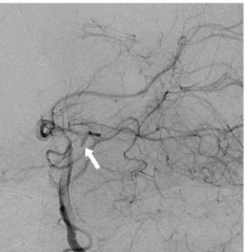
Fig. 3. Case 2. Digital subtraction angiography (DSA) of the basilar artery acquired 3 days after bleeding event, in the obli- que view demonstrate the cylinder-shape contrast stagnation in the upper basilar trunk at late arterial phase (arrow).
vascular abnormality (Fig. 2E). The patient received an external ventricular drain (EVD) for symptomatic hydrocephalus on posthemorrhage day 2. The pa- tient's clinical condition improved and her level of consciousness improved to alert immediate after EVD.
On the second day after EVD, her mentality suddenly deteriorated to semicoma. Immediate CT scan showed an increased amount of SAH, which suggested rebleeding. A repeat CTA after rebleeding did not show an abnormal vascular lesion. However, cerebral angiography at the same time confirmed a small cyl- inder like contrast stagnation from the upper basilar artery at late arterial phase, indicating pseudoaneur- ysm formation (Fig. 3). We inserted two Solitaire stents (4 × 20 mm, ev3, Irvine, CA, USA) for flow di- version and anticipating thrombus formation of the pseudoaneurysm. The patient's clinical course was un- eventful, however, one-month follow up CTA still showed a saccular lesion from the upper basilar artery. Therefore, we decided to perform DSA and intervention. DSA still showed contrast stagnation at late arterial phase on the upper basilar artery and su-
perselective microcatheter angiography confirmed a pseudoaneurysm arising from a small perforating ar- tery of the superior cerebellar artery (SCA) (Fig. 4).
Fig. 6. Case 2. Follow up digital subtraction angiography (DSA) done 11 months after hemorrhage confirmed no contrast stagnation or abnormal vascular lesion with patent stented basilar artery and superior cerebellar artery (SCA)s.
Due to concern about cerebellar infarction, we could not sacrifice the SCA. To facilitate thrombus for- mation of the pseudoaneurysm, we performed re- peated temporary balloon occlusion using a Sceptor XC balloon (4 × 10 mm, Microvention, Tustin, CA, USA) and achieved decreased contrast inflow to the pseudoaneurysm (Fig. 5). On follow-up at 11 months, the patient had no neurologic deficit, and she was able to live independently. Follow up DSA performed 11 months after hemorrhage confirmed no contrast stagnation and abnormal vascular lesion (Fig. 6).
DISCUSSION
In the current cases, we report on two patients with contrast extravasation from the basilar artery seen on CTA, but negative findings with immediate subsequent cerebral angiography. Incidence of patients with SAH of unknown etiology was approximately 10 to 30%.3)5)9)12) The precise cause of angiogram-negative SAH is unclear. However, patients with angiogram-negative SAH have a more favorable prognosis and lower re-
bleeding rate than those with other distributions of aneurysmal SAH.3)9)11)12)
DSA has been the gold standard method for identi- fication and diagnosis of intracranial aneurysms.
However, DSA has some weakness of being very in- vasive and takes time to performed. The risk of com- plications has been reported as 0.9% to 2.3% with per- manent deficit in 0.3%4) and 0.6% of cases occurring in the non-severe neurologic complications, include formation of hematoma, peripheral thromboembolic events, transient hypotension, arteriovenous fistulas, or infection.2)4)
Recently, CTA is useful for detection of intracranial aneurysms because it is less invasive and the proce- dure is easier to perform than DSA. Because contrast media for CTA is injected intravenously, it is less in- vasive than arterial injections required for DSA.7) Therefore, CTA is now used as the diagnostic method of first choice for detection of aneurismal SAH, vas- cular malformations, stenotic vessel disease, vaso- spasm, traumatic vessel disease, and for development of an occupational treatment plan.4)10)14) Sensitivity of
CTA for detection of intracranial aneurysms between 83.6% and 92.7% has been reported, and the specific- ity is between 77.2% and 98.9%.4) Due to the increased usefulness, sensitivity and specificity for detection of intracranial aneurysms, use of CTA has recently be- come more frequent. However, CTA has still not re- placed DSA as the standard method for detection of intracranial aneurysms. The disadvantages of CTA were difficulty in distinguishing small perforating ar- teries with a diameter less than 1 mm, difficulty in discriminating a infundibular dilatation of the arterial origin from an aneurysm, artifact of the adjacent vessel, display of venous systems imitating aneurysms, and extravasated blood such as hemorrhage surrounding the aneurysm imitating vascular structures.
Almost all extravasation occurs from ruptured aneurysms but has also been reported in association with other rare situations such as microangioma, in- farction with SAH due to obliteration of a perforating artery or increase in arterial pressure after the injection of contrast media.8)10) Another potentiality of extravasation is very small sized true aneurysmal SAH that is not large enough to be recognized. Extravasation on CTA has been reported previously, such as the "cap sign",
"corkscrew sign", "puddling" and the "spot sign". The
"cap sign" and the "corkscrew sign" are some kind pattern of extravasation. The "puddling" means multi- ple areas of contrast media on CTA and the "spot sign"
is enhancing foci within a hematoma mass associated with hematoma growth and poor prognosis.7)13)14) Nevertheless, sometimes DSA did not reveal a lesion that was initially detected on the CTA. Failure to de- tect an aneurysmal rupture by DSA that was detected on the initial CTA has been ascribed to a result of vasospasm, dissecting aneurysm, spontaneous throm- bosis of the aneurysm, microaneurysm, or error of interpretation.5)11) Aneurysms detected on CTA that do not present on initial DSA can become visible on repeat DSA. Some authors have previously reported discovery rates of repeat DSA of occult aneurysms overlooked on the initial DSA of 2 and 36%.5)6)11)
In the second case, we found a small cylinder like
vascular abnormality, which was confirmed by re- peated DSA after rebleeding. The proposed mecha- nism of this case could be as follows. First bleeding was due to rupture of a small perforating vessel aris- ing from the superior cerebellar artery and then spon- taneous thrombosis occurred. Therefore, we could not detect vascular abnormality on DSA despite detection of a saccular lesion on initial CTA. Subsequent re- bleeding into the hematoma could cause make pseu- doaneurysm formation. Therefore we were able to de- tect contrast stagnation on repeated DSA. Flow di- version through double stents and temporary balloon occlusion could facilitate thrombosis within the pseudoaneurym. Finally, we could achieve complete occlusion of the pseudoaneurysm arising from the SCA.
CONCLUSION
We reported on extremely rare cases of two patients who showed nonaneurysmal, angiogram-negative SAH in which supposed contrast extravasation on ini- tial CTA mimicked a basilar artery trunk aneurysm.
Based on our experience, if we encounter a patient with extravasation on CTA with negative DSA find- ings, repeated DSA is mandatory and superselective microangiography could reduce false-negative finding and improve understanding of pathophysiology.
Acknowledgments
This work was supported by a 2-year Research Grant of Pusan National University.
Disclosure
The authors report no conflict of interest concerning the materials or methods used in this study or the findings specified in this paper.
REFERENCES
1. Brisman JL, Song JK, Newell DW. Cerebral aneurysms.
N Engl J Med. 2006 Aug 31;355(9):928-39.
2. Cloft HJ, Joseph GJ, Dion JE. Risk of cerebral angiog-
raphy in patients with subarachnoid hemorrhage, cere- bral aneurysm, and arteriovenous malformation: a meta-analysis. Stroke. 1999 Feb;30(2):317-20.
3. Eskesen V, Sorensen EB, Rosenorn J, Schmidt K. The prognosis in subarachnoid hemorrhage of unknown etiology. J Neurosurg. 1984 Dec;61(6):1029-31.
4. Goddard AJ, Tan G, Becker J. Computed tomography angiography for the detection and characterization of in- tra-cranial aneurysms: current status. Clin Radiol. 2005 Dec;60(12):1221-36.
5. Inamasu J, Nakamura Y, Saito R, Horiguchi T, Kuroshima Y, Mayanagi K, et al. "Occult" ruptured cere- bral aneurysms revealed by repeat angiography: result from a large retrospective study. Clin Neurol Neurosurg.
2003 Dec;106(1):33-7.
6. Juul R, Fredriksen TA, Ringkjob R. Prognosis in sub- arachnoid hemorrhage of unknown etiology. J Neurosurg.
1986 Mar;64(3):359-62.
7. Nakatsuka M, Mizuno S, Uchida A. Extravasation on three-dimensional CT angiography in patients with acute subarachnoid hemorrhage and ruptured aneurysm.
Neuroradiology. 2002 Jan;44(1):25-30.
8. Ney JP. Midbrain stroke with angiogram-negative sub- arachnoid hemorrhage mimicking a perimesencephalic bleed.
J Stroke Cerebrovasc Dis. 2005 May-Jun;14(3):136-7.
9. Ronkainen A, Hernesniemi J. Subarachnoid haemorrhage of unknown aetiology. Acta Neurochir (Wien). 1992;119(1-4):29-34.
10. Ryu CW, Kim SJ, Lee DH, Suh DC, Kwun BD.
Extravasation of intracranial aneurysm during computed tomography angiography: mimicking a blood vessel. J Comput Assist Tomogr. 2005 Sep-Oct;29(5):677-9.
11. Stetson ND, Pile-Spellman J, Brisman JL. Contrast ex- travasation on computed tomographic angiography mim- icking a basilar artery aneurysm in angiogram-negative subarachnoid hemorrhage: report of 2 cases. Neurosurgery.
2012 Nov;71(5):E1047-52; discussion E1052.
12. Tatter SB, Buonanno FS, Ogilvy CS. Acute lacunar stroke in association with angiogram-negative subarachnoid hemorrhage. Mechanistic implications of two cases. Stroke.
1995 May;26(5):891-5.
13. Wada R, Aviv RI, Fox AJ, Sahlas DJ, Gladstone DJ, Tomlinson G, et al. CT angiography "spot sign" predicts hematoma expansion in acute intracerebral hemorrhage.
Stroke. 2007 Apr;38(4):1257-62.
14. Walker MT, Wattamwar A, Mellman D, Mo J. Active hemorrhage into a postresection cavity detected by neu- ro-CT angiography. AJNR Am J Neuroradiol. 2005 May;26(5):1163-5.