Push-out bond strengths of fiber-reinforced composite posts with various resin cements according to the root level
Hoon-Sang Chang1, DDS, PhD, Young-Sin Noh2, DDS, MSD, Yoon Lee3, DDS, PhD, Kyung-San Min4, DDS, PhD, Ji-Myung Bae5*, DDS, PhD
1Department of Conservative Dentistry, Dental Science Research Institute, School of Dentistry, Chonnam National University, Gwangju, Republic of Korea
2Department of Conservative Dentistry, College of Dentistry, Wonkwang University, Iksan, Republic of Korea
3Department of Conservative Dentistry, Wonju College of Medicine, Yonsei University, Wonju, Republic of Korea
4Department of Conservative Dentistry, School of Dentistry, Chonbuk National University, Jeonju, Republic of Korea
5Department of Dental Biomaterials and Institute of Biomaterials · Implant, College of Dentistry, Wonkwang University, Iksan, Republic of Korea
PURPOSE. The aim of this study was to determine whether the push-out bond strengths between the radicular dentin and fiber reinforced-composite (FRC) posts with various resin cements decreased or not, according to the coronal, middle or apical level of the root. MATERIALS AND METHODS. FRC posts were cemented with one of five resin cement groups (RelyX Unicem: Uni, Contax with activator & LuxaCore-Dual: LuA, Contax & LuxaCore- Dual: Lu, Panavia F 2.0: PA, Super-Bond C&B: SB) into extracted human mandibular premolars. The roots were sliced into discs at the coronal, middle and apical levels. Push-out bond strength tests were performed with a universal testing machine at a crosshead speed of 0.5 mm/min, and the failure aspect was analyzed. RESULTS.
There were no significant differences (P>.05) in the bond strengths of the different resin cements at the coronal level, but there were significant differences in the bond strengths at the middle and apical levels (P<.05). Only the Uni and LuA cements did not show any significant decrease in their bond strengths at all the root levels (P>.05); all other groups had a significant decrease in bond strength at the middle or apical level (P<.05). The failure aspect was dominantly cohesive at the coronal level of all resin cements (P<.05), whereas it was dominantly adhesive at the apical level. CONCLUSION. All resin cement groups showed decreases in bond strengths at the middle or apical level except LuA and Uni. [J Adv Prosthodont 2013;5:278-86]
KEY WORDS: Fiber reinforced-composite post; Resin cements; Bond strength; Root level
INTRODUCTION
Fiber-reinforced composite (FRC) posts are commonly used to retain core material in endodontically treated teeth with extensive loss of coronal tooth structure.1,2 FRC posts composed of glass or quartz fibers are white in color and offer better esthetics in the anterior regions.3-6 FRC posts offer a number of advantages over metal posts, such as favorable fracture patterns that are more likely to be restor- able because they are less rigid1,7 and easy removal of the post from the root canal in the case of endodontic retreat- ment.8,9 Furthermore, the bond strength between the FRC post and the resin core is known to be superior to that between the metal post and the resin core.10 As FRC posts become more clinically popular, there is an increasing demand for luting cement to be used for FRC post cemen-
Corresponding author:
Ji-Myung Bae
Department of Dental Biomaterials and Institute of Biomaterials · Implant, Dental College, Wonkwang University, 344-2 Shinyong-dong, Iksan, Jeonbuk, 570-749, Republic of Korea
Tel. 82638506859: e-mail, [email protected]
Received March 26, 2013 / Last Revision July 3, 2013 / Accepted July 11, 2013
© 2013 The Korean Academy of Prosthodontics
This is an Open Access article distributed under the terms of the Creative Commons Attribution Non-Commercial License (http://creativecommons.
org/licenses/by-nc/3.0) which permits unrestricted non-commercial use, distribution, and reproduction in any medium, provided the original work is properly cited.
This research was supported by Basic Science Research Program through the National Research Foundation of Korea (NRF) funded by the Ministry of Education (2013R1A1A2008051).
tation. In clinical studies, FRC posts most often failed due to adhesive failure between the post and the resin cement or between the dentin and the resin cement.1
Resin cements can be classified into conventional (total- etch) resin cements, adhesive (self-etch) resin cements, and self-adhesive resin cements.11 Because conventional resin cements do not bond to the tooth structure, they need the use of phosphoric acid followed by dentin bonding sys- tems. Conventional resin cements provide good esthetics and favorable compressive strength because their composi- tion is similar to that of the restorative resin composites.12 Adhesive resin cements bond to metallic oxide or zirconia as they contain reactive monomers such as 4-META, 10-MDP or Phenyl P,13,14 however, adhesive resin cements need a self-etching primer that is acidic and is not rinsed away to bond to the dentin. Most recently introduced self- adhesive resin cements are more convenient to use. They do not require a conditioning procedure and are less tech- nique sensitive.
Resin cements are also available in chemical-cure, light- cure, and dual-cure modes. Chemical-cured resin cements initiate polymerization when the base components are mixed with a catalyst. Light-cured resin cements are polym- erized when they are exposed to dental curing lights. The majority of available resin cements is dual-cured and polymerizes when both the base and catalyst components are mixed and also when they are exposed to a curing light source. Dual-cured resin cements offer extended working time and controlled polymerization,11 and their chemical activators ensure a high degree of conversion.15 However, most dual-cured resin cements still require light-curing pro- cedure given that they demonstrate inferior hardness when light-curing is not performed.9,16 When self- and dual-cure resin cements are used with some 6th or 7th generation adhe- sive (self-etching adhesive) systems, the residual acidic monomers of the adhesive react with initiators (tertiary amine) of the self- and dual-cure resin cements resulting in compromised polymerization of the resin cements.17 To counterbalance the acid-base incompatibility between the tertiary amines and the residual acidic monomers, a self- cure activator that is usually ternary catalyst should be used.17
Various factors affect the bond strength of resin cements including the moisture level inside the root canal, the degree of conversion of resin cement, the composition of resin matrix, the filler content of resin cement, the polym- erization shrinkage stress of resin cement filling the space between the post and the root canal wall, and the compati- bility between resin cements and adhesive systems.18-22
As FRC posts gain clinical popularity, a better evalua- tion of the bond strength between the FRC post and the radicular dentin is needed. Previous studies assessing bond strengths have been performed along the entire root canal wall.23,24 However, because the degree of conversion of dual-cured resin cements is affected by the curing light intensity, an evaluation of the bond strength according to root level would be an advantageous metric for clinicians.
The respective bond strengths of the various resin cements are another important metric for choosing appropriate resin cements for different clinical applications. It would be also interesting to observe the use of self-etching adhesive sys- tem with and without the self-cure activator in cementation of FRC post. The purpose of this study, therefore, was to evaluate the effect of the different resin cements and the root level of the coronal, middle, and apical third on the bond strengths between FRC posts and the radicular den- tin. Here, the null hypothesis to be tested was that the bond strengths of the FRC posts do not vary according to either resin cement or root level.
MATERIALS AND METHODS
Fifty intact human mandibular premolars extracted within 5 weeks were selected for use in the study. After removing the debris, the teeth were stored in distilled water that was changed every 2 days.25 The clinical crowns of the teeth were removed with a low speed microsaw (Diamond wheel, Topmet, Norderstedt, Germany) to achieve a uniform length of 14 mm. The root canal was then prepared with ProTaper rotary files (Dentsply Maillefer, Ballaigues, Switzerland) to F3, with 5.25% NaOCl used as an irrigant.
Gutta-percha cones (Dia-Pro, DiaDent, Cheongju, Korea) with resin sealer (AH-26, DeTrey Dentsply, Konstanz, Germany) were used to fill the canal based on the continu- ous wave condensation technique. The access cavities of the teeth were sealed with temporary sealing material (Caviton, GC, Tokyo, Japan) and stored in a sealed plastic box to maintain 100% relative humidity in 37℃ drying oven (FO-600 M, Jeio Tech, Kimpo, Korea) for 24 hours.
The root canal was enlarged and shaped with a low speed drill of size 2 (#2 D.T. DRILL, Bisco, Schaumburg, IL, USA) provided by the post manufacturer. A post space was prepared to a depth of 10 mm and at least 3 mm of gutta-percha was saved to ensure a clinically acceptable api- cal seal. The samples were randomly divided into five groups of ten teeth each and the FRC posts of size 2 (#2 D.T. LIGHT-POST, Bisco, Schaumburg, IL, USA) were inserted with the following resin cements: Uni - RelyX Unicem (3M ESPE, Seefeld, Germany); LuA - Contax with activator & LuxaCore-Dual (DMG, Hamburg, Germany);
Lu - Contax & LuxaCore-Dual (DMG, Hamburg, Germany); PA - Panavia F 2.0 (Kuraray Dental, Okayama, Japan); and SB - SuperBond C&B (Sun Medical, Shiga, Japan). To measure the filler loading of the resin cements, the specimens were prepared and combusted in an oven at 700℃ for 45 minutes, and the weight percentage (wt%) of the filler loading was calculated (Table 1).4,26
The cementation of the FRC posts into the root canals was performed according to the manufacturer’s instructions for each resin cement group as follows:
- Uni: The RelyX Unicem capsule was mixed with the RotoMix Capsule Mixing Unit (3M ESPE, Seefeld, Germany) according to manufacturer’s instructions and applied to post and root canal wall. The lentulo spiral was
not used according to the manufacturer’s instructions.
Light-curing was performed for 20 seconds after post inser- tion.
- LuA: Contax Primer was applied inside the root canal wall for 20 seconds and air-dried gently. Equal amounts of Contax Bond and Contax Activator were mixed for 5 sec- onds and applied inside the root canal wall for 20 seconds.
After air-drying the root canal, light polymerization was performed for 20 seconds. LuxaCore-Dual was then applied to the post and the root canal wall with a lentulo spiral and light-cured for 20 seconds after inserting the post.
- Lu: Contax Primer was applied to the root canal wall for 20 seconds and air-dried gently. Then, Contax Bond was applied for 20 seconds and air-dried. The post was then cemented just as for LuA.
- PA: Equal amounts of ED primer II A and B were mixed, applied into the root canal, and air-dried gently after
30 seconds. Equal amounts of Panavia F 2.0 paste A and B were mixed for 20 seconds, applied to the post and root canal wall using a lentulo spiral, and light cured with dental light-curing unit (Elipar FreeLight 2, 3M ESPE, Seefeld, Germany) for 10 seconds after inserting the post.
- SB: The Activator was applied inside the root canal wall for 10 seconds and washed out with tap water. The Monomer, Catalyst V and polymer were mixed according to the manufacturer’s instructions and were applied to the post and root canal wall with the lentulo spiral. The post was inserted and fixed for 7-8 minutes to allow for the chemical-curing of the resin cement.
The specimens were stored in distilled water for 24 hours at 37℃ and embedded in a chemical-cured acrylic resin (Ortho-Jet Acrylic, Lang Dental MFG, Wheeling, VA, USA) in a custom-made aluminum mold. The mold was placed on an aluminum base with a hole in the center. The Table 1. Classification of the groups according to the resin cement and the filler content that was measured by the combustion method
Resin cement RelyX Unicem LuxaCore-Dual LuxaCore-Dual Panavia F 2.0 SuperBond C&B
Group code Uni LuA Lu PA SB
Cement type Self-adhesive Adhesive
(Self-etch)
Adhesive (Self-etch)
Adhesive (Self-etch)
Adhesive (Self-etch)
Lot number 426489 639967 639967 A paste: 00423A
B paste: 00075A LT1
Conditioning method - Contax Primer Contax Primer ED primer Activator
Composition of primer Water, maleic acid, sodium fluoride
Water, maleic acid, sodium fluoride
10-MDP, HEMA, N-methacryloyl- 5aminosalicylic acid, sodium benzene sulfonate
Citric acid·H2O, ferric chloride·6H2O
Adhesive
- Contax Bond,
Contax Activator
Contax Bond Monomer
Catalyst V Composition of
adhesive
Contax Bond:
Refer to Lu group Contax Activator:
Hydrophilic and acidic Bis-GMA based resin matrix, benzoyl peroxide
Contax Bond:
Bis-GMA, methacrylic esters of polyalcohols, HEMA, catalyst
Monomer: MMA, 4-META Catalyst V: TBB, TBB-O
Composition of resin cement
Silica, glass, calcium hydroxide, methacrylated phosphoric ester, dimethacrylate, acetate
UDMA, aliphatic dimethacrylate, aromatic dimethacrylate
UDMA, aliphatic dimethacrylate, aromatic dimethacrylate
Barium glass powder, sodium fluoride, dimethacrylate, 10-MDP, silica filler
PMMA, pigments
Curing mode Dual-cure Dual-cure Dual-cure Dual-cure Self-cure
Filler (wt %) 68.74 ± 0.30 74.85 ± 0.40 74.85 ± 0.40 65.49 ± 0.52 0
* MDP: methacryloyloxydecyldihydrogenphosphate, HEMA: hydroxyethyl methacrylate, Bis-GMA: bisphenol A-glycidyl methacrylate, UDMA: urethane dimethacrylate, MMA: methyl methacrylate, 4-META: 4-methacryloxyethyl-trimellitic anhydride, TBB: Tri-butyl-borane, PMMA: polymethyl methacrylate
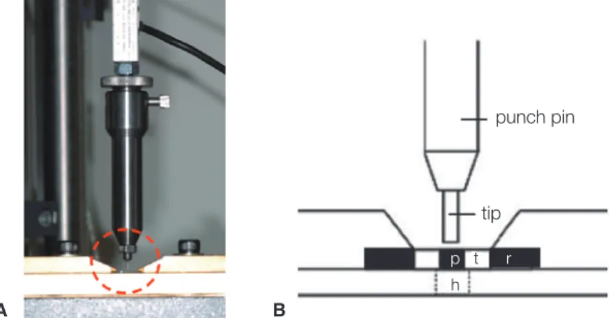
exposed part of the post was fixed to the hole in the alumi- num base which was the same size as the post diameter, such that the post was placed perpendicular to the tooth surface (Fig. 1). Using a low speed micro-saw, the embed- ded tooth specimens were cut into nine slices of 1 mm in thickness from the coronal to the apical direction to obtain three sliced specimens for the coronal, middle, and apical levels, respectively (Fig. 2). For the push-out bond strength test between the FRC post and the radicular dentin, a uni- versal testing machine (Z020, Zwickl, Ulm, Germany) was used with a 500 N load cell at a crosshead speed of 0.5 mm/min (Fig. 3). For the post loading, three different punch pins with diameters of 0.8 mm, 1.0 mm, and 1.2 mm were used for the apical, middle, and coronal samples, respectively. The posts inside the specimens were loaded on the apical to coronal direction.27 The maximum failure load was measured in newtons (N) and divided by the bonding surface calculated using a formula of conical frustrum to
obtain the bond strengths.20,21
After the push-out test, the failure aspect was observed at ×20 magnification by one examiner with an optical microscope (Axiotech Microscope, Carl Zeiss, Jena, Germany) and classified as either a cohesive failure (within the cement, post or dentin itself) or an adhesive failure (between the cement and tooth or between the post and cement). In the case of complex failure patterns, all distinct failure modes were noted. A representative failure aspect was observed at ×30 magnification with scanning electronic microscopy (JSM 6360, Jeol, Tokyo, Japan).
The data were statistically analyzed with SPSS 12.0 (SPSS GmbH, Munich, Germany). The effects of the resin cement and root level on the bond strengths was analyzed using a two-way ANOVA followed by post-hoc compari- sons with the Duncan test. The failure modes by root level were analyzed with a χ2 test. The probability level for statis- tical significance was set at α=.05.
Fig. 2. The FRC post luted with resin cement inside the root canal was embedded in acrylic resin and sectioned at 1 mm thickness producing 3 specimens from the coronal (a), middle (b), and apical areas (c), respectively.
At least 3 mm of gutta-percha was left to maintain the apical seal (t: tooth, r: acrylic resin).
a b c t
r
3 mm of Gutta-percha
Fig. 1. The cemented post in a tooth embedded in acrylic resin. The end of post was fixed into precisely fitted hole using an aluminum base (p: post, t: tooth, m: aluminum mold, b: aluminum base, h: fitting hole).
m
b
p t
h
A B
Fig. 3. Assembly of the push-out bond strength test. (A) photograph of the assembly installed on a universal testing machine, (B) a magnified schematic diagram of the circled area of (A) (p: post, t: tooth, r: acrylic resin, h: hole for the post being pushed-out on loading).
punch pin
tip p t r
h
RESULTS
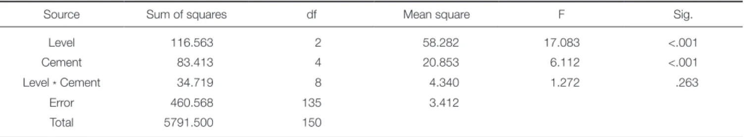
The bond strengths of resin cements according to the root level are shown in Table 2. A two-way ANOVA showed that the bond strength between the FRC post and the radic- ular dentin was significantly influenced by both the resin cement type (P<.001) and root level (P<.001), and there was no interaction between these two factors (Table 3). Of the resin cements, Lu and PA were found to have a signifi- cant decrease in their bond strengths at the apical level, whereas SB showed a significant decrease in the bond strength at the middle level (P<.05). In contrast, the bond strengths of LuA and Uni did not decrease significantly along the root length (P>.05). Of root level, the bond strengths did not differ significantly among the resin cements at coronal level (P>.05), but did differ significantly at the middle and apical levels (P<.05).
The failure aspects of the specimens showed an increase in adhesive failure and a decrease in cohesive failure moving
down the length of the canal from the coronal to the apical level (Table 4). Furthermore, adhesive failures at the apical level were mainly observed between the radicular dentin and the resin cement. At the coronal level, cohesive failures were dominant in all specimens, while at the middle level, cohesive failures were dominant for the Uni, LuA, and Lu cements (P<.05). At the apical level, PA, Lu, and SB had mostly adhesive failures (P<.05), while Uni and LuA had no significant difference between cohesive and adhesive fail- ures (P>.05). Scanning electron microscopic images of the failure aspects are shown in Fig. 4. A representative image of the adhesive failure between the radicular dentin and the resin cement is shown around the whole interface at apical level of PA (A), and a representative image of the adhesive failure between the FRC post and the resin cement is shown in middle level of SB (B). Both the adhesive failure between FRC post and resin cement and the cohesive fail- ure within resin cement are shown at middle level of Uni (C). The upper left corner is a ×300 magnification of the
Table 2. Push-out bond strengths of the FRC posts and the radicular dentin according to the resin cements used and the root level (unit: MPa)
Uni LuA Lu PA SB
Coronal 6.85 ± 2.45Aa 6.87 ± 1.76Aa 6.64 ± 1.24Aab 5.64 ± 1.31Aa 6.64 ± 1.27Aa Middle 6.78 ± 1.57Ca 7.26 ± 2.54BCa 7.95 ± 2.14ABCa 5.38 ± 1.56CDab 4.51 ± 2.37Db Apical 4.98 ± 2.15ABa 5.62 ± 0.95Aa 4.93 ± 1.45ABb 3.66 ± 1.38Bb 3.74 ± 2.47Bb The data were expressed as the mean ± standard deviation. Superscripts with the same capital letters in each row and the same small letters in each column are not significantly different at α=.05.
Table 3. Summary of a two-way ANOVA assessing the interaction of resin cement and root level on bond strength
Source Sum of squares df Mean square F Sig.
Level 116.563 2 58.282 17.083 <.001
Cement 83.413 4 20.853 6.112 <.001
Level * Cement 34.719 8 4.340 1.272 .263
Error 460.568 135 3.412
Total 5791.500 150
a. R Squared = .338 (Adjusted R Squared = .269)
Table 4. Percentage of adhesive failure and cohesive failure after the push-out bond strength test
Uni LuA Lu PA SB
Adhes Cohes Adhes Cohes Adhes Cohes Adhes Cohes Adhes Cohes
Coronal 12.0 88.0* 21.4 78.6* 17.1 82.9* 23.3 76.7* 20.7 79.3*
Middle 29.6 70.4* 17.9 82.1* 20.0 80.0* 47.8 52.2 42.1 57.9
Apical 53.3 46.7 47.1 52.9 72.2* 27.8 68.8* 31.3 85.7* 14.3
Adhes: Adhesive failure between the cement and dentin or between the post and cement.
Cohes: Cohesive failure within the cement, post, or dentin itself.
* = significantly different percentages between the adhesive and cohesive failures (α=.05).
circled area. The cohesive failures within the post, the resin cement and the dentin are shown at the coronal level of LuA (D).
DISCUSSION
Root canal environments are unfavorable for post cementa- tion with resin cements because of their high C-factor, which results in a high degree of polymerization shrinkage stress. The light exposure of resin cements to a curing light also limits the light-polymerization at deeper levels.
Therefore, to investigate the most appropriate resin cement to use in these circumstances, the push-out bond strength test was performed at three different root levels using five resin cements, and the failure aspect was observed. This study showed that the bond strengths were significantly affected by the resin cements used. Additionally, the bond
strengths significantly decreased for the specimens obtained in the apical area, except for the LuA and Uni groups, whose bond strengths did not decrease significantly at any root level. Therefore, the null hypothesis was thus over- ruled.
There are conflicting reports on the bond strengths between resin cements and radicular dentin, as it relates to different root levels. Higher bond strengths were reported for the apical third,20,28 while there are some reports with no significant differences among the different root levels29-31 and lower bond strengths at the apical third have also been reported.32-34 In this study, Lu, PA, and SB had lower bond strengths in the apical third. This finding could be exp- lained by the insufficient light-polymerization in the case of dual-cured resin cements due to the limited light exposure at the apical third. Insufficient light-polymerization could result in a low degree of conversion, even in the dual-cured Fig. 4. Representative SEM failure images shown at ×30 maginfication: (A) adhesive failure between dentin and cement at the apical level of PA, (B) adhesive failure between post and cement at the middle level of SB, (C) adhesive failure between post and cement and cohesive failure within cement at the middle level of Uni; the circled area is shown at
×300 in the upper left corner, (D) cohesive failure within post, cement and tooth at the coronal level of LuA (Δ: failure, p: post, c: cement, t: tooth).
A B
C D
resin cements.35
Considering the curing mode of resin cements, the bond strength of SB was expected to be affected less by the root level, because SB is self-cured resin cement that does not require light, unlike the other dual-cured resin cements.
However, the bond strength of SB decreased significantly below the middle third. This could be explained by factors other than the curing mode. SB is the only resin cement in this study that requires washing out the activator with water. The remnant water retained in the root canal even after the drying procedure might inhibit the hydrophobic resin cement from spreading over the radicular dentin, resulting in a decrease in the bond strength. Therefore, the difficulty with controlling the moisture in the root canals of SB group can be another explanation for lower bond strengths at the apical region of root canals.33 There is also a possibility of void inclusion during the mixing of the powder and liquid components for SB group, whereas oth- er groups adopt auto-mixing system with mixing tip.
Another factor could be the relatively short working time of the self-cured resin cement. Self-cured resin cement has limited working time compared to the light-cured and dual- cured resin cements. During procedures requiring sufficient working time, such as the luting of the post inside the root canal, the flow of the self-cured resin cement could decrease, which might result in the incomplete sealing of the resin cement at deep level of the root canal.36 Further- more, the filler content of the resin cement could also affect the bond strength. Filler-containing resin cements were found to have higher bond strengths than cements without fillers.37 The filler content of the resin cements used in this study were 74.9 ± 0.4 wt% for Lu and LuA, 68.7 ± 0.3 wt% for Uni, and 65.5 ± 0.5 wt% for PA, while SB did not contain fillers. From these results, it can be assumed that the residual water left in the root canal, hand mixing, short working time and no filler content of SB con- tributed to the lower bond strengths at the middle and api- cal root levels.
Both LuA and Lu group used LuxaCore-Dual resin cement, which has the highest filler content of the resin cements used in this study and is also commonly used as core material. The higher bond strength of LuA and Lu at the coronal and middle levels could be attributed to their higher level of filler loading. With increased filler loading, the flexural strength of the resin cement itself could be increased, and the polymerization shrinkage stress could be subsequently reduced.37 Therefore, the bond strength between the resin cement and the radicular dentin could also be increased.21,36 However, as filler loading of the resin cement increases, the flow of the resin cement also decreases. A balance should therefore be achieved between the filler loading and the flow of the resin cement. At the apical third, the bond strengths of LuA did not significantly decrease compared to the more coronal parts, whereas those of Lu decreased significantly. LuA and Lu group dif- fered only in the use of Contax Activator that is a self-cure activator.17 The Contax Activator includes hydrophilic and
acidic bis-GMA resin with benzoyl peroxide that is an initi- ator for the chemical-cured resins. Contax Primer is a self- etching primer. When this self-etching primer is used with self-cured or dual-cured resins such as LuxaCore, Contax Activator should be mixed into the Contax Bond to make it compatible with the self-cured or dual-cured resin. Based on the results of this study, Contax Activator attributed to maintain the bond strength at deeper levels of the root canal by making the bonding agent compatible with the res- in cement. Therefore, the use of Contax Activator is rec- ommended for the cementation of FRC posts with LuxaCore-Dual resin cement.
Uni also showed no significant differences in bond strengths at the different the root levels. RelyX Unicem is self-adhesive resin cement that does not require a pre-treat- ment step. It includes functional monomers, such as meth- acrylated phosphoric ester, that serve as a resin matrix as well as an etchant. After the phosphoric acid methacrylate decalcifies the tooth surface, the resin cement can infiltrate into the etched surface. Water is formed during the neutral- ization reaction of the phosphoric acid methacrylate, the basic fillers and the hydroxyapatite.20 The water is supposed to contribute to the wettability of the radicular dentin, resulting in increased bond strength.38 Its filler content is second to LuxaCore-Dual. This study did not show any sig- nificant difference between the adhesive resin cement and the self-adhesive resin cement.
In PA, the bond strengths decreased at the apical level.
Panavia F 2.0 is adhesive resin cement composed of self- etching primer that includes 10-methacryloyloxydecyl dihy- drogen phosphate (MDP). 10-MDP is an adhesive func- tional monomer that adheres well to base metals and zirco- nia and also acts as a weak etchant. When mixed with hydroxyethyl methacrylate (HEMA), 10-MDP demineraliz- es the apatite minerals of the dentin, allowing HEMA to infiltrate the space around the dentinal collagen networks to form a hybrid layer.39-41 However, the bond strength of 10-MDP to fiber post is lower than its bond strength to base metals or to zirconia.42 Therefore, PA was not found to be an effective luting material for use with FRC posts, especially at the apical third.
In terms of fracture aspects, adhesive failures increased from the coronal third to the apical third. Groups with higher bond strengths showed cohesive failure within the FRC post or the radicular dentin. However, specimens with lower bond strength at the apical third, such as PA, Lu and SB, tended to have more adhesive failures.24
Therefore, the bond strength between the FRC post and the radicular dentin was affected by the curing mode, the working time, the mixing method, the filler contents, the flow, the composition of the resin matrix, and the moisture control of the root canal. The degree of conversion of the resin cements, the shape of the root canal, the position of post in the root canal, and the space occupied by the resin cement between the post and the radicular dentin are also influential factors that require further study. The limitation of this study was that light-curing the resin cement through
the external root surface could not be controlled in the extracted teeth. Further studies will be needed for the sur- face treatment of FRC posts to increase bond strength between the FRC posts, the root canal dentin, and the resin cements.
Within the limitations of this in vitro study, the bond strengths between the radicular dentin and FRC posts decreased significantly at the middle or apical level except LuA and Uni. Therefore, clinicians should be concerned to treat the deeper level of the root canal to increase the bond strength with FRC posts.
CONCLUSION
According to the results of this study, the bond strength between the FRC post and the radicular dentin was signifi- cantly influenced by both the resin cement type used and the root level. Lu and PA were found to have a significant decrease in their bond strengths at the apical level, and SB was found to have a significant decrease in its bond strength below the middle level. A self-cure activator was effective to maintain the bond strength at deeper levels of the root canal for the cementation of FRC posts with LuxaCore-Dual resin cement. The bond strengths of LuA and Uni did not decrease significantly regardless of the root level. In conclusion, all resin cement groups showed decrease in bond strength at the middle or apical level except LuA and Uni.
REFERENCES
1. Schwartz RS, Robbins JW. Post placement and restoration of endodontically treated teeth: a literature review. J Endod 2004;30:289-301.
2. Sorensen JA, Engelman MJ. Ferrule design and fracture resis- tance of endodontically treated teeth. J Prosthet Dent 1990;
63:529-36.
3. Akkayan B, Gülmez T. Resistance to fracture of endodonti- cally treated teeth restored with different post systems. J Prosthet Dent 2002;87:431-7.
4. Bae JM, Kim KN, Hattori M, Hasegawa K, Yoshinari M, Kawada E, Oda Y. Fatigue strengths of particulate filler com- posites reinforced with fibers. Dent Mater J 2004;23:166-74.
5. Jung SH, Min KS, Chang HS, Park SD, Kwon SN, Bae JM.
Microleakage and fracture patterns of teeth restored with dif- ferent posts under dynamic loading. J Prosthet Dent 2007;
98:270-6.
6. Mezzomo E, Massa F, Libera SD. Fracture resistance of teeth restored with two different post-and-core designs cemented with two different cements: an in vitro study. Part I. Quintessence Int 2003;34:301-6.
7. Drummond JL, Toepke TR, King TJ. Thermal and cyclic loading of endodontic posts. Eur J Oral Sci 1999;107:220-4.
8. Mannocci F, Ferrari M, Watson TF. Intermittent loading of teeth restored using quartz fiber, carbon-quartz fiber, and zir- conium dioxide ceramic root canal posts. J Adhes Dent 1999;
1:153-8.
9. Viguie G, Malquarti G, Vincent B, Bourgeois D. Epoxy/car- bon composite resins in dentistry: mechanical properties re- lated to fiber reinforcements. J Prosthet Dent 1994;72:245-9.
10. Vallittu PK. Prosthodontic treatment with a glass fiber-rein- forced resin-bonded fixed partial denture: A clinical report. J Prosthet Dent 1999;82:132-5.
11. Duarte S Jr, Botta AC, Meire M, Sadan A. Microtensile bond strengths and scanning electron microscopic evaluation of self-adhesive and self-etch resin cements to intact and etched enamel. J Prosthet Dent 2008;100:203-10.
12. Krämer N, Lohbauer U, Frankenberger R. Adhesive luting of indirect restorations. Am J Dent 2000;13:60D-76D.
13. Özcan M, Mese A. Adhesion of conventional and simplified resin-based luting cements to superficial and deep dentin.
Clin Oral Investig 2012;16:1081-8.
14. Li N, Nikaido T, Takagaki T, Sadr A, Makishi P, Chen J, Tagami J. The role of functional monomers in bonding to enamel: acid-base resistant zone and bonding performance. J Dent 2010;38:722-30.
15. Ramos MB, Pegoraro TA, Pegoraro LF, Carvalho RM.
Effects of curing protocol and storage time on the micro- hardness of resin cements used to lute fiber-reinforced resin posts. J Appl Oral Sci 2012;20:556-62.
16. Sigemori RM, Reis AF, Giannini M, Paulillo LA. Curing depth of a resin-modified glass ionomer and two resin-based luting agents. Oper Dent 2005;30:185-9.
17. Ritter AV, Ghaname E, Pimenta LA. Dentin and enamel bond strengths of dual-cure composite luting agents used with dual-cure dental adhesives. J Dent 2009;37:59-64.
18. Bell AM, Lassila LV, Kangasniemi I, Vallittu PK. Bonding of fibre-reinforced composite post to root canal dentin. J Dent 2005;33:533-9.
19. Benetti AR, Asmussen E, Peutzfeldt A. Influence of curing rate of resin composite on the bond strength to dentin. Oper Dent 2007;32:144-8.
20. Bitter K, Meyer-Lueckel H, Priehn K, Kanjuparambil JP, Neumann K, Kielbassa AM. Effects of luting agent and ther- mocycling on bond strengths to root canal dentine. Int Endod J 2006;39:809-18.
21. Bitter K, Priehn K, Martus P, Kielbassa AM. In vitro evalua- tion of push-out bond strengths of various luting agents to tooth-colored posts. J Prosthet Dent 2006;95:302-10.
22. D’Arcangelo C, Cinelli M, De Angelis F, D’Amario M. The effect of resin cement film thickness on the pullout strength of a fiber-reinforced post system. J Prosthet Dent 2007;98:
193-8.
23. Mosharraf R, Baghaei Yazdi N. Comparative evaluation of effects of different surface treatment methods on bond strength between fiber post and composite core. J Adv Prosthodont 2012;4:103-8.
24. Khamverdi Z, Talebian R. Effect of ascorbic acid, ethanol and acetone on adhesion between the treated fiber posts and composite resin cores. J Adv Prosthodont 2012;4:187-91.
25. International Standards Organization. ISO/TS 11405:
2003(E) Dental materials - Testing of adhesion to tooth structure. ISO, Geneva, 2003.
26. Vallittu PK. Flexural properties of acrylic resin polymers re-
inforced with unidirectional and woven glass fibers. J Prosthet Dent 1999;81:318-26.
27. Carvalho CA, Breschi L, Navarro MF, Atta MT, Ferrari M.
Push-out bond strength and SEM evaluation of a new bond- ing approach into the root canal. J Appl Oral Sci 2012;20:
613-9.
28. Gaston BA, West LA, Liewehr FR, Fernandes C, Pashley DH. Evaluation of regional bond strength of resin cement to endodontic surfaces. J Endod 2001;27:321-4.
29. Foxton RM, Nakajima M, Tagami J, Miura H. Adhesion to root canal dentine using one and two-step adhesives with du- al-cure composite core materials. J Oral Rehabil 2005;32:97- 104.
30. Pereira PC, Melo RM, Chaves C, Galhano GA, Bottino MA, Balducci I. The adhesive system and root canal region do not influence the degree of conversion of dual resin cement. J Appl Oral Sci 2010;18:477-81.
31. Teixeira CS, Silva-Sousa YT, Sousa-Neto MD. Bond strength of fiber posts to weakened roots after resin restoration with different light-curing times. J Endod 2009;35:1034-9.
32. Mallmann A, Jacques LB, Valandro LF, Mathias P, Muench A.
Microtensile bond strength of light- and self-cured adhesive systems to intraradicular dentin using a translucent fiber post.
Oper Dent 2005;30:500-6.
33. Bouillaguet S, Troesch S, Wataha JC, Krejci I, Meyer JM, Pashley DH. Microtensile bond strength between adhesive cements and root canal dentin. Dent Mater 2003;19:199-205.
34. Perdigão J, Gomes G, Lee IK. The effect of silane on the bond strengths of fiber posts. Dent Mater 2006;22:752-8.
35. Acquaviva PA, Cerutti F, Adami G, Gagliani M, Ferrari M, Gherlone E, Cerutti A. Degree of conversion of three com- posite materials employed in the adhesive cementation of in- direct restorations: a micro-Raman analysis. J Dent 2009;37:
610-5.
36. Feilzer AJ, de Gee AJ, Davidson CL. Setting stresses in com- posites for two different curing modes. Dent Mater 1993;9:2- 5.
37. Kato H, Matsumura H, Atsuta M. Effect of etching and sandblasting on bond strength to sintered porcelain of un- filled resin. J Oral Rehabil 2000;27:103-10.
38. Radovic I, Monticelli F, Goracci C, Vulicevic ZR, Ferrari M.
Self-adhesive resin cements: a literature review. J Adhes Dent 2008;10:251-8.
39. Barkmeier WW, Cooley RL. Laboratory evaluation of adhe- sive systems. Oper Dent 1992;Suppl 5:50-61.
40. Staninec M, Kawakami M. Adhesion and microleakage tests of a new dentin bonding system. Dent Mater 1993;9:204-8.
41. Watanabe I, Nakabayashi N, Pashley DH. Bonding to ground dentin by a phenyl-P self-etching primer. J Dent Res 1994;73:
1212-20.
42. Gomes AL, Castillo-Oyagüe R, Lynch CD, Montero J, Albaladejo A. Influence of sandblasting granulometry and resin cement composition on microtensile bond strength to zirconia ceramic for dental prosthetic frameworks. J Dent 2013;41:31-41.