Copyright © 2019 The Korean Society for Bone and Mineral Research
This is an Open Access article distributed under the terms of the Creative Commons Attribution Non-Commercial Li- cense (http://creativecommons.org/licenses/by-nc/4.0/) which permits unrestricted non-commercial use, distribu- tion, and reproduction in any medium, provided the original work is properly cited.
Clinical Application of Bone Turnover Markers in Osteoporosis in Korea
So Young Park1, Seong Hee Ahn2, Jun-Il Yoo3, Youn-Jee Chung4, Yun Kyung Jeon5, Byung-Ho Yoon6, Ha Young Kim7, Seung Hun Lee8, Jehoon Lee9, Seongbin Hong2
1Division of Endocrinology and Metabolism, Department of Internal Medicine, Kyung Hee University Hospital, Seoul;
2Division of Endocrinology and Metabolism, Department of Internal Medicine, Inha University Hospital, Inha University School of Medicine, Incheon;
3Department of Orthopaedic Surgery, Gyeongsang National University Hospital, Jinju;
4Department of Obstetrics and Gynecology, Seoul St. Mary's Hospital, College of Medicine, The Catholic University of Korea, Seoul;
5Division of Endocrinology and Metabolism, Department of Internal Medicine, Pusan National University Hospital, Busan;
6Department of Orthopaedic Surgery, Inje University College of Medicine, Seoul Paik Hospital, Seoul;
7Division of Endocrinology, Department of Internal Medicine, Wonkwang University Sanbon Hospital, Wonkwang University School of Medicine, Gunpo;
8Division of Endocrinology and Metabolism, Asan Medical Center, University of Ulsan College of Medicine, Seoul;
9Department of Laboratory Medicine, College of Medicine, The Catholic University of Korea, Seoul, Korea
Bone turnover markers (BTMs) have important role in the management of osteoporosis.
Recently the clinical application of BTMs has achieved significant progress and measure- ment of BTMs give us better understanding of pathogenesis of osteoporosis. However, the use of BTMs is still insufficient in Korea. We summarized the available methods and standard interval of the BTMs in Korea. Also we reviewed published literatures on pre- analytical variability in the measurement of BTMs and provided recommendations for standardized sample handling and patient preparation for reducing those pre-analytical variabilities. The clinical application of BTMs in patients with chronic kidney disease who have a higher fracture risk than the general population is summarized.
Key Words: Biomarkers · Bone remodeling · Bone turnover markers · Chronic kidney dis- ease · Osteoporosis
INTRODUCTION
Osteoporosis is a major health problem worldwide. Osteoporosis is defined as a disease characterized by low bone mass and microarchitectural deterioration of bone tissue, leading to enhanced bone fragility and consequent increase in the risk of fracture.[1] The bone strength is determined by bone mass and bone quali- ty. Bone mass is expressed mainly in bone mineral density (BMD), and bone quali- ty is composed of microarchitecture, bone turnover rate, mineralization, and mi- crodamage accumulation.[2] The BMD measurement using dual energy X-ray ab- sorptiometry (DXA) is the most commonly used diagnostic criteria for osteoporo- sis.[3] Although BMD is used for treatment strategy determination and evaluation of bone loss rate or treatment response, it might not completely capture the os- Corresponding author
Seongbin Hong
Division of Endocrinology and Metabolism, Department of Internal Medicine, Inha University Hospital, Inha University School of Medicine, 100 Inha-ro, Michuhol-gu, Incheon 22212, Korea
Tel: +82-32-890-3597 Fax: +82-32-882-6578 E-mail: [email protected] Received: February 8, 2019 Revised: February 18, 2019 Accepted: February 19, 2019
No potential conflict of interest relevant to this article was reported.
teoporotic fracture risk. Also, changes in BMD are reflected at a slow rate.
In bone, bone turnover, which removes old bones by bone resorption, and forms new bones by bone formation, is continuously occurring.[4,5] The change of bone turnover rate could affect the bone quality. Bone turnover markers (BTMs) are an index reflecting the rate of bone turnover and samples to measure BTMs can be taken from urine and blood easily.[6] Considering the limitations of BMD and the characteristic of BTMs reflecting bone quality, interest in the clinical potential of BTM to predict fracture risk and mon- itor treatment is steadily increasing.
BTMs are classified as either bone formation markers or bone resorption makers. The BTMs have not yet to be prov- en to make diagnosis and treatment decision for osteopo- rosis. But, the clinical usefulness of BTMs to predict bone loss and fracture risk, and to monitor the response of os- teoporosis treatment have been shown in several studies.
[7-9] Also, the measurement of BTMs give us better under- standing of pathogenesis of osteoporosis. However, the val- ue of the BTMs might be influenced by physiological and pathological factors, and, in some cases, multiple method- ologies used for the same analyte. Theses strength and weak- ness of BTMs in clinical practice have been considered by several national societies and guidelines development groups and have resulted in differing recommendation.[10,11]
The use of BTMs is still insufficient in Korea. The Korean Society of Bone Mineral Society organized the bone turn- over working group to investigate on the application of BTMs and provide recommendations on their clinical use to physicians in Korea.
The aim of this paper is to investigate the available meth- ods and standard reference intervals of BTMs in Korea, and review the standardization of sampling to decrease the pre- analytical issue. The patients with chronic kidney disease (CKD) have a higher fracture risk than the general popula- tion. Bone disease related to advanced CKD features a large spectrum of clinical phenotype. The clinical application of BTMs in those population summarized separately.
THE AVAILABLE ASSAY METHODS FOR MEASUREMENT OF BTMS IN KOREA
1. Bone formation markersBone formation markers include osteocalcin, bone-spe-
cific alkaline phosphatase (BSALP), carboxy-terminal pro- peptide of type I collagen (P1CP), and aminoterminal pro- peptide of type I collagen (P1NP). BSALP and osteocalcin released by osteoblasts and plays a major role in bone min- eralization. P1CP and P1NP cleaved from procollagen type I during collagen synthesis. In Korea, osteocalcin and BSALP are most commonly used as bone formation markers. Sev- eral kits have been used to measure osteocalcin and BSALP depending on the assay methods. For osteocalcin, 1 radio- immunoassay (RIA; BGP [BRAHMS Diagnostica, Berlin, Ger- many]), 2 immunoradiometric assay (IRMA; DIAsource hOST- IRMA or CISBIO osteo-RIACT [DIAsource Immunoassays S.A., Nivelles, Belgium]) and 1 electrochemiluminescence assay (ECLIA; Elecsys N-MID Osteocalcin [Roche Diagnos- tics, Mannheim, Germany]) were used. For BSALP, 1 chemi- luminescence assay (CLIA; Beckman Coulter Inc., Sacra- mento, CA, USA) and 1 enzyme immunoassay (EIA; Micr- oVue BAP EIA [Quidel Corporation, San Diego, CA, USA]) were used. Also, it can be measure by electrophoresis assay (Spife ALP-20; Helena Laboratories, Beaumont, TX, USA). Al- though P1NP has not been widely used yet, P1NP can be measured by the ECLIA using Elecsys total P1NP (Roche).
In Korea, total P1NP (Roche Elecsys) is covered by insurance in osteoporotic patients since 2018. Reference interval and median value of P1NP in Korean population was publish- ed.[12]
2. Bone resorption markers
Bone resorption markers are serum C-terminal telopep- tides of type I collagen (CTX), urinary N-terminal telopep- tide of collagen type I (NTx), free and total pyridinoline (PYD) and free and total deoxypyridinoline (DPD). These are collagen cross-links.
In Korea, CTX, NTx, and DPD can be measured as bone resorption markers. In Korea, CTX is most commonly used as bone resorption markers. Several kits have been used to measure bone resorption markers depending on the assay methods. Currently for CTX, 1 automated immunoassay is available: Beta-CrossLaps Roche Elecsys (ECLIA [Roche Di- agnostics]). For NTx, enzyme-linked immunosorbent assay (ELISA) is used for urine and serum: OSTEOMARK (Alere Inc., Scarborough, ME, USA). CLIA (Ortho-Clinical Diagnos- tics, Inc., High Wycombe, UK) is also available. For DPD, EIA (Qulidelmetra, San Diego, CA, USA) and CLIA are available.
INTERPRETATION ON THE REFERENCE INTERVALS OF BTMS
The reference intervals of BTMs are useful for interpret- ing the results in osteoporosis patients. Several prospec- tive studies have reported the presence of increased BTMs have an additive effect on fracture risk in women.[7] The very high BTM values (>3 standard deviation above the mean of the reference values) during the initial assessment suggests other metabolic disease. However, sufficient con- sensus has not been achieved for cut-point of BTMs that increase fracture risk or assess response of treatment. It is considered necessary to establish reference intervals for different geographic area and ethnicities. Although refer- ence intervals of P1NP in Korean population were report- ed,[12] only reference of manufacture’s data is available for other BTMs (Table 1).
STANDARDIZATION OF PATIENT SAMPLE COLLECTION PROCEDURE
1. Importance of standardized patient sample collection procedure
The primary challenge to the adoption of many BTMs in
routine practice has been poor within-subject and between- lab reproducibility. For BTMs to be useful, the pre-analytical sources of variability, along with the underlying disease pro- cess, must be identified, minimized, and controlled through carefully standardized patient preparation and sample han- dling procedures.[13,14]
Although BTMs except CTX are relatively stable and very modestly affected by pre-analytical variables, several BTMs are frequently measured together on automated platforms within one sample.[15] Therefore, it is important that the conditions that ensure CTX integrity may be maintained when measuring BTMs. Moreover, serum P1NP has recent- ly been recommended as a reference BTM along with se- rum CTX.[16] Therefore, proper sample collection proce- dures for both CTX and P1NP are focused in this section.
2. Sources of pre-analytical variability sample handling
1) Sample type
Although either serum or plasma sample may be used for CTX and PINP measurements, EDTA plasma has the ad- vantage of superior sample stability for CTX compared with serum.[17-19] The same sample type such as serum or plas- ma should be used consistently when monitoring a patient.
2) Sample collection
CTX exhibits a circadian rhythm in blood. CTX level peaks during the early morning hours (2-5 a.m.) and reaches a nadir between 11 a.m. and 2 p.m. The only known modu- lator having a major effect on this circadian pattern is food intake. Overnight fasting markedly reduced the circadian variation of CTX. Therefore, blood samples for CTX mea- surement must be collected following an overnight fast during the morning between 7:30 a.m. and 10:00 a.m.[20- 22] Other BTMs have minimal circadian rhythm and are minimally affected by food intake.
3) Sample processing
Samples destined for CTX measurement should be fro- zen at ≤-20°C preferably within 2 hr of collection.[23] If measurement is to be made within 8 hr of phlebotomy, the sample may be stored at 4°C. Moderate hemolysis (0.5 g/dL) should be avoided for CTX measurement.[19,24]
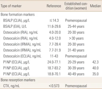
Table 1. The reference intervals and median values of bone turnover markers
Type of marker Reference Established con-dition (women) Median Bone formation markers
BSALP (CLIA), μg/L ≤14.3 Premenopausal BSALP (EIA), U/L 11.6-29.6 25-44 years Osteocalcin (RIA), ng/mL 4.0-20.0 20-30 years Osteocalcin (RIA), ng/mL 4.0-12.0 >30 years Osteocalcin (IRMA), ng/mL 7.7-39.4 20-30 years Osteocalcin (IRMA), ng/mL 7.7-31.9 31-40 years Osteocalcin (ECLIA), ng/mL 11-43 Premenopausal P1NP (ECLIA), μg/L 24.0-77.1 20-29 years 42.3 P1NP (ECLIA), μg/L 18.7-83.2 30-39 years 40.0 P1NP (ECLIA), μg/L 18.8-70.1 40-49 years 35.0 Bone resorption markers
CTX, ng/mL <0.573 Premenopausal
BSALP, bone-specific alkaline phosphatase; CLIA, chemiluminescence assay; EIA, enzyme immunoassay; RIA, radioimmunoassay; IRMA, immu- noradiometric assay; P1NP, aminoterminal propeptide of type I collagen;
ECLIA, electrochemiluminescence assay; CTX, C-terminal telopeptides of type I collagen.
4) Sample stability
CTX is more stable in EDTA plasma than in serum, regard- less of measurement methods.[23] For Roche package in- serts, CTX has stability of 24 hr at room temperature (RT;
20-25°C), 8 days at 2 to 8°C in EDTA plasma, and 6 hr at RT and 8 hr at 4°C in serum.[25] P1NP is more stable compared to CTX with stability of at least 24 hr at RT and 5 days at 4°C in both EDTA plasma and serum.[26] For long term storage, stability for 3 months for CTX and 6 months for P1NP at
≤-20°C is ensured for all methods. For longer term resear- ches, the recommendation is aliquots of samples at -70°C or below to allow to analysis of all samples in a single batch at a later time.[19,25,26]
5) Freeze/thaw cycle
Multiple freeze-thaw cycles are reported to be accept- able for CTX and P1NP.[19,24]
3. Sources of pre-analytical variability: patient related factors
The patient related factors are divided into controllable and uncontrollable factors. Controllable factors include the menstrual cycle, seasonal variation, and physical activities.
The optimal time to collect samples in pre-menopausal wom- en is the early-mid-follicular phase.[27] There is a minor but detectable seasonal variation for CTX in older adults and those with severe vitamin D deficit.[28,29] Intensive physical training (e.g., elite soccer players) moderately in- creases serum CTX and slightly decreases PINP. Vigorous exercise should be avoided the day prior to sampling.[30]
Uncontrollable factors include age, sex, pregnancy, geog- raphy, renal function, and specific diseases and medica- tions.[14]
USE IN PATIENTS WITH CKD
The patients with CKD have a higher fracture risk than the general population. In patients with CKD, secondary hyperparathyroidism, adynamic bone, hemodialysis asso- ciated amyloidosis, vitamin D deficiency, hypocalcemia, changes in the bone architecture, nutritional disturbance, and increase in oxidative stress could increase fracture risk.
[31] Because some BTMs are influenced by the renal func- tion, there are limitations to use BTMs to predict and evalu- ate the fracture risk in patients with CKD. The frequency of
monitoring serum calcium, phosphate, and parathyroid hormone (PTH) as well as BTMs are recommended to eval- uate the presence and magnitude of abnormalities, and the rate of progression of CKD.[32]
As bone formation markers, BSALP, P1NP, and P1CP are independent of the renal function status. But, osteocalcin is influenced by the renal function.[33,34] Combining a low PTH (<150 pg/mL) and a low BSALP (<27 IU/L) im- proved the specificity of diagnosing adynamic bone dis- ease in 103 dialysis patients with bone biopsy results.[35]
In the newer automated Ostase bone specific alkaline phos- phatase (ALP) assay, cut-off <20 IU/L is used. The Kidney Disease, Improving Global Outcomes (KDIGO) guidelines suggest that measurements of serum PTH or BSALP can be used to evaluate bone disease because markedly high or low values predict underlying bone turnover in patients with CKD G3a (estimated glomerular filtration rate, 45-59 mL/min/1.73m2)–G5D (end-stage renal disease patients who undergo chronic dialysis).[32] Since osteocalcin is cleared by kidney, its use in CKD patients is limited. The combina- tion of osteocalcin (<41 ng/L) and BSALP (<23 U/L) im- proved the positive predictive value for diagnosing ady- namic bone disease in a CKD-5 cohort to 77%.[33] The pro- portion of the monomeric form of P1NP is elevated in pa- tients with CKD, whereas the apparent concentration of P1NP is unaffected by glomerular filtration rate in kidney disease patients when an intact assay for P1NP is used.[34]
P1NP monomers are not cleared by conventional dialysis sessions and the least significant change is of 32% for the intact assay.[36]
As bone resorption markers, CTX is excreted by kidney and accumulates in CKD patients. CTX is cleared by dialysis and therefore predialysis sampling is required for longitu- dinal monitoring.[33] PYD, DPD and NTx are influenced by the renal function status. Therefore, the KDIGO guidelines recommended that the bone derived markers of collagen synthesis and breakdown should not be routinely measured in patients with CKD stages 3 to 5D.[32] Levels of tartrate- resistant acid phosphatase 5b correlates with PTH and ALP and are unaffected by renal function. Its use is limited by availability of automated assays.[33]
The limitations of our study is that few Korean data is avail- able. However, all available methods in Korea were surveyed.
This data will support upcoming study for standardization and reference intervals of BTMs can be used for decision
and monitoring of treatment.
CONCLUSIONS
BTMs could be used to predict the fracture risk predic- tion and monitor treatment response. In 2017, P1NP and CTX were flagged for standardization by the International Osteoporosis Foundation and International Federation of Clinical Chemistry and Laboratory Medicine. But, these BTMs are not widely used in clinical practice. The data on stan- dardized reference interval of PINP in Koreans are available.
Further researches for CTX assay in Koreans are needed.
The standardized patient preparation and sample handling procedures is important to decrease analytical sources of variability. In the patients with CKD, use of BTMs is limited.
When used in combination with PTH, BSALP can predict adynamic bone disease. This review support standardiza- tion and clinical use in the management of patients of os- teoporosis.
AUTHOR CONTRIBUTION
Conceptualization: Hong S, Park SY, Ahn SH, Yoo JI, Chung YJ, Jeon YK, Yoon BH, Kim HY, Lee SH, Lee J. Wrote first draft of manuscript: Hong S, Park SY, Ahn SH, Yoo JI, Chung YJ, Jeon YK, Yoon BH, Kim HY, Lee SH, Lee J. Comment and revise manuscript: Hong S, Park SY, Ahn SH, Yoo JI, Chung YJ, Jeon YK, Yoon BH, Kim HY, Lee SH, Lee J. Approved final version:
Hong S, Park SY, Ahn SH, Yoo JI, Chung YJ, Jeon YK, Yoon BH, Kim HY, Lee SH, Lee J.
REFERENCES
1. Peck WA. Consensus development conference: diagnosis, prophylaxis, and treatment of osteoporosis. Am J Med 1993;94:646-50.
2. Weinstein RS. True strength. J Bone Miner Res 2000;15:
621-5.
3. Blake GM, Fogelman I. Role of dual-energy X-ray absorpti- ometry in the diagnosis and treatment of osteoporosis. J Clin Densitom 2007;10:102-10.
4. Raggatt LJ, Partridge NC. Cellular and molecular mecha- nisms of bone remodeling. J Biol Chem 2010;285:25103-8.
5. Feng X, McDonald JM. Disorders of bone remodeling. Annu Rev Pathol 2011;6:121-45.
6. Delmas PD, Eastell R, Garnero P, et al. The use of biochemi- cal markers of bone turnover in osteoporosis. Committee of Scientific Advisors of the International Osteoporosis Foundation. Osteoporos Int 2000;11 Suppl 6:S2-17.
7. Garnero P, Hausherr E, Chapuy MC, et al. Markers of bone resorption predict hip fracture in elderly women: the EPI- DOS Prospective Study. J Bone Miner Res 1996;11:1531-8.
8. Garnero P, Sornay-Rendu E, Claustrat B, et al. Biochemical markers of bone turnover, endogenous hormones and the risk of fractures in postmenopausal women: the OFE- LY study. J Bone Miner Res 2000;15:1526-36.
9. Ross PD, Kress BC, Parson RE, et al. Serum bone alkaline phosphatase and calcaneus bone density predict fractures:
a prospective study. Osteoporos Int 2000;11:76-82.
10. Nishizawa Y, Nakamura T, Ohta H, et al. Guidelines for the use of biochemical markers of bone turnover in osteopo- rosis (2004). J Bone Miner Metab 2005;23:97-104.
11. Morris HA, Eastell R, Jorgensen NR, et al. Clinical usefulness of bone turnover marker concentrations in osteoporosis.
Clin Chim Acta 2017;467:34-41.
12. Yoo JI, Park AJ, Lim YK, et al. Age-related reference inter- vals for total collagen-I-N-terminal propeptide in healthy Korean population. J Bone Metab 2018;25:235-41.
13. Nishizawa Y, Ohta H, Miura M, et al. Guidelines for the use of bone metabolic markers in the diagnosis and treatment of osteoporosis (2012 edition). J Bone Miner Metab 2013;
31:1-15.
14. Szulc P, Naylor K, Hoyle NR, et al. Use of CTX-I and PINP as bone turnover markers: National Bone Health Alliance rec- ommendations to standardize sample handling and pa- tient preparation to reduce pre-analytical variability. Os- teoporos Int 2017;28:2541-56.
15. Bauer D, Krege J, Lane N, et al. National bone health alli- ance bone turnover marker project: current practices and the need for US harmonization, standardization, and com- mon reference ranges. Osteoporos Int 2012;23:2425-33.
16. Vasikaran S, Eastell R, Bruyere O, et al. Markers of bone turn- over for the prediction of fracture risk and monitoring of osteoporosis treatment: a need for international reference standards. Osteoporos Int 2011;22:391-420.
17. Christgau S, Rosenquist C, Alexandersen P, et al. Clinical evaluation of the serum CrossLaps one step ELISA, a new assay measuring the serum concentration of bone-derived degradation products of type I collagen C-telopeptides.
Clin Chem 1998;44:2290-300.
18. Garnero P, Borel O, Delmas PD. Evaluation of a fully auto- mated serum assay for C-terminal cross-linking telopep- tide of type I collagen in osteoporosis. Clin Chem 2001;47:
694-702.
19. Morovat A, Catchpole A, Meurisse A, et al. IDS iSYS auto- mated intact procollagen-1-N-terminus pro-peptide as- say: method evaluation and reference intervals in adults and children. Clin Chem Lab Med 2013;51:2009-18.
20. Qvist P, Christgau S, Pedersen BJ, et al. Circadian variation in the serum concentration of C-terminal telopeptide of type I collagen (serum CTx): effects of gender, age, meno- pausal status, posture, daylight, serum cortisol, and fast- ing. Bone 2002;31:57-61.
21. Redmond J, Fulford AJ, Jarjou L, et al. Diurnal rhythms of bone turnover markers in three ethnic groups. J Clin En- docrinol Metab 2016;101:3222-30.
22. Clowes JA, Hannon RA, Yap TS, et al. Effect of feeding on bone turnover markers and its impact on biological vari- ability of measurements. Bone 2002;30:886-90.
23. Stokes FJ, Ivanov P, Bailey LM, et al. The effects of sampling procedures and storage conditions on short-term stability of blood-based biochemical markers of bone metabolism.
Clin Chem 2011;57:138-40.
24. Rosenquist C, Fledelius C, Christgau S, et al. Serum Cross- Laps one step ELISA. First application of monoclonal anti- bodies for measurement in serum of bone-related degra- dation products from C-terminal telopeptides of type I col- lagen. Clin Chem 1998;44:2281-9.
25. Roche Diagnostics. Elecsys β-Crosslaps/serum (B-CTX in serum) immunoassay cobas package insert (V 14.1). Man- heim, DE: Roche Diagnostics; 2014.
26. Roche Diagnostics GmbH. Total PINP (2014) (total procol- lagen type I amino-terminal propeptide) immunoassay Cobas package insert (V11.0). Mannheim, DE: Roche Di- agnostics GmbH; 2014.
27. Gass ML, Kagan R, Kohles JD, et al. Bone turnover marker
profile in relation to the menstrual cycle of premenopaus- al healthy women. Menopause 2008;15:667-75.
28. Bhattoa HP, Nagy E, More C, et al. Prevalence and seasonal variation of hypovitaminosis D and its relationship to bone metabolism in healthy Hungarian men over 50 years of age: the HunMen Study. Osteoporos Int 2013;24:179-86.
29. Pasco JA, Henry MJ, Kotowicz MA, et al. Seasonal periodic- ity of serum vitamin D and parathyroid hormone, bone resorption, and fractures: the Geelong Osteoporosis Study.
J Bone Miner Res 2004;19:752-8.
30. Weiler R, Keen R, Wolman R. Changes in bone turnover markers during the close season in elite football (soccer) players. J Sci Med Sport 2012;15:255-8.
31. Nitta K, Yajima A, Tsuchiya K. Management of osteoporo- sis in chronic kidney disease. Intern Med 2017;56:3271-6.
32. Isakova T, Nickolas TL, Denburg M, et al. KDOQI US com- mentary on the 2017 KDIGO clinical practice guideline up- date for the diagnosis, evaluation, prevention, and treat- ment of chronic kidney disease-mineral and bone disor- der (CKD-MBD). Am J Kidney Dis 2017;70:737-51.
33. Chiang C. The use of bone turnover markers in chronic kidney disease-mineral and bone disorders. Nephrology (Carlton) 2017;22 Suppl 2:11-3.
34. Ueda M, Inaba M, Okuno S, et al. Clinical usefulness of the serum N-terminal propeptide of type I collagen as a mark- er of bone formation in hemodialysis patients. Am J Kid- ney Dis 2002;40:802-9.
35. Couttenye MM, D'Haese PC, Van Hoof VO, et al. Low serum levels of alkaline phosphatase of bone origin: a good mark- er of adynamic bone disease in haemodialysis patients.
Nephrol Dial Transplant 1996;11:1065-72.
36. Cavalier E, Delanaye P, Moranne O. Variability of new bone mineral metabolism markers in patients treated with main- tenance hemodialysis: implications for clinical decision making. Am J Kidney Dis 2013;61:847-8.