INTRODUCTION
Cyclooxygenase (COX) converts arachidonic acid into prosta- glandin H2, which is the rate-limiting step in prostaglandin (PG) formation including prostaglandin E2 (PGE2). At least two iso- forms of COX have been identified in mammalian cells: COX-1 is constitutively expressed, whereas COX-2 is inducible in re- sponse to growth factors, oncogenes, cytokines, and endotox- in.1 COX-2 expression is upregulated at sites of inflammation and in various carcinomas.2 Lipopolysaccharide (LPS), the main cell wall component of Gram-negative bacteria, activates in- flammatory responses through toll-like receptor 4 (TLR4), and LPS-activated macrophages show a high expression of COX-2.3
The immunomodulatory effects of PGE2 result largely from its
Suppression of Heme Oxygenase-1 by Prostaglandin E 2 -Protein Kinase A-A-Kinase Anchoring Protein Signaling Is Central for Augmented Cyclooxygenase-2 Expression in
Lipopolysaccharide-Stimulated RAW 264.7 Macrophages
Jae-Hyoung Lee,
1Nam-Hee Jung,
1Byoung-Hoon Lee,
1Sang-Hoon Kim,
1* Jin Hyun Jun
21Department of Internal Medicine, Eulji Hospital, Eulji University School of Medicine, Seoul, Korea
2Department of Bio-Medical Laboratory Science, College of Health Science, Eulji University, Suongnam, Korea
ability to increase intracellular cyclic AMP (cAMP) through the G protein-coupled E prostanoid (EP) receptors EP2 and EP4.4 Increases in intracellular cAMP serve as an important brake on excessive inflammatory activation in macrophages.5 The classi- cal cAMP effector is protein kinase A (PKA), which is composed of two regulatory (R) and two catalytic (C) subunits.5 In addition, scaffold proteins, termed A-kinase anchoring proteins (AKAP), Allergy Asthma Immunol Res. 2013 September;5(5):329-336.
http://dx.doi.org/10.4168/aair.2013.5.5.329 pISSN 2092-7355 • eISSN 2092-7363
Purpose: Prostaglandin (PG) E2 is an immunomodulatory lipid mediator generated mainly via the cyclooxygenase-2 (COX-2) pathway from arachi- donic acid at sites of infection and inflammation. A positive feedback loop of PGE2 on COX-2 expression is critical for homeostasis during toll-like re- ceptor (TLR)-mediated inflammatory processes. The mechanism of PGE2-regulated COX-2 expression remains poorly understood. The low-molecular- weight stress protein heme oxygenase-1 (HO-1) contributes to the anti-inflammatory, anti-oxidant and anti-apoptotic response against environmen- tal stress. Methods: We explored the involvement of HO-1 on PGE2 regulation of LPS-induced COX-2 expression in RAW 264.7 macrophages.
Results: LPS-induced COX-2 expression in RAW 264.7 macrophages was enhanced by exogenous PGE2 or cyclic AMP (cAMP) analogue and was suppressed by a COX inhibitor (indomethacin), a protein kinase A (PKA) inhibitor (KT5720), and A kinase anchoring protein (AKAP) disruptors (Ht31 and RIAD). This result suggests that the stimulatory effects of endogenous and exogenous PGE2 on COX-2 expression are mediated by a cAMP-PKA- AKAP-dependent pathway. The induction of HO-1 was observed in LPS-stimulated RAW 264.7 macrophages. This induction was suppressed by ex- ogenous PGE2 and enhanced by blockage of the endogenous PGE2 effect by the PKA inhibitor or AKAP disruptors. In addition, HO-1 induction by the HO activator copper protoporphyrin suppressed LPS-induced COX-2 expression, which was restored by the addition of exogenous PGE2. The induc- tion of HO-1 inhibited LPS-induced NF-κB p-65 nuclear expression and translocation. Conclusions: AKAP plays an important role in PGE2 regula- tion of COX-2 expression, and the suppression of HO-1 by PGE2-cAMP-PKA-AKAP signaling helps potentiate the LPS-induced COX-2 expression through a positive feedback loop in RAW 264.7 macrophages.
Key Words: Cyclooxygenase-2; heme oxygenase-1; lipopolysaccharide; prostaglandin E2; macrophages
This is an Open Access article distributed under the terms of the Creative Commons Attribution Non-Commercial License (http://creativecommons.org/licenses/by-nc/3.0/) which permits unrestricted non-commercial use, distribution, and reproduction in any medium, provided the original work is properly cited.
Correspondence to: Sang-Hoon Kim, MD, PhD, Department of Internal Medicine, Eulji Hospital, 14 Hangeulbiseok-gil, Nowon-gu, Seoul 139-711, Korea.
Tel: +82-2-970-8365; Fax: +82-2-970-8621; E-mail: [email protected] Received: September 30, 2012; Revised: December 13, 2012 Accepted: December 26, 2012
•There are no financial or other issues that might lead to conflict of interest.
involve the assembly of cAMP-dependent pathway compo- nents into multiprotein signaling complexes.6
COX is rapidly inactivated after the conversion of arachidonic acid into PGH2. Therefore, rapid synthesis of COX would be re- quired to sustain PG biosynthesis.7 A positive feedback loop be- tween PGE2 and COX-2 is established in various cells.8-10 Fur- thermore, there is some evidence that the canonical PGE2/EP2/
cAMP/PKA/CREB cascade plays a key role in the expression of COX-2 in activated macrophages.11 However, the mechanisms of PGE2 regulation of COX-2 expression remain poorly under- stood.
The low-molecular-weight stress protein heme oxygenase-1 (HO-1) provides an anti-inflammatory, anti-oxidant, and anti- apoptotic response against environmental stress.12-14 Induction of HO-1 attenuates LPS-induced COX expression through sup- pression of the nuclear factor (NF)-κB pathway.15-17 Despite the critical role of HO-1 in the anti-inflammatory process, little is known about its role in the positive feedback loop of COX-2.
Here, we identify an autocrine amplification loop for COX-2 ex- pression composed of PGE2, cAMP, PKA, and AKAP in RAW 264.7 macrophages. Furthermore, suppression of HO-1 by cAMP-PKA-AKAP signaling plays an important role in the posi- tive feedback loop through NF-κB signaling.
MATERIALS AND METHODS Chemical and reagents
Trypan blue, Dulbecco’s Modified Eagle’s Medium (DMEM), fetal bovine serum (FBS), streptomycin, penicillin, and trypsin were purchased from Gibco BRL (Grand Island, NY, USA). COX- 2, NF-κB p65 antibody, anti-mouse IgG, and anti-rabbit IgG horseradish peroxidase antibody were from Cell Signaling (Boston, MA, USA). The HO-1 antibody, PKA inhibitor KT5720, copper protoporphyrin (CoPP), and zinc protoporphyrin (ZnPP) were purchased from Enzo Life Sciences (Victoria, BC, USA).
The β-actin antibody was from Abcam (San Francisco, CA, USA).
Dibutyryl cAMP (dbcAMP: N6,2′-O-dibutyryladenosine 3′:5′- cAMP) was from Calbiochem (San Diego, CA, USA). The RII/
AKAP disruptor peptide Ht31 was obtained from Promega (Madison, WI, USA). The RI/AKAP disruptor peptide RIAD was purchased from Anaspec (San Jose, CA, USA). PGE2, indometh- acin, Escherichia coli (055:B5) LPS, sucrose, aprotinin, phenyl- methylsulfonyl fluoride (PMSF), dithiothreitol (DTT), sodium dodecyl sulfate (SDS), barium chloride (BaCl), and benzene were obtained from Sigma (St. Louis, MO, USA). All other chem- icals utilized in this study were of highest purity available from commercial sources.
Cells culture
The murine macrophage cell line RAW 264.7 was obtained from American Type Culture Collection (ATCC). Cells were cul- tured in DMEM, supplemented with 100 IU/mL penicillin, 100
μg/mL streptomycin, and 10% heat-inactivated FBS, at 37°C and 5% CO2 in a humidified chamber. The medium was changed at 3 days. The cells were allowed to reach confluence, and sub- cultured using trypsin.
Western blotting
RAW 264.7 macrophages were washed with cold PBS (pH 7.4) and harvested. The cells were homogenized in nine volumes of 0.3 M sucrose, 0.26 unit/mo aprotinin, 0.1 mM PMSF, and 1 mM DTT, using a Teflon-potter homogenizer with 10 strokes at 900- 1,000 rpm. The protein samples (30 μg) were boiled at 100°C for 10 min, and subjected to 10% SDS-polyacrylamide gel electro- phoresis (SDS-PAGE). Protein content was measured using the Bradford method (Bio-Rad protein assay). The separated pro- teins were transferred to a polyvinylidene difluoride (PVDF) membrane (Immunobilon-P: Millipore, Bedford, MA, USA).
After transfer, membranes were blocked with a solution of 5%
non-fat dried milk in Tris-buffered saline (TBS) containing 10 mM Tris (pH 8.0) and 150 mM NaCl for 1 h and incubated with the respective primary antibodies (COX-2, 1:1,000; HO-1, 1:1,000; NF-κB p65, 1:1,000; β-actin, 1:10,000) overnight at 4°C.
After washing three times with TBS containing 0.05% Tween 20 (TBST), the membranes were hybridized with horseradish per- oxidase-conjugated secondary antibodies (1:1,000) for 1 h at room temperature and developed with an enhanced chemilu- minescence detection kit (Amersham Pharmacia Biotech.
Buckinghamshire, UK). Protein bands were visualized on X-ray film. Relative band densities were determined by densitometric analysis, and the ratio of COX-2 and HO-1 to β-actin was calcu- lated. In all instances, density values of bands were corrected by subtracting the background values.
Nuclear protein extraction
RAW 264.7 macrophages (3×10⁶ cells/plate) were plated in 100 mm plates. Then, cells were treated with 1 μg/mL LPS in the presence or absence of a tested compound for 1 h, and washed with ice-cold PBS buffer and kept on ice for 1 min. The suspension was mixed with buffer A (10 mM HEPES, pH 7.5, 10 mM KCl, 0.1 mM EGTA, 0.1 mM EDTA, 1 mM DTT, 0.5 mM PMSF, 5 mg/mL aprotinin, 5 mg/mL pepstatin, and 10 mg/mL leupeptin) and lysed via three freeze-thaw cycles. Cytosolic fractions were obtained by centrifugation at 12,000×g for 20 min at 4°C. The pellets were resuspended in buffer B (20 mM HEPES, pH 7.5, 0.4 M NaCl, 1 mM EGTA, 1 mM EDTA, 1 mM DTT, 1 mM PMSF, 5 mg/mL aprotinin, 5 mg/mL pepstatin, and 10 mg/mL leupeptin), kept on ice for 40 min, and centrifuged at 14,000×g for 20 min at 4°C. Levels of NF-κB p65 subunit in the nuclear protein extract were determined by Western blotting analysis as described above.
HO activity
The activity of HO was measured spectrophotometrically by
quantifying the release of bilirubin into the culture media. RAW 264.7 macrophages (2×10⁶ cells/plate) were incubated for varying periods of time, and 0.5 mL each culture supernatant was collected. Then, 250 mg BaCl and 0.75 mL benzene were added and the samples were vortexed for 50-60 sec and centri- fuged (30 min at 13,000×g). The upper benzene layers were collected and the absorbance at 450 nm with a reference wave- length at 600 nm was measured with a spectrophotometer (ex- citation coefficient of 27.3 mM-1.cm-1).
NF-κB immunofluorescence
RAW 264.7 cells on cover slips were fixed with 4% paraformal- dehyde solution in PBS (pH 8.4; 15 min, room temperature) and permeabilized using 0.1% Triton X-100 solution in PBS (30 min, room temperature). The non-specific binding sites were blocked with 4% bovine serum albumin (BSA) in PBS for 30 min at room temperature and the NF-κB p65 subunit was de- tected by incubation with rabbit polyclonal antibody (1:50 dilu- tion) for 1 h. Cells were washed three times and incubated with FITC-conjugated anti-donkey-IgG secondary antibody (1:100;
Invitrogen) for 1 h. Moreover, all cells were stained with a proper dilution of 4′,6′-diamidino-2-phenylindole (DAPI) for 2 min to counter-stain DNA in the nuclei. After extensive washings with PBS, coverslips were mounted onto the glass slides, and cells were photographed with fluorescence microscopy to deter- mine NF-κB translocation. To analyze NF-κB activity, the re- gions of interest (ROI) were drawn around the cell and nucleus.
Mean pixel intensity for each ROI was obtained using image soft- ware (i-Solution Lite, iMTechnology, Korea). The following ratio was calculated for each cell to indicate the relative levels of anti-
body staining in the cell and nucleus: ratio=(nuclear intensity- background intensity)/(cell intensity-background intensity).
Statistical analysis
Data is expressed as means±SD of at least 3 independent ex- periments. Statistical analysis was performed using SigmaPlot software version 9.0 (SigmaPlot Software Inc., San Diego, CA, USA). All results were analyzed by one-way ANOVA followed by a multiple comparison test, Scheffe’s test. A P value of <0.05 was considered statistically significant.
RESULTS
PGE2-cAMP-PKA-AKAP signaling involved in LPS-induced COX-2 expression in RAW 264.7 macrophages
To determine the role of endogenous prostanoids in LPS-in- duced COX-2 expression in RAW 264.7 macrophages, cells were challenged with LPS (1 μg/mL) in the absence or presence of the nonselective COX inhibitor indomethacin (5 μΜ). Indo- methacin significantly reduced the LPS-induced expression of COX-2 protein (Fig. 1A), implicating that an endogenous pros- tanoid potentiates COX-2 expression. LPS-induced COX-2 ex- pression was enhanced by pretreatment of macrophages with PGE2, which partially restored the inhibitory effects of indometh- acin (Fig. 1A). This suggests that PGE2 involves the expression of COX-2 enzyme in response to LPS by modulating intracellular signaling events.
Diaz-Munoz et al.9 reported that EP2 signaling is required for the upregulation of COX-2 in activated RAW 264.7 macrophages.
Because EP2 generally increases intracellular cAMP, we investi-
COX-2 β-actin
COX-2 expression (% of LPS)
- - + - + Indo PGE2
LPS 200180
160140 120100 8060 4020 0
* *
COX-2 β-actin
COX-2 expression (% of LPS)
- - Ht31 RIAD LPS 120
100 80 60 40 20
* COX-2
β-actin
COX-2 expression (% of LPS)
- - + - + KT5720 PGE2
LPS 160
140 120 100 80 60 40 20 0
* COX-2
β-actin
COX-2 expression (% of LPS)
- - cAMP LPS 200
150
100 50
0
*
A B C D
Fig. 1. The cyclic AMP/protein kinase A/A-kinase anchoring protein (cAMP/PKA/AKAP) axis is responsible for prostaglandin E2 (PGE2)-potentiated lipopolysaccharide (LPS)-induced COX-2 expression in RAW 264.7 macrophages. (A) Cells were pretreated with the COX inhibitor indomethacin (Indo, 5 μM) for 30 min followed by PGE2
(1 μM) for 10 min and incubated with LPS (1 μg/mL) for another 24 h. (B) Cells were incubated with dibutyryl cAMP (1 μM) for 10 min followed by LPS for 24 h. (C) Cells were pretreated with PKA inhibitor KT5720 (1 μM) for 30 min followed by PGE2 for 10 min before incubation for 24 h with LPS. (D) Cells were pretreated for 20 min with the AKAP/PKA RII-specific disruptor peptide Ht31 (25 μM) or the AKAP/PKA RI-specific disruptor RIAD (25 μM) followed by LPS for 24 h. In all experiments, cell lysates (30 μg protein) were subjected to Western blot analysis of COX-2 and β-actin, upper. Results from one experiment of three are shown. Relative expres- sion of COX-2 was determined by densitometric analysis of immunoblots from three different experiments, normalized for β-actin expression, and expressed as per- cent of LPS alone, lower. The data are the means±S.E. values of three independent experiments, each performed in duplicate. *P<0.05.
gated whether the elevation of the second messenger could augment COX-2 expression. The cAMP analog dibutyryl AMP (1 μM) enhanced LPS-induced COX-2 expression in a manner similar to PGE2 itself (Fig. 1B). In macrophages, cAMP works mainly by activating PKA.5 To delineate whether PKA involves TLR4-induced COX-2 expression, we first treated cells with LPS in the presence or absence of a PKA-specific inhibitor (KT5720, 1 μM) and then added PGE2 to confirm that PKA is indeed re- sponsible for the potentiation of COX-2 expression. As shown in Fig. 1C, the PKA inhibitor attenuated LPS-induced COX-2 ex- pression. Indeed, KT5720 also blocked the potentiating effects of exogenous PGE2. Taken together, these results suggest that cAMP-dependent PKA is responsible for the amplification of COX-2 expression by TLR4. PKA is a holoenzyme that consists of two regulatory (R) subunits, RI and RII, and two catalytic subunits. The R subunits of PKA are important for localization inside the cell. AKAPs bind to both regulatory subunits and serve as scaffold proteins to target PKA for specific microdo- mains. To determine whether AKAPs are involved in the PKA- dependent enhancement of LPS-induced COX-2 expression, we disrupted the potential PKA RI-AKAP interaction (RIAD peptide) and the PKA RII-AKAP interaction (Ht31 peptide).
Cells were treated with PKA RI-specific AKAP disruptor peptide (RIAD, 25 μM) and PKA RII-specific AKAP disruptor peptide (Ht31, 25 μM). Both disruptors attenuated LPS-induced COX-2 expression, although RIAD had a relatively stronger effect on LPS-induced COX-2 expression (Fig. 1D). No additive effect was observed when the two disruptors were used together (data not shown). These data suggest that the enhanced expression of LPS-induced COX-2 by endogenous cAMP signaling depends on AKAPs and the presence of multiple signaling pathways af- ter the interaction between AKAPs and both PKAs.
LPS-induced COX-2 expression is regulated by HO-1 induction in RAW 264.7 macrophages
In response to stress such as hypoxia, cytokines, and infection, HO-1 is induced to have an anti-oxidant and anti-inflammato- ry function. HO-1 regulates cellular contents with suppression of LPS-induced inflammation.13 To determine the effects of HO-1 on LPS-induced COX-2 expression in RAW 264.7 macro- phages, we examined the effect of the HO inhibitor ZnPP (20 μM) or the HO activator CoPP (10 μM) at 6 or 24 h post-treat- ment. As shown in Fig. 2, enhancement of HO-1 expression by CoPP treatment suppressed LPS-induced COX-2 expression at both time points. In contrast, inhibition of HO-1 expression by the HO inhibitor ZnPP augmented LPS-induced COX-2 expres- sion. Notably, COX-2 expression was not influenced by HO-1 expression level in the absence of LPS. This suggests that induc- tion of HO-1 can inhibit the intracellular signaling events asso- ciated with cellular expression of COX-2 enzyme in response to LPS.
Suppression of LPS-induced HO-1 expression via the PGE2- PKA-AKAP signaling pathway in RAW 264.7 macrophages
HO-1 is induced by TLR4 ligation.18 Because induction of HO-1 inhibits LPS-induced COX-2 expression, we hypothe- sized that PGE2-PKA-AKAP signaling could suppress LPS-in- duced HO-1 expression and cause positive feedback augmen- tation of COX-2 expression. As shown in Fig. 3, the HO-1 activa- tor CoPP significantly reduced LPS-induced COX-2 expression, and exogenous PGE2 restored CoPP-inhibited responses to LPS at 6 h. Exogenous PGE2 attenuated LPS-induced HO-1 expres- sion and modestly blocked the potentiating effects of exoge- nous CoPP on LPS-induced HO-1. This suggests that suppres- sion of LPS-induced HO-1 expression by the addition of PGE2 is responsible for the amplification of COX-2 in response to liga- tion of TLR4. To delineate the role of endogenous PGE2-PKA- AKAP signaling on LPS-induced HO-1 expression, we chal- lenged cells with LPS in the presence or absence of a PKA in- hibitor (KT5720) or AKAP disruptors (Ht31 or RIAD). The PKA inhibitor enhanced LPS-induced HO-1 expression, and these potentiating effects were blocked by exogenous PGE2 (Fig. 4A).
To determine whether AKAPs are involved in the PKA-depen- dent suppression of LPS-induced HO-1, we disrupted the po- tential PKA RI-AKAP interactions with RIAD peptide and RII- AKAP interactions with Ht31 peptide. Both AKAP disruptors augmented LPS-induced HO-1 expression to an extent similar to that observed with the PKA inhibitor KT5720 (Fig. 4B). To confirm that PKA is indeed responsible for blocking HO-1 ex- pression, we measured bilirubin generated from HO activity. In the presence of KT5720, bilirubin increased markedly, and this effect was restored by exogenous PGE2 (Fig. 5). Taken together, these data clearly demonstrate that suppression of HO-1 ex- pression via PGE2-PKA-AKAP signaling potentiates LPS-induced COX2 expression in RAW 264.7 macrophages.
Inhibition of NF-κB translocation and expression by the induction of HO-1 in RAW 264.7 macrophages
TLR4 responses are mainly mediated via NF-κB activation.19,20 Fig. 2. Heme oxygenase-1 (HO-1) activation results in decreased LPS-induced COX-2 expression. Cells were pretreated with the HO activator copper proto- porphyrin (CoPP, 10 μM) or the HO inhibitor zinc protoporphyrin (ZnPP, 20 μM) for 10 min followed by LPS (1 μg/mL) or vehicle for 6 h (left) and 24 h (right). Cell lysates at the indicated time points were subject to Western blot analysis of COX-2, HO-1, and β-actin. The blots are representative of three independently performed experiments.
COX-2
β-actin HO-1
LPS
- ZnPP CoPP- ZnPP CoPP - ZnPP CoPP - ZnPP CoPP LPS
6 hr 24 hr
To evaluate whether induction of HO-1 involves LPS-induced NF-κB activation, we stimulated cells for 1 h with LPS, then ob- served NF-κB p65 expression in the nuclear fraction via Western blotting. As shown in Fig. 6A, NF-κB p65 proteins translocated to the nucleus only with LPS treatment, and PGE2 augmented the p65 activation via TLR4 ligation. In addition, the HO-1 acti- vator CoPP suppressed NF-κB p65 expression, and exogenous PGE2 partially restored its effect. These findings were confirmed by assessing the nuclear translocation of NF-κB p65 subunit by immunofluorescence microscopy and by quantifying the nu- clear colocalization of p65 with DAPI. LPS enhanced p65 nu- clear translocation in macrophages; however, LPS-induced p65 translocation was attenuated with the HO-1 activator CoPP (Fig.
6B and C). Taken together, these results demonstrate that the induction of HO-1 has an inhibitory effect on LPS-induced COX-2 expression through the attenuation of LPS-induced p65 translocation.
DISCUSSION
This study elucidates the role of HO-1 in the positive feedback regulation of COX-2 expression by TLR4 stimulation and char- acterizes some of the molecular mechanisms involved in this phenomenon. Our results show that 1) endogenous and exoge- nous PGE2 enhance COX-2 expression in macrophages stimu- lated with the TLR4 ligand LPS; 2) PKA-AKAP signaling has an important role in PGE2 potentiation of LPS-induced COX-2 ex- pression; 3) suppression of HO-1 expression by LPS-induced endogenous PKA-AKAP signaling is essential to augment COX- 2 expression; 4) induction of HO-1 attenuates LPS-induced COX-2 expression through the inhibition of LPS-induced NF-κB p65 activation and translocation. Suppression of HO-1 by en- dogenous PKA signaling is involved in amplifying the expres- sion of COX-2 induced by LPS. The proposed mechanism by which PGE2, cAMP, PKA, AKAP, and HO-1 interrelate in poten- tiating LPS-induced COX-2 expression is depicted in the model Fig. 3. Exogenous PGE2 suppresses LPS-induced HO-1 expression. Cells were
pretreated with CoPP (10 μM) for 10 min followed by PGE2 (1 μM) for 10 min and then incubated with LPS (1 μg/mL) for 6 h. Lysates were subjected to West- ern blot analysis for COX-2, HO-1, and β-actin. Results from one experiment of three are shown. Relative expression levels of COX-2 and HO-1 were determined by densitometry analysis of immunoblots from three different experiments, nor- malized for β-actin expression, and expressed as percent of LPS alone. Data are the means±SEM of three independent experiments. *P<0.05.
COX-2 HO-1 β-actin
COX-2 expression (% of LPS)
160 140 120 100 80 60 40 20 0
* *
- - + - + PGE2
LPS CoPP
HO-1 expression (% of LPS)
350 300 250 200 150 100 50 0
* *
Fig. 4. PKA/AKAP interaction is required for PGE2-suppressed effects on LPS-in- duced HO-1 expression. (A) Cells were pretreated with the PKA inhibitor KT5720 (1 μM) for 30 min followed by PGE2 for 10 min before incubation for 6 h with LPS. (B) Cells were pretreated with Ht31 (25 μM) or RIAD (25 μM) for 20 min and then incubated with LPS (1 μg/mL) for 6 h. In all experiments, lysates were subjected to Western blot analysis for HO-1 and β-actin. Results from one ex- periment of three are shown (upper). Relative expression of COX-2 and HO-1 were determined by densitometry analysis of immunoblots from three different experiments, normalized for β-actin expression, and expressed as percent of LPS alone (lower). Data represent the means±SEM of three separate experi- ments. *P<0.05.
COX-2 HO-1 β-actin
COX-2 expression (% of LPS)
120 100 80 60 40 20 0
*
HO-1 expression (% of LPS)
350 300 250 200 150 100 50 0
*
- - HTt31 RIAD LPS COX-2
HO-1 β-actin
COX-2 expression (% of LPS)
250 200 150 100 50 0
*
*
HO-1 expression (% of LPS)
350 300 250 200 150 100 50 0
*
*
- - + - + PGE2
LPS KT5720
A B
illustrated in Fig. 7.
In this study, we extended the exploration of the mechanisms by which PGE2 modulates LPS-induced COX-2 expression by focusing on its ability to modulate HO-1 expression and activi- ty. A role for PGE2 in the positive feedback regulation of COX-2 expression has been suggested by studies documenting en-
hanced COX-2 expression by autonomous EP2-cAMP-PKA sig- naling in various cells, such as renal medullary cells,11 mono- cytes,10 and macrophages.9 However, there is a paucity of infor- mation about the involvement of various AKAPs.
To examine these issues, our first approach was to inhibit macrophages with a PKA inhibitor and AKAP disruptors and to Fig. 5. Inhibition of PKA induces an increase in HO-1 activity. Cells were pre-
treated with the PKA inhibitor KT5720 (1 μM) for 20 min followed by PGE2 (1 μM) or vehicle for 10 min and then incubated with LPS for 6 h. Supernatants were harvested, and HO-1 activity was measured as described in the Materials and Methods. Data represent the means±SEM of three separate experiments.
*P< 0.05.
Bilirubin (nM)
Activity 250
200
150
100
50
0
* *
- - + - + PGE2
LPS KT5720
Fig. 6. Enhancement of HO-1 activity suppresses NF-κB signaling. (A) Cells were pretreated with vehicle control, or the HO activator CoPP (10 μM) for 10 min and then treated with PGE2 (1 μM) for 10 min before the addition of LPS (1 μg/mL). After 1 h, cells were harvested and the nuclear proteins were extracted as described in the Material and Methods. Immunoblotting of nuclear extracts was performed using anti-nuclear factor (NF)-κB p65. Results are representative of those from three ex- periments. *P<0.05. (B) Immunostaining for nuclear p65 localization in cells after 1 h exposure to: control, LPS only, LPS plus PGE2 (1 μM), LPS plus PGE2 plus CoPP, and LPS plus CoPP. Images depict nuclear translocation of NF-κB (arrows at green nuclei depict the translocation of NF-κB). (C) NF-κB immunofluorescence density determined by normalization with respect to translocation fluorescence. The nonfluorescent control cells were treated with second antibody only. Data are summa- rized and expressed as the means±SEM of three independent experiments. *P<0.05.
NF-κB p65 β-actin
- - + - + PGE2
LPS CoPP
NF-
κB p65 expression (% of LPS) 140 120 100 80 60 40 20 0
* *
- - + - + PGE2
LPS CoPP
NF-
κB immunofluorescence density (% of LPS) 140 120 100 80 60 40 20 0
* *
Control
400×
400×
400×
400×
400×
400×
LPS+PGE2
LPS+PGE2+CoPP
LPS
LPS+CoPP
Secondantibody FITC, 1:100
A B C
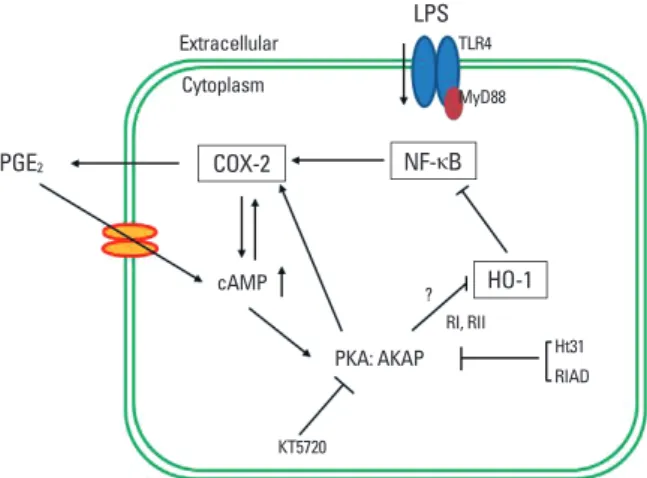
Fig. 7. Schematic representation of the proposed mechanism by which PGE2
signaling amplifies COX-2 expression through an autocrine manner in LPS-stim- ulated macrophages. Upon LPS binding to the toll-like receptor 4 (TLR4), the NF- κB signaling pathway becomes activated, leading to an increase in COX-2 ex- pression, which in turn activates cAMP/PKA/AKAP through the production of PGE2. The PKA-AKAP complex blocks HO-1 activity, which results in enhanced translocation of NF-κB p65 to the nucleus.
LPS
PGE2 COX-2
HO-1 NF-κB Extracellular
cAMP
PKA: AKAP Cytoplasm
TLR4 MyD88
RI, RII
?
KT5720
Ht31 RIAD
assess their individual effects on COX-2 expression. Our results indicate that both PKA RI- and RII-AKAP disruptors inhibit LPS-induced COX-2 expression. This finding is of interest be- cause the PKA RI- or RII-AKAP complex provides a means for spatial and substrate specificity of PKA signaling. RII-AKAP me- diated the PGE2 suppression of LPS-induced tumor necrosis factor alpha (TNF-α) and macrophage inflammatory protein (MIP)-1α, whereas RI-AKAP provided PGE2 enhancement of LPS-induced granulocyte stimulating factor (G-CSF) in RAW 264.7 macrophages.21 Likewise, PGE2 potentiated LPS-induced nitric oxide via RI-interacting AKAPs.22 Taken together, our re- sults suggest that various AKAPs may involve LPS-induced COX-2 expression through different signaling pathways. In ad- dition, PKA RI and RII AKAP disruptors exhibited no additive effects on LPS-induced HO-1 expression (data not shown). This result could be explained by the likely convergence of signaling pathways downstream from PKA RI or RII and AKAP binding.
We investigated the specific role of HO-1 in the positive feed- back loop of COX-2 expression induced by LPS. HO-1 was cho- sen because it represents a major inducible cellular and tissue defense protein against oxidative stress including LPS.12 In ad- dition, induction of HO-1 generally inhibits COX-2 expression, and HO-1 involvement in LPS-induced COX-2 expression has been reported in various cells such as J774 macrophages,23 mouse brain endothelial cells,16 and human umbilical vein en- dothelial cells.24 In this study, HO-1 protein expression was ob- served at 6 h in macrophages after binding with TLR4 ligand.
Furthermore, our results with the HO-1 inhibitor and activator confirmed that LPS-induced COX-2 expression was attenuated by HO-1 induction. However, the relative importance of HO-1 in positive signaling loops of COX-2 expression and the molec- ular mechanism evoked by PKA in these processes are unclear.
Previous reports have proposed that cAMP-PKA signaling is involved in HO-1 induction.25-27 In contrast, our data suggest that suppression of HO-1 by cAMP-PKA signaling is critical for the positive feedback loop of LPS-induced COX-2 expression because a magnifying effect on LPS-induced HO-1 expression was observed in the presence of a PKA inhibitor or AKAP dis- ruptors. These observations are indicative of modulatory effects of cAMP-PKA on HO-1 induction that are context-dependent.
LPS activates NF-κB through TLR4 and leads to the expression of COX-2.28 In the present study, we verified that NF-κB expres- sion and translocation were inhibited by HO-1 induction, sug- gesting that the effect of this small molecule precedes the inhi- bition of COX-2 expression in LPS-activated macrophages.
Our data show that PKA RI and RII AKAPs involve feedback enhancement of LPS-induced COX-2 expression. However, we did not identify the responsible AKAPs because little is known about AKAP-related COX-2 expression induced by LPS stimu- lation. Additional work is necessary to clarify this issue.
In conclusion, this study describes the important role that PGE2-PKA-AKAP signaling plays in enhancing COX-2 expres-
sion induced by LPS through positive feedback in RAW 264.7 macrophages. Furthermore, the attenuation of LPS-induced HO-1 expression achieved by PKA-AKAP signaling contributes to the subsequent augmentation of LPS-induced COX-2 expres- sion through the magnification of LPS-induced NF-κB activa- tion and nuclear translocation in RAW 264.7 macrophages.
Whereas previous studies have established separately that PGE2
enhances COX-2 expression and that PGE2 inhibits LPS-induced HO-1 expression in macrophages, our study directly links these two phenomena for the first time.
ACKNOWLEDGMENTS
We thank Dr. Marc Peters-Golden (University of Michigan Health Systems), and Carlos H. Serezani (Indiana University School of Medicine) for scientific input. Special thanks for Dr.
You-Young Kim for thoughtful support. This research was sup- ported by EMBRI Grants 2010-SN-04 from Eulji University.
REFERENCES
1. Hwang D. Modulation of the expression of cyclooxygenase-2 by fatty acids mediated through toll-like receptor 4-derived signaling pathways. FASEB J 2001;15:2556-64.
2. Harris RE. Cyclooxygenase-2 (cox-2) blockade in the chemopre- vention of cancers of the colon, breast, prostate, and lung. Inflam- mopharmacology 2009;17:55-67.
3. Ren W, Hu L, Hua F, Jin J, Wang Y, Zhu L. Myeloid differentiation protein 2 silencing decreases LPS-induced cytokine production and TLR4/MyD88 pathway activity in alveolar macrophages. Im- munol Lett 2011;141:94-101.
4. Vancheri C, Mastruzzo C, Sortino MA, Crimi N. The lung as a privi- leged site for the beneficial actions of PGE2. Trends Immunol 2004;
25:40-6.
5. Serezani CH, Ballinger MN, Aronoff DM, Peters-Golden M. Cyclic AMP: master regulator of innate immune cell function. Am J Respir Cell Mol Biol 2008;39:127-32.
6. Jarnaess E, Taskén K. Spatiotemporal control of cAMP signalling processes by anchored signalling complexes. Biochem Soc Trans 2007;35:931-7.
7. Bos JL. Epac proteins: multi-purpose cAMP targets. Trends Biochem Sci 2006;31:680-6.
8. Hinz B, Brune K, Pahl A. Prostaglandin E(2) upregulates cyclooxy- genase-2 expression in lipopolysaccharide-stimulated RAW 264.7 macrophages. Biochem Biophys Res Commun 2000;272:744-8.
9. Díaz-Muñoz MD, Osma-García IC, Fresno M, Iñiguez MA. In- volvement of PGE2 and the cAMP signalling pathway in the up- regulation of COX-2 and mPGES-1 expression in LPS-activated macrophages. Biochem J 2012;443:451-61.
10. Hinz B, Brune K, Pahl A. Cyclooxygenase-2 expression in lipopoly- saccharide-stimulated human monocytes is modulated by cyclic AMP, prostaglandin E(2), and nonsteroidal anti-inflammatory drugs. Biochem Biophys Res Commun 2000;278:790-6.
11. Steinert D, Küper C, Bartels H, Beck FX, Neuhofer W. PGE2 poten- tiates tonicity-induced COX-2 expression in renal medullary cells in a positive feedback loop involving EP2-cAMP-PKA signaling. Am
J Physiol Cell Physiol 2009;296:C75-87.
12. Paine A, Eiz-Vesper B, Blasczyk R, Immenschuh S. Signaling to heme oxygenase-1 and its anti-inflammatory therapeutic poten- tial. Biochem Pharmacol 2010;80:1895-903.
13. Ryter SW, Alam J, Choi AM. Heme oxygenase-1/carbon monoxide:
from basic science to therapeutic applications. Physiol Rev 2006;86:
583-650.
14. Chin BY, Otterbein LE. Carbon monoxide is a poison... to microbes!
CO as a bactericidal molecule. Curr Opin Pharmacol 2009;9:490- 500.
15. Suh GY, Jin Y, Yi AK, Wang XM, Choi AM. CCAAT/enhancer-bind- ing protein mediates carbon monoxide-induced suppression of cyclooxygenase-2. Am J Respir Cell Mol Biol 2006;35:220-6.
16. Shih RH, Yang CM. Induction of heme oxygenase-1 attenuates li- popolysaccharide-induced cyclooxygenase-2 expression in mouse brain endothelial cells. J Neuroinflammation 2010;7:86.
17. Leung PO, Wang SH, Lu SH, Chou WH, Shiau CY, Chou TC. Simv- astatin inhibits pro-inflammatory mediators through induction of heme oxygenase-1 expression in lipopolysaccharide-stimulated RAW264.7 macrophages. Toxicol Lett 2011;207:159-66.
18. Waltz P, Carchman EH, Young AC, Rao J, Rosengart MR, Kaczo- rowski D, Zuckerbraun BS. Lipopolysaccaride induces autophagic signaling in macrophages via a TLR4, heme oxygenase-1 depen- dent pathway. Autophagy 2011;7:315-20.
19. Rhee SH, Hwang D. Murine TOLL-like receptor 4 confers lipopoly- saccharide responsiveness as determined by activation of NF kap- pa B and expression of the inducible cyclooxygenase. J Biol Chem 2000;275:34035-40.
20. Chhikara M, Wang S, Kern SJ, Ferreyra GA, Barb JJ, Munson PJ, Danner RL. Carbon monoxide blocks lipopolysaccharide-induced gene expression by interfering with proximal TLR4 to NF-kappaB signal transduction in human monocytes. PLoS One 2009;4:e8139.
21. Wall EA, Zavzavadjian JR, Chang MS, Randhawa B, Zhu X, Hsueh
RC, Liu J, Driver A, Bao XR, Sternweis PC, Simon MI, Fraser ID. Sup- pression of LPS-induced TNF-alpha production in macrophages by cAMP is mediated by PKA-AKAP95-p105. Sci Signal 2009;2:ra28.
22. Kim SH, Serezani CH, Okunishi K, Zaslona Z, Aronoff DM, Peters- Golden M. Distinct protein kinase A anchoring proteins direct prostaglandin E2 modulation of Toll-like receptor signaling in alve- olar macrophages. J Biol Chem 2011;286:8875-83.
23. Olszanecki R, Kurnyta M, Biedroń R, Chorobik P, Bereta M, Mar- cinkiewicz J. The role of heme oxygenase-1 in down regulation of PGE2 production by taurine chloramine and taurine bromamine in J774.2 macrophages. Amino Acids 2008;35:359-64.
24. Liu XH, Pan LL, Yang HB, Gong QH, Zhu YZ. Leonurine attenuates lipopolysaccharide-induced inflammatory responses in human endothelial cells: involvement of reactive oxygen species and NF- kappaB pathways. Eur J Pharmacol 2012;680:108-14.
25. Immenschuh S, Kietzmann T, Hinke V, Wiederhold M, Katz N, Muller-Eberhard U. The rat heme oxygenase-1 gene is transcrip- tionally induced via the protein kinase A signaling pathway in rat hepatocyte cultures. Mol Pharmacol 1998;53:483-91.
26. Kim HJ, Tsoy I, Park MK, Lee YS, Lee JH, Seo HG, Chang KC. Iron released by sodium nitroprusside contributes to heme oxygenase-1 induction via the cAMP-protein kinase A-mitogen-activated pro- tein kinase pathway in RAW 264.7 cells. Mol Pharmacol 2006;69:
1633-40.
27. Park MK, Kang YJ, Ha YM, Jeong JJ, Kim HJ, Seo HG, Lee JH, Chang KC. EP2 receptor activation by prostaglandin E2 leads to induction of HO-1 via PKA and PI3K pathways in C6 cells. Biochem Biophys Res Commun 2009;379:1043-7.
28. Nakao S, Ogata Y, Shimizu-Sasaki E, Yamazaki M, Furuyama S, Sug- iya H. Activation of NFkappaB is necessary for IL-1beta-induced cy- clooxygenase-2 (COX-2) expression in human gingival fibroblasts.
Mol Cell Biochem 2000;209:113-8.