Delayed Hemorrhage of the Hepatic Artery Caused by Biliary
Stenting after Concurrent Chemoradiotherapy
동시항암화학방사선요법 후 담도 스텐트에 의해 발생한 지연성 간동맥 출혈
Joon Ho Cho, MD , Hyoung Nam Lee, MD*
Department of Radiology, Soonchunhyang University Cheonan Hospital, Cheonan, Korea
Neoadjuvant concurrent chemoradiotherapy has been increasingly used to obtain secondary resectability for locally advanced pancreatic cancers. Although most patients require biliary de- compression, only a few studies have investigated the safety of biliary stenting with chemora- diotherapy. Herein, we report a rare case of delayed hemorrhage of the hepatic artery caused by biliary stenting after chemoradiotherapy. The serial follow-up CT demonstrated that the bili- ary stent was approaching the right hepatic artery and eventually caused acute angulation and indentation. Diagnostic catheter angiography revealed contrast extravasation at the right he- patic artery, and endovascular embolization was performed. This report highlights the rele- vance of anatomical deformation after chemoradiotherapy, which can result in fatal complica- tions. Indentation of the hepatic artery caused by biliary stents should be recognized as a warning sign of vascular injury.
Index terms Stents; Pancreatic Neoplasms; Embolization, Therapeutic; Neoadjuvant Therapy;
Chemoradiotherapy
INTRODUCTION
The prognosis of patients with pancreatic cancer is poor, with a current overall 5-year survival of only 7%. At presentation, 40% of patients have distant metastases, and 30%
of patients are classified as having locally advanced pancreatic cancer (LAPC) (1). LAPC can be defined as nonmetastatic unresectable pancreatic cancer, which has one of the following features such as extensive peripancreatic lymph node involvement, encase-
Received May 2, 2019 Revised September 13, 2019 Accepted November 13, 2019
*Corresponding author Hyoung Nam Lee, MD Department of Radiology, Soonchunhyang University Cheonan Hospital, 31 Suncheonhyang 6-gil, Dongnam-gu, Cheonan 31151, Korea.
Tel +82-41-570-3515 Fax +82-41-579-9026 E-mail
radiology2010.hnl@gmail.com This is an Open Access article distributed under the terms of the Creative Commons Attribu- tion Non-Commercial License (https://creativecommons.org/
licenses/by-nc/4.0) which permits unrestricted non-commercial use, distribution, and reproduc- tion in any medium, provided the original work is properly cited.
ORCID iDs Joon Ho Cho https://
orcid.org/0000-0001-7222-6621 Hyoung Nam Lee
https://
orcid.org/0000-0002-2135-9384
ment of superior mesenteric vein or portal venous confluence, or involvement of superior mesenteric artery, celiac axis, inferior vena cava, or aorta (2). Palliative management has been the standard treatment for LAPC because of difficulty in achieving complete surgical resection and subsequent poor clinical outcomes. However, neoadjuvant concurrent chemo- radiotherapy (CCRT) is increasingly applied to obtain secondary resectability (1). Although most patients with LAPC require biliary decompression to safely receive chemotherapeutic agents, there have been only a few reports concerning the safety of self-expandable metal stent (SEMS) with CCRT (3-5). Herein, we report a rare case of delayed hepatic artery hemor- rhage caused by biliary stent after CCRT.
CASE REPORT
A 55-year-old man was admitted for jaundice and weight loss (6 kg over 6 months). The lab- oratory results were as follows: total bilirubin, 23.8 mg/dL (direct bilirubin 21.8 mg/dL); alka- line phosphatase, 801 IU/L; and gamma-glutamyl transferase, 1310 IU/L. These results sug- gested obstructive jaundice. Abdominal CT and MRI revealed a 2.8 cm mass at the uncinated process of the pancreas with biliary obstruction (Fig. 1A). The involvement of the superior mesenteric vein and the 1st and 2nd jejunal branches of the superior mesenteric artery were identified. Endoscopic biliary decompression was performed with an 8.5-F × 5 cm plastic biliary stent (ST-2 Soehendra Tannenbaum Biliary Stent; Cook Medical, Bloomington, IN, USA). The patient was diagnosed with stage III LAPC, and subsequent neoadjuvant CCRT was scheduled.
After induction chemotherapy with gemcitabine, the patient underwent intensity-modulat- ed radiation therapy (Fig. 1B). The total prescribed dose was 4500 cGy, 25 times (180.0 cGy/frac- tion) in one month. However, the neoadjuvant CCRT failed to achieve resectability. After one month of CCRT, the plastic stent was exchanged with a 10 mm × 6 cm metal stent (ComVi Stent;
Taewoong Medical, Seoul, Korea). Additional palliative chemotherapy with gemcitabine and erlotinib was initiated.
After 9 cycles of outpatient chemotherapy (each cycle lasting 3 weeks, total 10 months), the patient was readmitted due to abrupt onset anemia with 50–100 g of hematochezia. Two units of packed red blood cells raised the hemoglobin from 5.7 to 8.1 g/dL. The initial vital signs were as follows: blood pressure, 90/60; heart rate, 72/min; respiratory rate, 16/min; and temper- ature, 36.4°C. Dual-phase CT of the abdomen, pancreas and biliary tract revealed acute angu- lation of the right hepatic artery compressed by the biliary stent without definite contrast ex- travasation (Fig. 1C). A retrospective review of follow-up CT scans at three, six, eight, and ten months after completion of CCRT demonstrated that progressive approximation of SEMS with hepatic artery, eventually result in acute angulation and indentation of the hepatic artery. The endoscopic evaluation revealed bleeding through the opening of the choledochoduodenal fis- tula. Injection of diluted epinephrine (1:10000 dilution) was failed to achieve hemostasis. The follow-up vital signs suggest a continuous blood loss: blood pressure, 80/50; heart rate, 128/
min; respiratory rate, 18/min; and temperature 36.1°C. The patient was referred to the interven- tional clinic for diagnostic angiography and further therapeutic embolization if needed.
Emergent common hepatic angiography using a 5-F catheter (Rosch Hepatic; Cook Medical)
revealed contrast extravasation at the proximal right hepatic artery, which corresponded to the angulated segment on the CT scan (Fig. 1D). Even though coaxial microcatheter system (2.2-F Progreat; Terumo, Tokyo, Japan) was used, it was unable to navigate through the angulated seg- ment to restore distal circulation. A limited embolization was performed with two detachable microcoils (5–6 mm Concerto; Medtronic, Sunnyvale, CA, USA) (Fig. 1E). The follow-up CT af- ter 3 days revealed no evidence of pseudoaneurysm or hepatic infarction. The hematochezia subsided, and the hemoglobin level recovered to 9.1 g/dL after 5 days. After one month, the patient was transferred to another hospital in stable condition.
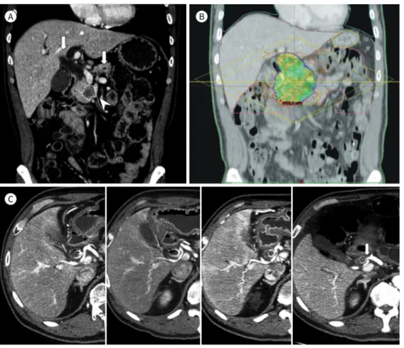
Fig. 1. Delayed hepatic artery hemorrhage caused by biliary stent after concurrent chemoradiotherapy in a 55-year-old man with pancreatic head cancer.
A. Coronal reformatted contrast-enhanced CT reveals heterogeneously enhancing cancer of the pancreatic head (dotted line) with the double duct sign (arrows) and involvement of the superior mesenteric artery (ar- rowhead).
B. Intensity-modulated radiation therapy plan shows dose distributions of the radiotherapy field in the cor- onal view.
C. Contrast-enhanced abdominal CT performed 3, 6, 8, and 10 months after the completion of radiotherapy demonstrate that the biliary stent is approaching the right hepatic artery and eventually caused acute vas- cular angulation and indentation (arrowhead). An abrupt focal narrowing segment (arrow) is noted at the proximal right hepatic artery adjacent to the biliary stent without definite evidence of contrast leakage.
C
A B
DISCUSSION
Most patients with pancreatic cancer develop biliary obstructions and require biliary de- compression. SEMS is preferred over plastic stents to maintain long-term patency for patients with LAPC (6). Delayed (> 30 days) hemorrhage is one of the serious complications after placement of SEMS and constant erosion of the arterial wall by the radial force was thought to be one of the possible cause (7). In this report, we offer insight into the exacerbating effect of CCRT for delayed hemorrhage after SEMS placement.
The pathogenesis of stent migration and hepatic artery injury after CCRT in the present case is unclear. However, two most plausible mechanisms are as follows; 1) direct injury of arterial wall and bile duct, induced by chemoradiotherapy and 2) stent position change caused by radiation-induced fibrosis (RIF). According to the studies assessing the toxicity of chemo- radiotherapy, direct damages to arterial wall, pancreas and biliary systems have been de- scribed (4, 8).
To the best of our knowledge, there has been no report that chemotherapy resulted in stent migration. On the other hand, the distance of intersessional stent position change during ra- diotherapy was reported to be up to 4 cm in previous study, focused on the use of biliary stents as a targeting surrogate for the pancreatic cancer (3). There is no established explanation how radiotherapy affected stent migration, but RIF may play a role in anatomical change (9). RIF is one of the major late complication of radiotherapy and usually occurs 4 to 12 months after treatment, which is a comparable period with the present case report (9). When considering the field of radiotherapy, RIF appears to have caused progressive approximation of bile duct to the hepatic artery (Fig. 1C).
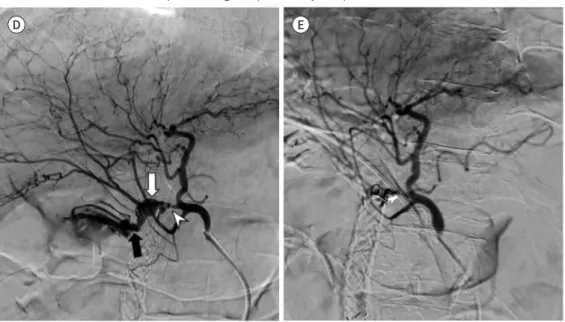
Fig. 1. Delayed hepatic artery hemorrhage caused by biliary stent after concurrent chemoradiotherapy in a 55-year-old man with pancreatic head cancer.
D. Common hepatic angiography reveals massive contrast extravasation at the proximal portion of the right hepatic artery (white arrow), which corresponds to the abrupt focal narrowing segment on CT (arrowhead).
There is direct contrast leakage into the duodenum via the choledochoduodenal fistula (black arrow).
E. Selective embolization of the proximal right hepatic artery was performed with two microcoils.
D E
According to previous studies, pseudoaneurysm or extravasation near or inside the biliary stent on CT angiography was the characteristic finding which suggest hemorrhage in most cases (7). On the other hand, there was no direct CT finding of hemorrhage in present case, so it was difficult to recognize the warning signs despite periodic imaging workups. The bleed- ing might have been intermittent due to extrinsic compression by SEMS. Retrospective care- ful review of follow-up images identified progressive approximation of SEMS to the hepatic artery. The process of anatomical deformation was too slow to be recognized without inten- sive attention to the anatomy included in the radiotherapy field.
The endovascular treatment method in the present case was not optimal because only prox- imal embolization was achieved with microcoils. The guidewire continued to penetrate into the disrupted right hepatic artery through the mesh of the SEMS. The use of n-butyl cyanoac- rylate was also considered, but the proximal segment from the hepatic confluence was too short to achieve precise handling. Since retrograde flow may persist from intrahepatic or ex- trahepatic collateral pathway, additional embolization is often required (10). In our report, active extravasation from retrograde flow was not evident during follow-up studies.
In summary, we reported a rare case of delayed hepatic artery hemorrhage caused by bili- ary stent after CCRT. This case report highlights the importance of clinicians paying careful attention to patients with biliary stents who are undergoing chemoradiotherapy. Anatomical deformation after chemoradiotherapy is one of the potential causes of unexpected life-threat- ening complications. Indentation of the hepatic artery caused by biliary stents should be rec- ognized as a warning sign of impending vascular rupture.
Author Contributions
Writing—original draft, all authors; and writing—review & editing, all authors.
Conflicts of Interest
The authors have no potential conflicts of interest to disclose.
REFERENCES
1. Huguet F, Girard N, Guerche CS, Hennequin C, Mornex F, Azria D. Chemoradiotherapy in the management of locally advanced pancreatic carcinoma: a qualitative systematic review. J Clin Oncol 2009;27:2269-2277 2. Willett CG, Czito BG, Bendell JC, Ryan DP. Locally advanced pancreatic cancer. J Clin Oncol 2005;23:4538-
4544
3. Chu KY, Eccles CL, Brunner TB. Endobiliary stent position changes during external-beam radiotherapy. J Med Imaging Radiat Sci 2015;46:57-64
4. Tsuji Y, Yoshimura H, Uto F, Tamada T, Iwata K, Tamamoto T, et al. Physical and histopathological assess- ment of the effects of metallic stents on radiation therapy. J Radiat Res 2007;48:477-483
5. Lee JK, Kwack WK, Lee SH, Jung JH, Kwon JH, Han IW, et al. Effect of external beam radiotherapy on pa- tency of uncovered metallic stents in patients with inoperable bile duct cancer. Hepatobiliary Pancreat Dis Int 2014;13:423-427
6. Boulay BR, Parepally M. Managing malignant biliary obstruction in pancreas cancer: choosing the appro- priate strategy. World J Gastroenterol 2014;20:9345-9353
7. Hyun D, Park KB, Hwang JC, Shin BS. Delayed, life-threatening hemorrhage after self-expandable metallic biliary stent placement: clinical manifestations and endovascular treatment. Acta Radiol 2013;54:939-943 8. Torrisi JM, Schwartz LH, Gollub MJ, Ginsberg MS, Bosl GJ, Hricak H. CT findings of chemotherapy-induced
toxicity: what radiologists need to know about the clinical and radiologic manifestations of chemotherapy toxicity. Radiology 2011;258:41-56
9. Straub JM, New J, Hamilton CD, Lominska C, Shnayder Y, Thomas SM. Radiation-induced fibrosis: mecha- nisms and implications for therapy. J Cancer Res Clin Oncol 2015;141:1985-1994
10. Charnsangavej C, Chuang VP, Wallace S, Soo CS, Bowers T. Angiographic classification of hepatic arterial collaterals. Radiology 1982;144:485-494
동시항암화학방사선요법 후 담도 스텐트에 의해 발생한 지연성 간동맥 출혈
조준호 · 이형남*
국소진행성 췌장암 환자에서 이차적 근치절제 가능성을 얻기 위해 수술 전 동시항암화학방 사선요법이 점차 많이 이용되고 있다. 대부분의 환자들에게 담도의 감압이 필요함에도 불구 하고, 담도 스텐트와 동시항암화학방사선요법을 함께 시행하는 안전성에 대한 연구는 많지 않다. 저자들은 동시항암화학방사선요법 후 담도 스텐트에 의해 발생한 지연성 간동맥 출혈 의 드문 증례를 보고하는 바이다. 추적 관찰 CT에서 담도 스텐트와 우간동맥과의 거리가 점점 가까워졌고, 결국 스텐트가 간동맥을 압박하여 간동맥에 심한 굴곡이 생겼다. 카테터 혈관조 영술에서 우간동맥에 조영제의 혈관 외 유출이 발견되어 혈관 내 색전술을 이용하여 치료하 였다. 본 증례 보고는 동시항암화학방사선요법 후 발생하는 해부학적 변형이 심각한 합병증의 원인이 될 수도 있음을 강조하고 있다. 간동맥이 담도 스텐트에 의해 압박되는 소견은 혈관 손상의 경고 신호로서 주의가 필요하다.
순천향대학교 천안병원 영상의학과