© 2016 The Korean Academy of Medical Sciences.
This is an Open Access article distributed under the terms of the Creative Commons Attribution Non-Commercial License (http://creativecommons.org/licenses/by-nc/4.0) which permits unrestricted non-commercial use, distribution, and reproduction in any medium, provided the original work is properly cited.
pISSN 1011-8934 eISSN 1598-6357
Factors Related to Radiation Exposure during Lumbar Spine Intervention
Fluoroscopy guidance is useful to confirm anatomical landmark and needle location for spine intervention; however, it can lead to radiation exposure in patients, physicians, and medical staff. Physicians who used fluoroscopy should be cognizant of radiation exposure and intend to minimize radiation dose. We retrospectively reviewed three lumbar spine intervention procedures (nerve root block, medial branch block, and facet joint block) at our institution between June and December, 2014. We performed 268 procedures on 220 patients and found significant difference in radiation dose between two groups classified by performing physicians. The physician who controlled the fluoroscopy unit directly used significantly shorter fluoroscopy (6 seconds) that resulted in a smaller radiation dose (dose area product [DAP] 0.59 Gy∙cm2) than the physician supervising the radiographer controlling the fluoroscopy unit (72 seconds, DAP 5.31 Gy∙cm2, P < 0.001). The analysis indicates that the difference in fluoroscopy time depends on whether a physician or a radiographer controls the fluoroscopy unit.
Keywords: Back Pain; Radiography, Interventional; Fluoroscopy; Radiation Dosage;
Radiographic Magnification Moon Hyung Choi, Byung Gil Choi,
Seung Eun Jung, and Jae Young Byun Department of Radiology, Seoul St. Mary’s Hospital, College of Medicine, The Catholic University of Korea, Seoul, Korea
Received: 28 April 2015 Accepted: 1 September 2015 Address for Correspondence:
Byung Gil Choi, MD
Department of Radiology, Seoul St. Mary’s Hospital, College of Medicine, The Catholic University of Korea, 222 Banpo-daero, Seocho-gu, Seoul 06591, Korea
E-mail: [email protected]
http://dx.doi.org/10.3346/jkms.2016.31.S1.S55 • J Korean Med Sci 2016; 31: S55-58
INTRODUCTION
Back pain is a common problem that afflicts 70%-85% of people in their lifetime and 15%-45% annually (1). Patients hesitate to undergo surgery; in addition, surgical treatment is recommend- ed to only indicated patients due to various sources of pain and inconstant surgical results (2,3). These causes increased use of nonsurgical and conservative treatment such as spine interven- tion to avoid surgery. Many interventional procedures such as epidural steroid injection, facet joint block, medial branch block, nerve root block, and sacroiliac joint injection are used to re- lieve back pain (4).
Most interventional procedures use X-ray to identify imaging landmarks and confirm the precise location for percutaneously inserted needle under fluoroscopy or C-arm guidance (5). “As low as reasonably achievable (ALARA)” principles should be respected when X-ray is used because excessive radiation to a patient or a physician can cause radiation injury or stochastic effect such as neoplasm and genetic mutation (6,7). Several ar- ticles reported radiation dose for spine intervention (8-12) and some revealed factors that affect radiation dose; however, no study has investigated the effect of the fluoroscopy unit control- ler. Either the physician or radiographer can control the fluo- roscopy unit in fluoroscopy-guided procedures. The aim of this study was to evaluate a difference of fluoroscopy time and radi- ation dose depending on medical staff operating the fluorosco-
py unit. We also evaluated other factors related to radiation dose in spine interventions including patients’ body habitus, a type of procedure, and image magnification.
MATERIALS AND METHODS
We performed 500 spine interventional procedures in the same fluoroscopy unit at our institution between June and Decem- ber, 2014. This study included the three most common inter- ventional procedures of nerve root block, medial branch block, and facet joint block performed at the lumbar spine; however, we excluded procedures without a proper dose report or two or more different types of procedures done simultaneously. Even- tually 268 procedures in 220 patients were included in this study;
86 procedures were performed on 78 men and 182 procedures on 142 women. The mean age of patients was 63 years with range of 22-93 years and the mean body mass index (BMI) was 24.5 kg/m2 with range of 16.5-39.5 kg/m2.
All procedures were performed by two physicians with three- year (physician A) and one-year experience (physician B). When physician A performed the procedures, the fluoroscopy unit was controlled by a radiographer in the control room according to the physician’s instructions; however, physician B controlled the unit directly without assistance in the examination room.
They used the same fluoroscopic unit (Axiom Artis, Siemens Healthcare, Erlangen, Germany). Procedures at two or more SPECIAL ARTICLE
Choi MH, et al. • Radiation Exposure in Spine Intervention
S56 http://jkms.org http://dx.doi.org/10.3346/jkms.2016.31.S1.S55 levels of the lumbar spine were done simultaneously on some
patients and some patients underwent procedures several times.
We recorded treatment related factors such as multiplicity of treated region, procedure type, and physician who performed the procedure. We collected radiation dose data measured by the built-in dose area product (DAP) meter and fluoroscopy time from radiation dose reports generated by the fluoroscopy unit. We also reviewed the fluoroscopic images of all procedures to evaluate if X-ray beam collimation or image magnification was used. Treatment responses for procedures were collected from medical records to evaluate outcomes. A lack of detailed medical records on pain assessment required that treatment responses be divided into improvement or no improvement in symptoms. Our institution does not use spot images for spine intervention; therefore, the number of spot images was not re- corded.
Descriptive statistics including median, range and interquar- tile interval of fluoroscopy time and DAP (Gy∙cm2) were calcu- lated. We used nonparametric analysis as the data did not ap- proximate a normal distribution. Spearman correlation test as- sessed the correlation of radiation dose and patient’s BMI as well as radiation dose and fluoroscopy time. We classified all procedures into two groups by the performing physician; Group A and B by physician A and B, respectively. A χ2 test compared the frequency of several factors between two groups. A Mann- Whitney U-test compared fluoroscopy time and radiation dose between two groups as well as analyzed differences in fluoros- copy time and radiation dose according to multiplicity of the treated regions, response after treatment in each group, and a difference in radiation dose according to magnification. A Krus- kal-Wallis H test compared differences of fluoroscopy time and radiation dose among the type of procedures. A P value < 0.05 was considered statistically significant.
Ethics statement
The institutional review board at our hospital approved this study
(IRB No. KC15RISI0140) and informed consent was waived due to the retrospective design of the study.
RESULTS
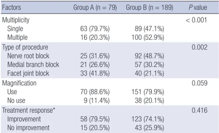
We analyzed 268 procedures by physician A (n = 79) and physi- cian B (n = 189). The most common procedure was a nerve root block. Physician B performed significantly more procedures at multiple levels of the spine. Pain relief was indicated for 181 pro- cedures, no pain relief for 58 procedures, and no medical re- cords of treatment response for 29 procedures. There was no significant difference in frequency of symptom improvement between two groups (P = 0.416). Table 1 summarizes the num- ber of procedures according to multiple factors.
The mean DAP and fluoroscopy time of all procedures were 3.3 ± 5.4 Gy∙cm2 (range, 0.05-37.01 Gy∙cm2) and 39.1 ± 59.08 seconds (range, 1-457 seconds), respectively. In all procedures, fluoroscopy time was significantly and positively correlated to radiation dose (correlation coefficient = 0.886, P < 0.001) (Fig.
1). BMI, known to affect radiation dose, did not show a signifi- cant correlation to radiation dose (correlation coefficient = 0.108, P = 0.143).
There were significant differences in radiation dose and fluo- roscopy time between two groups. Group A, by physician A
Table 1. Treatment related factors in lumbar spine interventions in each group Factors Group A (n = 79) Group B (n = 189) P value Multiplicity
Single Multiple
63 (79.7%) 16 (20.3%)
89 (47.1%) 100 (52.9%)
< 0.001
Type of procedure Nerve root block Medial branch block Facet joint block
25 (31.6%) 21 (26.6%) 33 (41.8%)
92 (48.7%) 57 (30.2%) 40 (21.1%)
0.002
Magnification Use
No use 70 (88.6%)
9 (11.4%) 151 (79.9%) 38 (20.1%)
0.059
Treatment response*
Improvement
No improvement 58 (79.5%)
15 (20.5%) 123 (74.1%) 43 (25.9%)
0.416
n, number of procedures; parenthesis, percentage in each group. *There was no medi- cal record about treatment response in 29 patients.
Table 2. Comparison of fluoroscopy time and dose area product (DAP) according to variables
Time & dose Group A* Group B* P value
Fluoroscopy time (sec) 72.0 (33.8-114.5) 6.0 (3.0-20.3) < 0.001 DAP (Gy ∙ cm2) 5.31 (1.72-11.28) 0.59 (0.26-1.33) < 0.001 Data presented as median (interquartile interval). *Group A included procedures by a physician who instructed radiographers to control the fluoroscopy unit. Group B in- cluded procedures by physician B who controlled the unit directly. Much less fluoros- copy time and DAP were recorded when the physician controlled the unit directly.
Fig. 1. Patient radiation dose versus fluoroscopy time. Scatter plots show a relation- ship between radiation dose and fluoroscopy time. There is a positive correlation be- tween fluoroscopy time and radiation dose (correlation coefficient: 0.886, P < 0.001).
DAP (Gy∙cm2)
Fluoroscopy time (sec)
0 100 200 300 400 500 40
30
20
10
0
Choi MH, et al. • Radiation Exposure in Spine Intervention
http://jkms.org S57
http://dx.doi.org/10.3346/jkms.2016.31.S1.S55
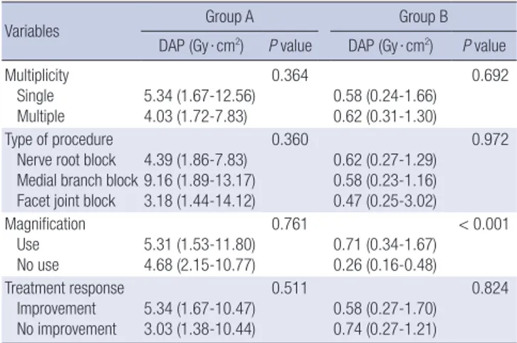
who did not control the fluoroscopy unit, showed a significantly longer fluoroscopy time and higher radiation dose than Group B (P < 0.001) (Table 2). There was significant difference in fluo- roscopy time according to type of procedure in Group A; how- ever, there was no difference in radiation dose in both groups.
Magnification caused a significantly higher radiation dose in Group B. There was no significant difference in radiation dose in each group according to multiplicity of treated regions and treatment response. Tables 3 and 4 summarizes fluoroscopy time and radiation dose (= DAP) according to all evaluated fac- tors in two groups.
DISCUSSION
The routine use of X-ray in fluoroscopy-guided minimally inva- sive procedures has made radiation dose exposure an impor- tant issue. Analysis in this study indicated that fluoroscopy time and radiation dose were significantly decreased when the phy- sician controlled the fluoroscopy unit directly comparing with when the physician supervising the radiographer controlling the fluoroscopy unit. Additionally radiation dose increased when image magnification is applied.
The results showed significant differences in fluoroscopy time and radiation dose between two physicians that corresponded to those of a previous study (8). However, a previous study indi- cated that more radiation dose and more fluoroscopy time were recorded for physicians who had less procedural experience. In our study, the less experienced physician (physician B) used less X-ray for procedures than the more experienced physician (physician A). An important difference between two groups was that different personnel controlled the fluoroscopy unit.
Physician A allowed a radiographer to control the unit and phy- sician B controlled the unit without assistance. When a physi- cian controlled the fluoroscopy unit, they could immediately turn on or off the machine without delay and fluoroscopy time could be shortened. Our study showed the direct control of the fluoroscopy unit by a physician was more influential in decreas-
ing fluoroscopy time than the physicians’ experience. It is also important to note that physician B performed procedures prop- erly and that there was no difference in treatment responses between two groups.
Two physicians seemed to have different level of awareness about radiation exposure that made a potent difference in fluo- roscopy time and radiation dose. This supposition was support- ed the fact that most images during physician A’s procedures contained the physician’s hands and those during physician B’s procedures did not. It is imperative that physicians who con- duct fluoroscopy-guided examinations make efforts to prevent radiation from directly contacting their hands. Awareness about radiation exposure itself was well-known as an important factor to decrease radiation dose (13,14).
Procedures with image magnification caused a significantly higher radiation dose than without magnification in Group B.
This result showed that magnification caused more radiation exposure. Magnification is necessary when a small area is ex- amined and a detailed structure is required; however, image magnification should be used selectively. Unlike Group B, there was no significant difference in Group A for radiation dose in examinations with or without magnification. It could be explain- ed by that fluoroscopy time had a stronger effect than magnifi- cation on the radiation dose for Group A.
Fluoroscopy time was different according to type of proce- dure in Group A, but radiation dose was similar. A precise com- parison of the results of our study and previous studies was im- possible as previous studies included different procedures (8,12);
however, the results suggest a commonality in the type of spine intervention that helps determine fluoroscopy time. A differ- ence in fluoroscopy time could be explained by the familiarity or difficulty of the procedures.
Radiation exposure from spot images is distinct and addition- al to that from fluoroscopy; therefore, spot images during fluo- roscopic procedures increased radiation exposure. Prior efforts Table 3. Comparison of fluoroscopy time (sec) according to variables in two groups
Variables
Group A Group B
Fluoroscopy time
(sec) P value Fluoroscopy time (sec) P value Multiplicity
Single Multiple
71.0 (32.5-121.5) 73.0 (44.0-100.0)
0.734
6.0 (3.0-20.0) 6.0 (3.0-24.0)
0.632
Type of procedure Nerve root block Medial branch block Facet joint block
50.0 (32.0-91.0) 89.5 (69.25-146.25) 71.0 (18.0-134.0)
0.043
7.0 (4.0-19.25) 5.0 (2.0-20.0) 6.5 (2.25-30.0)
0.336
Treatment response Improvement
No improvement 73.0 (34.5-104.0) 62.0 (14.5-132.0)
0.946
6.5 (3.0-28.5) 3.0 (2.0-16.0)
0.585
Data presented as median (interquartile interval).
Table 4. Comparison of DAP (Gy ∙ cm2) according to variables in two groups
Variables Group A Group B
DAP (Gy ∙ cm2) P value DAP (Gy ∙ cm2) P value Multiplicity
Single
Multiple 5.34 (1.67-12.56) 4.03 (1.72-7.83)
0.364
0.58 (0.24-1.66) 0.62 (0.31-1.30)
0.692
Type of procedure Nerve root block Medial branch block Facet joint block
4.39 (1.86-7.83) 9.16 (1.89-13.17) 3.18 (1.44-14.12)
0.360
0.62 (0.27-1.29) 0.58 (0.23-1.16) 0.47 (0.25-3.02)
0.972
Magnification Use No use
5.31 (1.53-11.80) 4.68 (2.15-10.77)
0.761
0.71 (0.34-1.67) 0.26 (0.16-0.48)
< 0.001
Treatment response Improvement
No improvement 5.34 (1.67-10.47) 3.03 (1.38-10.44)
0.511
0.58 (0.27-1.70) 0.74 (0.27-1.21)
0.824
Data presented as median (interquartile interval).
Choi MH, et al. • Radiation Exposure in Spine Intervention
S58 http://jkms.org http://dx.doi.org/10.3346/jkms.2016.31.S1.S55 have been made to reduce radiation dose by the use of last im-
age hold in other fluoroscopic examinations (15,16). In our in- stitution, a spot image was not taken from all spine interven- tions. The experience in our institution indicated that preferable outcomes could be obtained after spine interventional proce- dures without spot images. We believed that common spine in- terventional procedures should be performed without spot im- ages if the fluoroscopy unit has a last image hold function.
Our study has several limitations. First, we could not perform a cross comparison due to the retrospective design of this study.
There is a possibility that differences between two physicians might be caused by individual preferences; however this is doubt- ful due to the significant differences between the two methods of a fluoroscopy unit controlled by physician directly and that controlled by another medical staff. Second, only two physi- cians performed all interventional procedures. Individual skill and experience could relate to radiation dose and fluoroscopy time rather than awareness. The physician who used fluorosco- py for a longer time had more experience and indicated no dif- ference in treatment response; therefore, it seemed more rea- sonable that fluoroscopy time and radiation dose differences were due to the physician’s habit and awareness. Third, we could not evaluate the effect of X-ray collimation to radiation dose due to the small number of procedures that used a not-colli- mated X-ray beam.
In conclusion, there is a significant difference of radiation dose depending on whether a physician or a radiographer op- erates the fluoroscopy unit. This can be explained by that direct control of the fluoroscopy unit can lead to shorten fluoroscopy time. In addition, image magnification increases radiation dose.
The selective use of fluoroscopy and non-use of magnification does depend upon the physician, therefore, physicians should perform procedures using fluoroscopy carefully in order to min- imize radiation exposure.
DISCLOSURE
The authors have no potential conflicts of interest to disclose.
AUTHOR CONTRIBUTION
Conception and design of the study: Choi BG, Jung SE. Data collection and analysis: Choi BG, Choi MH. Writing and revi- sion: Choi MH. Supervision of study: Byun JY. Manuscript ap- proval: all authors.
ORCID
Moon Hyung Choi http://orcid.org/ 0000-0001-5962-4772 Byung Gil Choi http://orcid.org/0000-0002-2950-2069
Seung Eun Jung http://orcid.org/0000-0003-0674-5444 Jae Young Byun http://orcid.org/0000-0002-0038-3860
REFERENCES
1. Andersson GB. Epidemiological features of chronic low-back pain. Lan- cet 1999; 354: 581-5.
2. Deyo RA. Back surgery--who needs it? N Engl J Med 2007; 356: 2239-43.
3. Chou R, Baisden J, Carragee EJ, Resnick DK, Shaffer WO, Loeser JD. Sur- gery for low back pain: a review of the evidence for an American Pain So- ciety Clinical Practice Guideline. Spine 2009; 34: 1094-109.
4. Fenton DS, Czervionke LF. Image-guided spine intervention. 1st ed. Phila- delphia, PA: Saunders; 2003, p9-50.
5. Iannuccilli JD, Prince EA, Soares GM. Interventional spine procedures for management of chronic low back pain-a primer. Semin Intervent Radiol 2013; 30: 307-17.
6. Hricak H, Brenner DJ, Adelstein SJ, Frush DP, Hall EJ, Howell RW, McCol- lough CH, Mettler FA, Pearce MS, Suleiman OH, et al. Managing radia- tion use in medical imaging: a multifaceted challenge. Radiology 2011;
258: 889-905.
7. Jones AK, Balter S, Rauch P, Wagner LK. Medical imaging using ionizing radiation: optimization of dose and image quality in fluoroscopy. Med Phys 2014; 41: 014301.
8. Zhou Y, Singh N, Abdi S, Wu J, Crawford J, Furgang FA. Fluoroscopy radia- tion safety for spine interventional pain procedures in university teaching hospitals. Pain Physician 2005; 8: 49-53.
9. Botwin KP, Thomas S, Gruber RD, Torres FM, Bouchlas CC, Rittenberg JJ, Rao S. Radiation exposure of the spinal interventionalist performing fluo- roscopically guided lumbar transforaminal epidural steroid injections.
Arch Phys Med Rehabil 2002; 83: 697-701.
10. Manchikanti L, Cash KA, Moss TL, Pampati V. Radiation exposure to the physician in interventional pain management. Pain Physician 2002; 5:
385-93.
11. Botwin KP, Freeman ED, Gruber RD, Torres-Rames FM, Bouchtas CG, Sanelli JT, Hanna AF. Radiation exposure to the physician performing flu- oroscopically guided caudal epidural steroid injections. Pain Physician 2001; 4: 343-8.
12. Hanu-Cernat DE, Duarte R, Raphael JH, Mutagi H, Kapur S, Senthil L.
Type of interventional pain procedure, body weight, and presence of spi- nal pathology are determinants of the level of radiation exposure for fluo- roscopically guided pain procedures. Pain Pract 2012; 12: 434-9.
13. Frederick-Dyer KC, Faulkner AR, Chang TT, Heidel RE, Pasciak AS. On- line training on the safe use of fluoroscopy can result in a significant de- crease in patient dose. Acad Radiol 2013; 20: 1272-7.
14. Yu SK, Cheung YK, Chan TL, Kung CM, Yuen MK. Reduction of radiation dose to patients undergoing barium enema by dose audit. Br J Radiol 2001; 74: 162-5.
15. Fefferman NR, Sabach AS, Rivera R, Milla S, Pinkney LP, Strubel NA, Babb J. The efficacy of digital fluoroscopic image capture in the evaluation of vesicoureteral reflux in children. Pediatr Radiol 2009; 39: 1179-87.
16. Delichas M, Psarrakos K, Molyvda-Athanassopoulou E, Giannoglou G, Sioundas A, Hatziioannou K, Papanastassiou E. Radiation exposure to cardiologists performing interventional cardiology procedures. Eur J Ra- diol 2003; 48: 268-73.