PGHN
Original Article
Changing Prevalence of Helicobacter pylori Infections in Korean Children with Recurrent Abdominal Pain
Kyung Mi Jang, Byung-Ho Choe, Jae Young Choe, Suk Jin Hong, Hyo Jung Park, Mi Ae Chu, Seung Man Cho and Jung Mi Kim
Department of Pediatrics, Kyungpook National University School of Medicine, Daegu, Korea
Purpose: The aim of this study is to investigate the changing prevalence rate of Helicobacter pylori infection in children, of different age groups, with recurrent abdominal pain over a 10-year period.
Methods: Children with recurrent abdominal pain who visited the pediatric outpatient clinic at university hospital were screened for H. pylori. Children were divided into 3 age categories of 4-5, 6-11, and 12-16 years. To study the changes in the annual prevalence rates of H. pylori infection, the study period was divided into 3 time periods: 2004-2007, 2008-2010, and 2011-2014. Urea breath test was performed for all children aged 4-16 years, with a cut-off value of 4.0‰ for children aged ≥6 years and 7‰ for children aged <6 years.
Results: A total of 2,530 children (1,191 boys) with a mean age of 10.0±3.0 years (range, 4.0-16.9 years) were included in the study. The total prevalence of H. pylori infection was 7.4% (187/2,530). The prevalence rate of H. pylori infection in children with recurrent abdominal pain was 8.0% (70/873) in 2004-2007, 7.7% (51/666) in 2008-2010, and 6.7%
(66/991) in the 2011-2014. Nevertheless, a significant difference was observed in the prevalence rate between chil- dren <12 years old and ≥12 years of age (p=0.018).
Conclusion: The prevalence of H. pylori infection in Korean children with recurrent abdominal pain was 7.4%, showing no significant decrease in the last 11 years; however, the prevalence rate in children <12 years old was significantly lower than that in those ≥12 years old.
Key Words: Helicobacter, Functional gastrointestinal disorder, Breath test, Endoscopy, Child
Received:September 22, 2014, Revised:October 13, 2014, Accepted:October 21, 2014
Corresponding author: Byung-Ho Choe, Department of Pediatrics, Kyungpook National University School of Medicine, 130, Dongdeok-ro, Jung-gu, Daegu 700-721, Korea. Tel: +82-53-200-5704, Fax: +82-53-425-6683, E-mail: [email protected]
Copyright ⓒ 2015 by The Korean Society of Pediatric Gastroenterology, Hepatology and Nutrition
This is an openaccess article distributed under the terms of the Creative Commons Attribution NonCommercial License (http://creativecommons.org/licenses/by-nc/3.0/) which permits unrestricted noncommercial use, distribution, and reproduction in any medium, provided the original work is properly cited.
INTRODUCTION
Helicobacter pylori is a gram-negative bacterium present in more than 50% of the world’s population;
and it is one of the most common pathogen in chil-
dren worldwide [1]. Although H. pylori infection is linked to the development of chronic gastritis and peptic ulcer in children with long exposure to in- fection [2], most of the infected children remain asymptomatic.
The prevalence of H. pylori differs according to in age, region, and race. The prevalence of H. pylori in- fection increases with age and has been reported to be variable in both developed and developing coun- tries, linking the prevalence of H. pylori infection with the socioeconomic status [3]. In addition, H. py- lori first infection is usually acquired during early childhood, rarely spontaneously eradicated, suggest- ing that changes in childhood prevalence can sensi- tively reflect changes in the overall prevalence.
According to Malaty et al.’s study [4-6], the rate of H.
pylori infection is 60-80%, showing a significant dif- ference in Saudi Arabia, India, and Vietnam, against the developed countries such as the United States, Australia, or France, with infection rates of 20-25%
in the 20s-30s even in 1991. Similarly, Malaty and several other studies [4-6] have reported the follow- ing prevalence rates for children in different coun- tries in 2006: 35% in Russia, 20% in China and Poland, 12% in South Korea and the United States, and less than 10% in France, Belgium, and Finland.
Although H. pylori infection rates show a declining trend worldwide, high rates of infection are still present in developing countries. In line with these evidences, the prevalence rate of H. pylori in children in the 2000s has been considered to be lower than the rate in the 1990s; however, there is almost no re- search on the prevalence rates in Korean children in the last 10 years. In this study, we investigated changes in the prevalence rates in children with re- current abdominal pain using the urea breath test (UBT), currently the most accurate screening modality.
MATERIALS AND METHODS
Materials
The UBT was performed for children with re- current abdominal pain who visited the pediatric outpatient clinics at Kyungpook National University Hospital and Kyungpook National University Children’s Hospital from January 2004 to May 2014.
Recurrent abdominal pain by the Rome II criteria, es- tablished in 1999, was defined as abdominal pain in
children aged 4-16 years that interferes with daily life for more than 3 months [7].
If children tested positive by the UBT (UBiT-IR 300; Otsuka Electronics Co., Ltd., Osaka, Japan), en- doscopy was recommended for confirmation of H.
pylori; however, endoscopy was not performed when UBT results were not more than 3‰ point over the cut-off value due to the high number of false-positives.
In these cases under 12-year-old, observation was recommended. Though positive by UBT, both a neg- ative CLO test result and a negative histopathological examination results by endoscopy were regarded as negative for H. pylori.
Patients with abdominal pain who presented with peptic ulcer or gastrointestinal bleeding were ex- cluded from this study as well as patients for whom the test was performed owing to other indications such as iron deficiency anemia, idiopathic thrombo- cytopenia purpura, growth retardation, or chronic urticarial.
Methods
A retrospective cohort of 2,530 children who un- derwent the UBT and presented with recurrent ab- dominal pain was studied for determining the preva- lence of H. pylori by age and year. Not only identifying UBT titers, we established endoscopic remarks, per- formed the CLO test, and analyzed histopathological findings when endoscopy was done.
Changes in the prevalence of H. pylori infection were determined according to year, gender, and age.
The children were divided into 3 age groups: 4-5, 6-11, and 12-16 years. To determine the changes in the annual prevalence of H. pylori infection, the study period was classified into 3 time periods: 2004-2007, 2008-2010, and 2011-2014.
Prevalence trends were compared between the groups of children aged 4-11 years and 12-16 years, assuming that the prevalence will be further de- creased in younger children.
In order to reduce false positive UBT results, a 4.0‰
cut-off value was applied to children ≥6 years and a 7‰ cut-off value to children <6 years [8]. Since en- doscopy results performed in patients with a UBT tit-
Table 2. The Prevalence Helicobacter pylori Infections in Children Aged 4-11 and 12-16 Years
Year Age (y)
p-value
4-11 12-16
2004-2007 2008-2010 2011-2014
Mean prevalence of each age group
7.1 (44/622) 6.2 (28/450) 6.2 (40/641) 6.5 (112/1,713)
10.4 (26/251) 10.7 (23/216) 7.4 (26/350) 9.2 (75/817)
0.13 0.06 0.51 0.018 Values are presented as percentage (number/total number).
Table 1. The Prevalence of Helicobacter pylori Infections in Children with Recurrent Abdominal Pain according to Age Groups (p=0.04) and Time Periods (p=0.27) by the Urea Breath Test (UBT)
Year Age (y)
Mean prevalence of each period
4-5 6-11 12-16
2004-2007 2008-2010 2011-2014
Mean prevalence of each age group
7.6 (9/118) 7.1 (6/84) 7.6 (10/131) 7.5 (25/333)*
6.9 (35/504) 6.0 (22/366) 5.9 (30/510) 6.3 (87/1,380)*
10.4 (26/251) 10.7 (23/216) 7.4 (26/350) 9.2 (75/817)*
8.0 (70/873)† 7.7 (51/666)† 6.7 (66/991)† 7.4 (187/2,530) Values are presented as percentage (number/total number).
UBT cut-off value: <7‰ for those aged <6 years and <4‰ for those aged 6-16 years.
*p=0.04 (each age group), †p=0.27 (each period).
er near the cut-off value was shown to be negative, endoscopy was only performed for children aged 4-11 who had 3‰ points higher than the cut-off value. Prevalence of H. pylori infection was de- termined again using cut-off values that had been modified to 10.0‰ for those aged 4-5 years; 7.0‰, for those aged 6-11 years; and 4.0‰, for those aged 12-16 years to further reduce the false positive rates.
Statistical analysis
IBM SPSS Statistics ver. 21.0 (IBM Co., Armonk, NY, USA) and the general linear model were used for statistical analysis. A p-value of ≤0.05 was consid- ered statistically significant.
RESULTS
A total of 2,530 children (1,191 boys, 1,339 girls;
mean age 10.0±3.0 years; range, 4.0-16.9 years) un- derwent the UBT, who had presented with recurrent abdominal pain from January 2004 to May 2014.
Prevalence rate by different age group and time periods
The total infection prevalence rate was 7.4%
(187/2,530) with an overall prevalence of 7.6% and 7.2% in boys and girls, respectively; there was no dif- ference in the prevalence in boys and girls over time.
The prevalence rate in children was 8.0% in 2004-2007, 7.7% in 2008-2010, and 6.7% in 2011-2014, showing no significant difference among the time periods.
The prevalence rate in the different age groups was 7.5% for those aged 4-5 years, 6.3% for those aged 6-11 years, and 9.2% for those aged 12-16 years, which showed a significant difference (p=0.04) (Table 1). No significant relation was observed be- tween increasing age groups and prevalence.
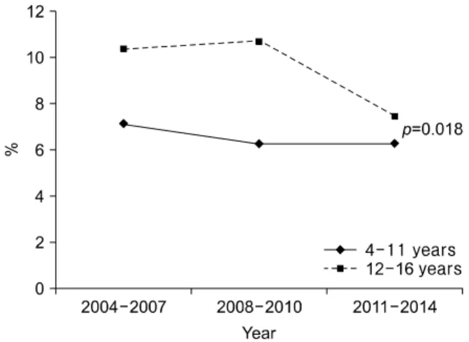
Nevertheless, the prevalence of H. pylori infection was 6.5% for those aged 4-11 years and 9.2% for those aged 12-16 years, demonstrating that the prev- alence in children aged <12 years was significantly lower than that in children aged ≥12 years (p=0.018) (Table 2, Fig. 1).
Table 3. The Prevalence of Helicobacter pylori Infections according to Age Group (p<0.01) and Time Period (p=0.24) by the Modified Cut-Off Values
Year Age (y)
Mean prevalence of each period
4-5 6-11 12-16
2004-2007 2008-2010 2011-2014
Mean prevalence of each age group
6.8 (8/118) 1.2 (1/84) 4.6 (6/131) 4.5 (15/333)*
5.4 (27/504) 5.2 (19/366) 5.1 (26/510) 5.2 (72/1,380)*
10.4 (26/251) 10.7 (23/216) 7.4 (26/350) 9.2 (75/817)*
7.0 (61/873)† 6.5 (43/666)† 5.8 (58/991)† 6.4 (162/2,530) Values are presented as percentage (number/total number).
*p<0.01 (each age group), †p=0.24 (each period).
Fig. 1. Changes in the prevalence of Helicobacter pylori infections in Korean children with recurrent abdominal pain according to age and time period. Prevalence of H. pylori infection was not significantly decreased according to time period. But prevalence of H. pylori infections in children aged 4-11 years was signi- ficantly lower than that in those aged 12-16 years (p=0.018).
Prevalence rate according to modified cut-off value
After endoscopy, 1 child <6 years old and 4 chil- dren aged 6-16 years old with a titer not more than 3‰ points over the cut-off value had negative find- ings for H. pylori, CLO test, and biopsy. Ten children
<6 years old and 15 children ≥6 years were tempo- rarily excluded for reanalysis by applying modified cut-off value. When cut-off values were modified, the prevalence rates were 7.0% for the period 2004-2007, 6.5% for the period 2008-2010, and 5.8%
for the period 2011-2014, which showed no sig- nificant difference over time (p=0.27). However, a statistically significant decrease in the prevalence was observed in the younger group with prevalence
values of 4.6% for those aged 4-5 years when com- pared to 5.3% for those aged 6-11 and 9.2% for 12-16 years (p<0.01; Table 3).
DISCUSSION
The socioeconomic status, numbers of infected family and the personal hygiene are thought to be important factors affecting the prevalence of H. pylori infection in childhood [9]. In developing countries, the infection begins in children <10 years old [10].
If it is not treated, since natural eradication is rare [11], childhood infections persist until adulthood, suggesting a relationship between a high prevalence in adults and previous infection in their childhood.
Korean prevalence rates of H. pylori in the late 1990s are between the rate values of developed and developing countries. The prevalence rate of older adults is similar to that of underdeveloped countries while children’s prevalence close to that of devel- oped countries [12]. In addition, an adult study re- ported that the prevalence of H. pylori was sig- nificantly decreased from 1998 to 2005 [13], prob- ably due to the economic development of the country as well as the improved sanitary conditions.
H. pylori is transmitted from person to person, with infection within families being the main route of transmission [14]. According to a recent report, even in developing countries, the onset of infection occurs usually in childhood within the family, under- estimating the importance of other infection routes, such as the fecal-oral transmission. Furthermore, ac- cording to Rocha et al. [15], H. pylori-infected moth-
ers can act as a strong independent factor to their children. In this study, PCR analysis of the mother’s saliva revealed that 78% of mothers of H. pylori-in- fected children were infected with H. pylori [16]. This suggests an oral-oral transmission, suggesting that contact between the mother and child is the major source of infection [17]. As a result, the analysis of the risk factors associated with H. pylori infection in- dicates that the socioeconomic status in adult age has only a limited effect, whereas is related in chil- dren as well as living standards, such as mother’s ed- ucation, drinking water hygiene, and family income.
Differences in prevalence according to the socio- economic status, and the housing conditions during their childhood period have been identified as im- portant factors related to the infection rate in adults and children [18].
Here, we suggest that high prevalence of H. pylori in Koreans in the 1980s and 1990s was mainly owing to the underdeveloped state of the socioeconomic status, environment, and sanitation, as well as be- cause of the mouth-to-mouth transmission between mother and child, mainly associated with the masti- cation culture in the weaning food period. According to a study conducted in 2005 with 15,916 healthy people >16 years old, the prevalence of H. pylori was 29.3% for those in their 20s, 49.1% for those in their 30s, 57.8% for those in their 40s, and 61.5% for those in their 50s, pointing to a significant increase in prev- alence in those in their 40s and 50s [19].
In our study, there was a significant difference in prevalence in patients ≥12 years old and <12 years old. Thus, when the age of the mother with children
≥12 years old is estimated to be over 40s, she would have been over 30s in 2005. Therefore, 49.1% of mother’s infection rate affects children; it was thought to reflect the current prevalence in children aged ≥12 years, relatively low prevalence in 20s in 2005 affects decrease of prevalence in children <12 years. Thus, infection rate in children can be ex- pected to decrease further if the maternal infection rates are lowered along with a decrease in the rate of mouth-to-mouth transmission by disappearing mas- tication culture in weaning food period.
A reduction in H. pylori prevalence of domestic in the 2000s when compared to the 1990s has already been reported [20,21]. Nevertheless, these studies did not demonstrate a significant decrease in the prevalence in Korean children in the last 11 years.
First, these results can be linked to the country’s economic slow growth. As reported by the Bank of Korea in 2014, the changes in the economic growth and development of our country increased sharply in the 80s and 90s. However, the economic growth has stunted after the late 90s. Thus, due to the rapid eco- nomic and environment growth, and improvements in hygiene in the 80s and 90s, the prevalence of H. py- lori decreased rapidly. Nonetheless, there was no sig- nificant decrease in H. pylori infection in the last 11 years probably due to the economic depression in the late 90s. Secondly, public awareness is increased over the last 10 years, that mouth-to-mouth transmission by mastication culture during weaning period is one of the routes of H. pylori infection transmission.
Moreover, the decrease in the H. pylori infection rate in children during the last decade was lower due to the increasing maternal age.
The use of endoscopic biopsy for CLO tests and his- topathologic examination is the most accurate pro- cedure in the diagnosis of H. pylori infection. However, unlike adults, endoscopy is not always recom- mended for children due to the possible complica- tions during examination and the psychological bur- den of the patient and their parents, so that there would be an increased risk of selection bias for preva- lence study. The use of H. pylori immunoglobulins is an alternative method with high sensitivity and spe- cificity in adults; however, because of very low sensi- tivity in children, this method is not used for diag- nosis of H. pylori infection in children [22]. On the other hand, the UBT is a non-invasive test that is commonly used easily and safely without the need of experienced personnel, in contrast to endoscopy. The UBT has been used in a relatively accurate way to de- termine treatment success, since it has a high sensi- tivity and specificity. Here, we tried to estimate the prevalence rate of H. pylori in patients with recurrent abdominal pain, based on the findings that recurrent
abdominal pain in children has no direct association with H. pylori [23]. Our study conducted the UBT for screening patients with recurrent abdominal pain, followed by endoscopy in cases where screening re- sults were positive. Thus, we were able to examine the change in the prevalence rate of H. pylori in- fection in children, while improving the test accu- racy and minimizing the use of endoscopy.
Applying UBT for children has an interpreting week point because of the higher false positive rate in the younger children. In a previous study, children
<6 years of age showed a 8.3% false positive rate af- ter UBT, showing 10 times higher values than the rate of 0.85% in children ≥6 years old [24]. Yang et al. [8] suggested a 7.0‰ cut-off value for those <6 years old in order to reduce false positive rates.
In this study, the prevalence of H. pylori infection in children with recurrent abdominal pain was stat- istically similar when compared to that in normal children (p=0.74) [25]. An endoscopic biopsy study by Lee et al. [26] among children revealed a preva- lence of 11.3% for the period of 2003-2005 and 10.8%
for the period of 2006-2008. These results were com- parable to our study results, with a prevalence of 8.01% for the period 2004-2007 (p=0.28).
During our study, no significant decrease in H. py- lori prevalence infection was observed in children with recurrent abdominal pain. However, when comparing children >12 and ≤12 years old, the prevalence of H. pylori was significantly lower in chil- dren <12 years. Therefore, we predict a reduction of H. pylori prevalence in the future. According to Fig. 1, the age difference between the 2 groups is about 7 years. The prevalence of H. pylori in the group aged 4-12 years can be estimated to be 10% in the late 1990s; since H. pylori is not naturally eradicated, we assume that the prevalence in children aged 4-12 years was decreased from 10% in the late 1990s (7 years before 2004-2007) to 6% currently. Further, we raised the cut-off value in order to reduce the false positive rate based on early experience that children who present with values 3‰ points higher than the cut-off value tested negative for H. pylori after endoscopy. Hence, when applying the raised cut-off
value, the prevalence increased significantly in the higher age groups.
This study has some limitations. Our study was performed in a single institution. Hence, the preva- lence of H. pylori in Korean children may not be rep- resentative of the whole population. In addition, re- cent studies have reported a lack of association be- tween H. pylori infection and abdominal pain [23,27]; however, our study included children with recurrent abdominal pain and cannot accurately re- flect the prevalence in normal children.
Here, we performed a study to target children with functional gastrointestinal disorders, presenting as recurrent abdominal pain. During the last 11 years, we found that the rate of H. pylori infection in chil- dren <12 years was significantly lower than that in children ≥12 years old. The study results will help to predict the decrease in the prevalence rate of H. pylori in Korean children in the future. In addition, they may be used as the basis for a national multicenter study.
REFERENCES
1. Sherman P, Czinn S, Drumm B, Gottrand F, Kawakami E, Madrazo A, et al. Helicobacter pylori infection in chil- dren and adolescents: Working Group Report of the First World Congress of Pediatric Gastroenterology, Hepatology, and Nutrition. J Pediatr Gastroenterol Nutr 2002;35(Suppl 2):S128-33.
2. Seo JK, Chi JG, Kim EC. Gastrofiberscopic findings and Helicobacter pylori gastritis in children with recurrent abdominal pain. J Korean Pediatr Soc 1992;35:1646-56.
3. Marshall BJ. Helicobacter pylori. Am J Gastroenterol 1994;89(8 Suppl):S116-28.
4. Malaty HM. Epidemiology of Helicobacter pylori infection. Best Pract Res Clin Gastroenterol 2007;21:
205-14.
5. Malaty HM, Kim JG, Kim SD, Graham DY. Prevalence of Helicobacter pylori infection in Korean children: in- verse relation to socioeconomic status despite a uni- formly high prevalence in adults. Am J Epidemiol 1996;143:257-62.
6. Malaty HM, Paykov V, Bykova O, Ross A, Graham DP, Anneger JF, et al. Helicobacter pylori and socio- economic factors in Russia. Helicobacter 1996;1:82-7.
7. Apley J, Naish N. Recurrent abdominal pains: a field
survey of 1,000 school children. Arch Dis Child 1958;
33:165-70.
8. Yang HR, Ko JS, Seo JK. Does the diagnostic accuracy of the 13C-urea breath test vary with age even after the application of urea hydrolysis rate? Helicobacter 2008;
13:239-44.
9. Mitchell HM. The epidemiology of Helicobacter pylori.
Curr Top Microbiol Immunol 1999;241:11-30.
10. Mégraud F, Brassens-Rabbé MP, Denis F, Belbouri A, Hoa DQ. Seroepidemiology of Campylobacter pylori in- fection in various populations. J Clin Microbiol 1989;
27:1870-3.
11. Valle J, Kekki M, Sipponen P, Ihamäki T, Siurala M.
Long-term course and consequences of Helicobacter py- lori gastritis. Results of a 32-year follow-up study.
Scand J Gastroenterol 1996;31:546-50.
12. Kim JH, Kim HY, Kim NY, Kim SW, Kim JG, Kim JJ, et al; Korea H. pylori Study Group, South Korea.
Seroepidemiological study of Helicobacter pylori in- fection in asymptomatic people in South Korea. J Gastroenterol Hepatol 2001;16:969-75.
13. Do MY, Lee YC, Choi CH, Kim SJ, Mun CS, Moon HJ, et al. The changes in prevalence and the related factors of Helicobacter pylori infection in Korean health check-up subjects during 8 years. Korean J Gastroenterol 2009;53:76-83.
14. Kivi M, Tindberg Y, Sörberg M, Casswall TH, Befrits R, Hellström PM, et al. Concordance of Helicobacter py- lori strains within families. J Clin Microbiol 2003;41:
5604-8.
15. Rocha GA, Rocha AM, Silva LD, Santos A, Bocewicz AC, Queiroz Rd Rde M, et al. Transmission of Helicobacter pylori infection in families of preschool-aged children from Minas Gerais, Brazil. Trop Med Int Health 2003;8:987-91.
16. Sinha SK, Martin B, Gold BD, Song Q, Sargent M, Bernstein CN. The incidence of Helicobacter pylori acquis- ition in children of a Canadian First Nations community and the potential for parent-to-child transmission.
Helicobacter 2004;9:59-68.
17. Konno M, Fujii N, Yokota S, Sato K, Takahashi M, Sato K, et al. Five-year follow-up study of mother-to-child transmission of Helicobacter pylori infection detected by a random amplified polymorphic DNA finger-
printing method. J Clin Microbiol 2005;43:2246-50.
18. Bures J, Kopácová M, Koupil I, Vorísek V, Rejchrt S, Beránek M, et al; European Society for Primary Care Gastroenterology. Epidemiology of Helicobacter pylori infection in the Czech Republic. Helicobacter 2006;11:
56-65.
19. Yim JY, Kim N, Choi SH, Kim YS, Cho KR, Kim SS, et al. Seroprevalence of Helicobacter pylori in South Korea. Helicobacter 2007;12:333-40.
20. Lim SH, Kwon JW, Kim N, Kim GH, Kang JM, Park MJ, et al. Prevalence and risk factors of Helicobacter pylori infection in Korea: nationwide multicenter study over 13 years. BMC Gastroenterol 2013;13:104.
21. Seo JK, Ko JS, Choi KD. Serum ferritin and Helicobacter pylori infection in children: a sero-epi- demiologic study in Korea. J Gastroenterol Hepatol 2002;17:754-7.
22. de Oliveira AM, Rocha GA, Queiroz DM, Mendes EN, de Carvalho AS, Ferrari TC, et al. Evaluation of en- zyme-linked immunosorbent assay for the diagnosis of Helicobacter pylori infection in children from different age groups with and without duodenal ulcer. J Pediatr Gastroenterol Nutr 1999;28:157-61.
23. Sherman PM, Macarthur C. Current controversies as- sociated with Helicobacter pylori infection in the pedia- tric population. Front Biosci 2001;6:E187-92.
24. Kindermann A, Demmelmair H, Koletzko B, Krauss- Etschmann S, Wiebecke B, Koletzko S. Influence of age on 13C-urea breath test results in children. J Pediatr Gastroenterol Nutr 2000;30:85-91.
25. Ko JS, Chung JY, Bae SH, Kim EJ, Seo JK. Relation be- tween recurrent abdominal pain and Helicobacter pylo- ri infection and the role of CagA and VacA in pediatric Helicobacter pylori infection. Korean J Gastroenterol 2001;37:167-72.
26. Lee SY, Ko JS, Seo JK. Changes in the prevalence of bi- opsy-proven Helicobacter pylori infection in Korean children with functional recurrent abdominal pain over the last 18 years. Korean J Pediatr Gastroenterol Nutr 2009;12:150-5.
27. Macarthur C. Helicobacter pylori infection and child- hood recurrent abdominal pain: lack of evidence for a cause and effect relationship. Can J Gastroenterol 1999;13:607-10.