The effect of periodontal and prosthodontic therapy on glycemic control in patients with diabetes
Hak-Ki Kim1, Yong-Gun Kim1, Jin-Hyun Cho2, Sang-Kyu Lee3*, Jae-Mok Lee1*
1Department of Periodontology, Kyungpook National University School of Dentistry, Daegu, Republic of Korea
2Department of Prosthodontics, Kyungpook National University School of Dentistry, Daegu, Republic of Korea
3College of Pharmacy and Research Institute of Pharmaceutical Sciences, Kyungpook National University, Daegu, Republic of Korea
PURPOSE. To evaluate the effect of periodontal and prosthodontic therapy on glycated hemoglobin A(HbA1c) level in patients with diabetes. MATERIALS AND METHODS. This is a retrospective study of 70 patients suffering from diabetes who visited the Kyungpook National University Hospital between January 2016 and May 2018.
Patients underwent medical evaluation for their routine check-up, which includes laboratory test for HbA1c levels. Among the 70 patients, 35 patients also visited Kyungpook National University Dental Hospital during the same period to receive periodontal and prosthodontic therapy, while the other 35 patients did not receive such therapy. The HbA1c levels were compared before and after periodontal and prosthodontic therapy. Comparisons between groups and within groups were performed using independent t-test. RESULTS. The HbA1c levels in the group who have received periodontal and prosthodontic therapy decreased from 7.2 to 6.7 (P=.001). The HbA1c levels in the control group decreased from 7.2 to 7.1 (P=.580). The difference in changes between the two patient groups was statistically significant (P=.011). CONCLUSION. Periodontal and prosthodontic therapy can be effective on glycemic control in patients with diabetes. [J Adv Prosthodont 2019;11:247-52]
KEYWORDS: Diabetes mellitus; Glycated hemoglobin A; Periodontal diseases; Prosthodontics
INTRODUCTION
Diabetes mellitus and periodontal disease are two common diseases that are in close relation to one another.1 Diabetes mellitus alters glucose tolerance or impairs metabolism of lipids and carbohydrates.2 It is considered as a manifestation of a persistent chronic low-grade inflammation.3 Periodontal disease is a chronic inflammatory condition that causes an
imbalance in anabolic and catabolic processes, ultimately resulting in resorption of alveolar bone or tooth-supporting tissues.4
Previous clinical studies5,6 provided evidence on a mutu- al relationship between diabetes mellitus and periodontal disease.In diabetes, hyperglycemia is a potential risk factor for periodontitis.7-9 In periodontitis, a systematic review by Borgnakke et al.10 suggested that severe periodontitis was related to significantly elevated serum levels of HbA1c.
Previous research1 has given effort to clarify the underly- ing mechanisms between diabetes mellitus and periodontal disease. Gingival crevicular fluid is a serum transudate origi- nating from the gingival plexus of blood vessels.11 It has been shown that elevated serum levels of inflammatory mediators and cytokines such as tumor necrosis factor-alpha (TNF-a) in diabetic patients correlated with increased levels of these mediators and cytokines in gingival crevicular flu- id.12 Systemic infection of gram negative organisms such as Porphyromonas gingivalis, Tannerella forsythensis, Prevotella interme- dia or their products may promote an elevated inflammatory state and increased levels of serum inflammatory markers.13 Serum markers of inflammation such as C-reactive protein (CRP), IL-6, and fibrinogen are significantly higher in
Corresponding author:
Jae-Mok Lee
Department of Periodontology, Kyungpook National University School of Dentistry, 2177 Dalgubeol-daero, Jung-gu, Daegu 41566, Republic of Korea Tel. +82536007522: e-mail, [email protected]
Sang-Kyu Lee
College of Pharmacy and Research Institute of Pharmaceutical Sciences, Kyungpook National University, Daehak-ro 80, Buk-gu, Daegu 41566, Republic of Korea
Tel. +82539508571: e-mail, [email protected]
Received February 27, 2019 / Last Revision October 29, 2019 / Accepted October 30, 2019
© 2019 The Korean Academy of Prosthodontics
This is an Open Access article distributed under the terms of the Creative Commons Attribution Non-Commercial License (http://creativecommons.
org/licenses/by-nc/4.0) which permits unrestricted non-commercial use, distribution, and reproduction in any medium, provided the original work is properly cited.
patients with periodontitis, than in patients without peri- odontitis.14-16 Gram-negative infections in periodontal dis- ease may also engender insulin resistance and exacerbate metabolic control in patients with diabetes.17
In view of these relations between diabetes mellitus and periodontal disease, it has been speculated that treatment of periodontal diseases can improve metabolic control of dia- betes.18 According to previous studies,19,20 the magnitude of the reduction in HbA1c ranged from 0.27% to 0.48% 3 - 4 months after periodontal treatment. Despite periodontal treatment seeming to ameliorate metabolic control, evidence was not sufficient to significant associate periodontal treat- ment and metabolic control in patients with diabetes.21 According to a consensus report, there is a lack of data to manifest that this effect is maintained over 6 months fol- lowing periodontal treatment.22
Prosthodontic therapy, such as restoration, implant pros- thesis, and removable partial denture, have been reported to cause dental problems on abutment teeth.23 Plaques can be accumulated on teeth used as abutment, thus becoming a risk for infection and deteriorate metabolic control for patients with diabetes. However, there was not enough evi- dence to conclude whether these changes on abutment teeth could be mitigated when periodontal therapy was combined.
Therefore, in this retrospective study, we sought to com- pare the changes in glycemic control between patients receiv- ing periodontal and prosthodontic therapy and patients not receiving the therapy over 6 month period.
MATERIALS AND METHODS
70 diabetic patients who visited the Kyungpook National University Hospital to monitor their HbA1c levels between January 2016 and May 2018 were analyzed in this study. Among the 70 patients, 35 patients who also visited Kyungpook National University Dental Hospital to receive periodontal and prosthodontic therapy during the same period was des- ignated as the treatment group, and the other 35 patients who did not received such therapy was designated as the control group.
The treatment group was further divided to subgroups according to the type of periodontal therapy: active peri- odontal therapy and supportive periodontal therapy. The
treatment group was also classified based on whether prosthodontic therapy was carried out or not (with or with- out prosthodontic therapy). Active periodontal therapy encompassed oral hygiene instruction, full mouth scaling, subgingival curettage, extraction and flap operation per- formed under local anesthesia. Supportive periodontal ther- apy consisted of oral hygiene instructions, full mouth scal- ing, and root plaining. The mean interval between visits for supportive periodontal therapy was 3.5 months. Prosthodontic therapy consisted of implant prosthodontics, single and bridge restorations, occlusal adjustment, and removable par- tial denture. Patients in the treatment group received at least one of these therapies.
In the treatment group, data such as the type of peri- odontal therapy, the presence or absence of prosthodontic therapy, and the number of teeth lost, were collected. The number of teeth lost was calculated by subtracting the num- ber of remaining teeth from 32 teeth.
A statistical analysis was performed using a SPSS soft- ware (IBM SPSS Statistics for Windows, Version 23.0. IBM, Armonk, NY, USA). Comparison of HbA1c levels before and after therapy within groups, and difference in extent of HbA1c reduction between groups were performed using independent t-test. Correlation between level of HbA1c before therapy and the number of teeth lost was investigated through simple linear regression analysis. It was considered to be statistically significant when P value is below 0.05.
RESULTS
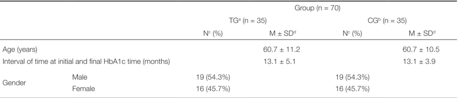
Clinical characteristics are summarized in Table 1. At base- line, the two groups had similar mean values for age, gender distribution, and the time interval between the initial and final HbA1c levels.
The HbA1c levels in the treatment group decreased from 7.2 to 6.7 (P = .001) following periodontal and prosth- odontic therapy, and this change was statistically significant (Fig. 1). During the similar time frame, the HbA1c levels in the control group also decreased from 7.2 to 7.1 (P = .580) but this change was not statistically significant. Furthermore, the difference in changes between the two groups was sta- tistically significant (P = .011, Fig. 1).
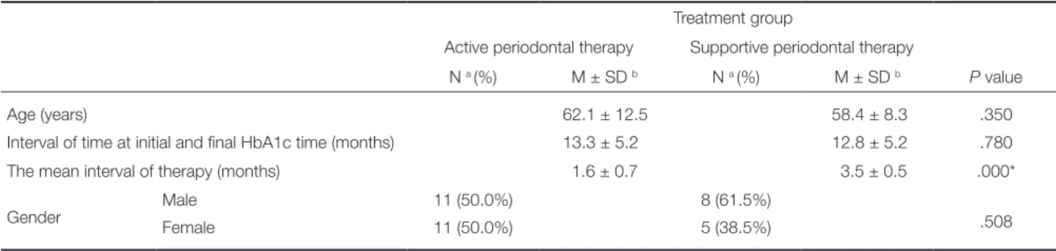
Clinical characteristics of two subcategories of the treat-
Table 1. Clinical characteristics of patients of treatment and control groups
Group (n = 70)
TGa (n = 35) CGb (n = 35)
Nc (%) M ± SDd Nc (%) M ± SDd
Age (years) 60.7 ± 11.2 60.7 ± 10.5
Interval of time at initial and final HbA1c time (months) 13.1 ± 5.1 13.1 ± 3.9
Gender Male 19 (54.3%) 19 (54.3%)
Female 16 (45.7%) 16 (45.7%)
a Treatment group, b Control group, c Number of patients, d Mean ± standard deviation.
Table 2. Clinical characteristics of two subcategories of the treatment group according to the type of periodontal therapy Treatment group
Active periodontal therapy Supportive periodontal therapy
N a (%) M ± SD b N a (%) M ± SD b P value
Age (years) 62.1 ± 12.5 58.4 ± 8.3 .350
Interval of time at initial and final HbA1c time (months) 13.3 ± 5.2 12.8 ± 5.2 .780
The mean interval of therapy (months) 1.6 ± 0.7 3.5 ± 0.5 .000*
Gender Male 11 (50.0%) 8 (61.5%)
Female 11 (50.0%) 5 (38.5%) .508
*Significant difference at the 0.05 level.
a Number of patients, b Mean ± standard deviation.
ment group according to the type of periodontal therapy are summarized in Table 2. The two groups were well matched for age, gender distribution, and the time interval between the initial and final HbA1c levels. The only statisti- cal difference between the two groups was the mean inter- val of therapy; the mean interval of active periodontal ther- apy group was 1.6 ± 0.7 months, whereas that of support- ive periodontal therapy group was 3.5 ± 0.5 months.
The HbA1c levels in the active periodontal therapy group decreased from 7.6 to 6.9 (P = .003), and this change was statistically significant (Fig. 2). During a similar time frame, the HbA1c levels in the supportive periodontal ther-
apy group also decreased from 6.5 to 6.3 (P = .194), but this change was not statistically significant. Furthermore, the dif- ference in changes between the two groups was statistically significant (P = .029, Fig. 2). The initial HbA1c level of the active periodontal therapy group and supportive periodontal therapy group was 7.6 and 6.5, respectively, and this differ- ence was statistically significant (P = .016).
Clinical characteristics of two subcategories of the treat- ment group based on whether or not prosthodontic therapy was carried (with or without prosthodontic therapy) were summarized in Table 3. The two groups differed in age, gen- der distribution, and the time interval between the initial and
Table 3. Clinical characteristics of two subcategories of the treatment group based on whether prosthodontic therapy was carried out or not (with or without prosthodontic therapy)
Treatment group Periodontal with
prosthodontic therapy
Periodontal without prosthodontic therapy
N a (%) M ± SD b N a (%) M ± SD b P value
Age (years) 64.1 ± 8.5 59.0 ± 12.2 .201
Interval of time at initial and final HbA1c time (months) 12.6 ± 5.5 13.3 ± 5.0 .680
Gender Male 5 (41.7%) 14 (60.9%)
Female 7 (58.3%) 9 (39.1%) .279
a Number of patients, b Mean ± standard deviation.
Fig. 1. Comparison of HbA1c reduction between treat-
ment and control group before and after therapy. Fig. 2. Comparison of HbA1c reduction between active and supportive periodontal group before and after therapy.
7.6 7.4 7.2 7.0 6.8 6.6 6.4 6.2
8.0 7.8 7.6 7.4 7.2 7.0 6.8 6.6 6.4 6.2 6.0 Initial HbA1c (mean)
Final HbA1c (mean)
Initial HbA1c (mean) Final HbA1c (mean)
*
P < .001† P < .05
*
P < .05 Treatment group Control group Active periodontal Supportive periodentaltherapy therapy
final HbA1c levels, but this difference was not statistically significant.
The HbA1c levels in the periodontal with prosthodontic therapy group decreased from 7.9 to 7.0 (P = .025), and this change was statistically significant (Fig. 3). During a similar time frame, the HbA1c levels in the periodontal without prosthodontic therapy group also decreased from 6.8 to 6.6 (P = .014), and this change was statistically significant (Fig.
3). However, the difference in changes between the two groups was not statistically significant.
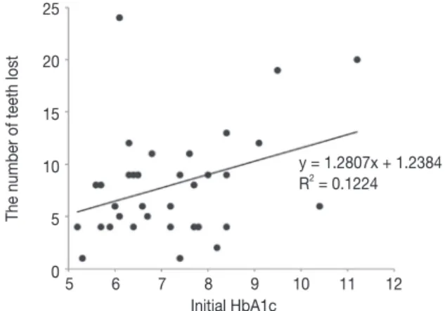
The simple linear regression model showed that there was a positive correlation between initial HbA1c and the number of teeth loss in the treatment group, which was sta- tistically significant (P = .039, Fig. 4).
DISCUSSION
There were previously published studies24-26 investigating the relationship between periodontal therapy and HbA1c levels.
Moeintaghavi et al.27 noted a significant reduction of HbA1c level (0.74%) in the treatment group after 3 months. Sun et al.28 also observed a reduction of HbA1c level (0.5%) in the treatment group after 3 months. Our results supported those of previous studies, showing a reduction of HbA1c level from 7.2 to 6.7 (0.5%) after periodontal and prosth- odontic therapy.
The mechanism by which HbA1c decreases after peri- odontal and prosthodontic therapy can be explained by the regulation of TNF-alpha (Tumor Necrosis Factor-alpha) levels. In diabetic patients, elevated levels of TNF-alpha can engender insulin resistance and worsen glycemic control2. Therefore, in diabetic patients, decreasing TNF-alpha levels through periodontal and prosthetic therapy can restore insulin sensitivity and improve glycemic control. In a study29 of 13 type 2 diabetic subjects with periodontitis, periodontal therapy decreased TNF-alpha levels. In another study30 using rat model, the expression of interleukin-1beta, cyclo-
oxygenase 2, and TNF-alpha tended to be elevated by trau- ma from occlusion, and thereby occlusal stabilization through prosthodontic therapy was expected to decrease TNF-alpha levels.
Recent randomized clinical trial27 examined the effect of periodontal treatment on the glycemic control and showed a reduction in HbA1c after 3 to 4 months of follow-up. There are insufficient data to show that this effect is maintained after 6 months or more.5 Within the limitation of retrospec- tive study, we found a significant reduction in the HbA1c level (0.5%) in the treatment group after 13.1 months in average.
We noticed that active periodontal therapy was more effective in reducing HbA1c level than supportive periodon- tal therapy. This result could be due to the difference in mean interval of therapy. Active periodontal therapy had much shorter time interval than supportive periodontal therapy. We also noticed that the initial HbA1c level of sup- portive periodontal therapy group was much lower than that of active periodontal therapy group. Supportive periodontal therapy group had already been treated with active peri- odontal therapy before the initiation of supportive peri- odontal therapy, thus having lower initial HbA1c level from the beginning.
Prosthodontic therapy such as implant prosthesis, resto- ration, and removable partial denture could change the eco- logical system of the oral cavity possibly raising plaque accumulation at teeth.31 This could lead to bacteremia in the oral cavity and could exacerbate metabolic in diabetic patients. However, our results showed that periodontal and prosthodontic therapy group showed significant reduction in the HbA1c level, even more than periodontal therapy alone. This indicated that when periodontal therapy was performed together, prosthodontic therapy did not worsen glycemic control in patients with diabetes.
Polak and Shapira32 suggested a biological pathway that the elevated pro-inflammatory factors in the gingiva of Fig. 3. Comparison of HbA1c reduction between peri-
odontal with prosthodontic and periodontal without prosthodontic group before and after therapy.
Fig. 4. Simple linear regression analysis showing the cor- relation between level of HbA1c before therapy and the number of teeth lost.
8.4 8.2 8.0 7.8 7.6 7.4 7.2 7.0 6.8 6.6 6.4 6.2
Periodontal with Periodontal without prosthodontic therpy prosthodontic therpy
Initial HbA1c (mean) Final HbA1c (mean)
*
P < .05The number of teeth lost
25
20
15
10
5
0
Initial HbA1c
5 6 7 8 9 10 11 12 y = 1.2807x + 1.2384 R2 = 0.1224
patients with poorly controlled serum glucose may aggra- vate periodontitis. In this study, we examined the relation- ship between initial HbA1c and teeth loss rates in treatment group. Our study showed positive correlation between ini- tial HbA1c and teeth loss rates and this result was statistical- ly significant. This indicates that the severity of diabetes may affect the progression of periodontitis.
The retrospective design of this study presents inherent limitations. Not all HbA1c levels of patients were measured at the same time after periodontal and prosthodontic thera- py. Another limitation is the inability to examine TNF-alpha levels after periodontal and prosthodontic treatment. Further studies are needed to examine TNF-alpha and C-Reactive Protein levels after periodontal and prosthetic treatment through randomized controlled design.
CONCLUSION
There is strong evidence for an association between diabetes mellitus and periodontal disease. Diabetes mellitus increases the risk of periodontitis, and periodontal and prosthodontic therapy improves glycemic control.
ORCID
Hak-Ki Kim https://orcid.org/0000-0002-1597-1518 Yong-Gun Kim https://orcid.org/0000-0002-2793-7667 Jin-Hyun Cho https://orcid.org/0000-0002-2453-9372 Sang-Kyu Lee https://orcid.org/0000-0002-7568-2902 Jae-Mok Lee https://orcid.org/0000-0002-0291-6114 REFRENCES
1. Sonnenschein SK, Meyle J. Local inflammatory reactions in patients with diabetes and periodontitis. Periodontol 2000 2015;69:221-54.
2. Mealey B. Diabetes and periodontal diseases. J Periodontol 1999;70:935-49.
3. Xie W, Du L. Diabetes is an inflammatory disease: evidence from traditional Chinese medicines. Diabetes Obes Metab 2011;13:289-301.
4. Taiyeb-Ali TB, Raman RP, Vaithilingam RD. Relationship be- tween periodontal disease and diabetes mellitus: an Asian per- spective. Periodontol 2000 2011;56:258-68.
5. Chávarry NG, Vettore MV, Sansone C, Sheiham A. The rela- tionship between diabetes mellitus and destructive periodontal disease: a meta-analysis. Oral Health Prev Dent 2009;7:107-27.
6. Lalla E, Papapanou PN. Diabetes mellitus and periodontitis: a tale of two common interrelated diseases. Nat Rev Endocrinol 2011;7:738-48.
7. Emrich LJ, Shlossman M, Genco RJ. Periodontal disease in non-insulin-dependent diabetes mellitus. J Periodontol 1991;62:123-31.
8. Lalla E, Cheng B, Lal S, Tucker S, Greenberg E, Goland R, Lamster IB. Periodontal changes in children and adolescents with diabetes: a case-control study. Diabetes Care 2006;29:295- 9.
9. Taylor GW, Burt BA, Becker MP, Genco RJ, Shlossman M, Knowler WC, Pettitt DJ. Non-insulin dependent diabetes mellitus and alveolar bone loss progression over 2 years. J Periodontol 1998;69:76-83.
10. Borgnakke WS, Ylöstalo PV, Taylor GW, Genco RJ. Effect of periodontal disease on diabetes: systematic review of epidemio- logic observational evidence. J Clin Periodontol 2013;40:S135- 52.
11. Barros SP, Williams R, Offenbacher S, Morelli T. Gingival crevic- ular fluid as a source of biomarkers for periodontitis. Periodontol 2000 2016;70:53-64.
12. Salvi GE, Yalda B, Collins JG, Jones BH, Smith FW, Arnold RR, Offenbacher S. Inflammatory mediator response as a po- tential risk marker for periodontal diseases in insulin-depen- dent diabetes mellitus patients. J Periodontol 1997;68:127-35.
13. Wu T, Trevisan M, Genco RJ, Falkner KL, Dorn JP, Sempos CT. Examination of the relation between periodontal health status and cardiovascular risk factors: serum total and high density lipoprotein cholesterol, C-reactive protein, and plasma fibrinogen. Am J Epidemiol 2000;151:273-82.
14. Naguib G, Al-Mashat H, Desta T, Graves DT. Diabetes pro- longs the inflammatory response to a bacterial stimulus through cytokine dysregulation. J Invest Dermatol 2004;123:87-92.
15. Noack B, Genco RJ, Trevisan M, Grossi S, Zambon JJ, De Nardin E. Periodontal infections contribute to elevated sys- temic C-reactive protein level. J Periodontol 2001;72:1221-7.
16. Loos BG, Craandijk J, Hoek FJ, Wertheim-van Dillen PM, van der Velden U. Elevation of systemic markers related to cardiovascular diseases in the peripheral blood of periodonti- tis patients. J Periodontol 2000;71:1528-34.
17. Mealey BL, Oates TW; American Academy of Periodontology.
Diabetes mellitus and periodontal diseases. J Periodontol 2006;77:1289-303.
18. Stewart JE, Wager KA, Friedlander AH, Zadeh HH. The ef- fect of periodontal treatment on glycemic control in patients with type 2 diabetes mellitus. J Clin Periodontol 2001;28:306- 10.
19. Li Q, Hao S, Fang J, Xie J, Kong XH, Yang JX. Effect of non-surgical periodontal treatment on glycemic control of patients with diabetes: a meta-analysis of randomized con- trolled trials. Trials 2015;16:291.
20. Teshome A, Yitayeh A. The effect of periodontal therapy on glycemic control and fasting plasma glucose level in type 2 di- abetic patients: systematic review and meta-analysis. BMC Oral Health. 2016;17:31.
21. Mauri-Obradors E, Merlos A, Estrugo-Devesa A, Jané-Salas E, López-López J, Viñas M. Benefits of non-surgical peri- odontal treatment in patients with type 2 diabetes mellitus and chronic periodontitis: A randomized controlled trial. J Clin Periodontol 2018;45:345-53.
22. Sanz M, Ceriello A, Buysschaert M, Chapple I, Demmer RT, Graziani F, Herrera D, Jepsen S, Lione L, Madianos P, Mathur M, Montanya E, Shapira L, Tonetti M, Vegh D. Scientific evi- dence on the links between periodontal diseases and diabetes:
Consensus report and guidelines of the joint workshop on peri- odontal diseases and diabetes by the International Diabetes Federation and the European Federation of Periodontology. J
Clin Periodontol 2018;45:138-49.
23. Pretzl B, Kaltschmitt J, Kim TS, Reitmeir P, Eickholz P. Tooth loss after active periodontal therapy. 2: tooth-related factors. J Clin Periodontol 2008;35:175-82.
24. Madianos PN, Koromantzos PA. An update of the evidence on the potential impact of periodontal therapy on diabetes outcomes. J Clin Periodontol 2018;45:188-95.
25. Mauri-Obradors E, Jané-Salas E, Sabater-Recolons Mdel M, Vinas M, López-López J. Effect of nonsurgical periodontal treatment on glycosylated hemoglobin in diabetic patients: a systematic review. Odontology 2015;103:301-13.
26. Wang TF, Jen IA, Chou C, Lei YP. Effects of periodontal therapy on metabolic control in patients with type 2 diabetes mellitus and periodontal disease: a meta-analysis. Medicine (Baltimore) 2014;93:e292.
27. Moeintaghavi A, Arab HR, Bozorgnia Y, Kianoush K, Alizadeh M. Non-surgical periodontal therapy affects metabolic con- trol in diabetics: a randomized controlled clinical trial. Aust Dent J 2012;57:31-7.
28. Sun WL, Chen LL, Zhang SZ, Wu YM, Ren YZ, Qin GM.
Inflammatory cytokines, adiponectin, insulin resistance and metabolic control after periodontal intervention in patients with type 2 diabetes and chronic periodontitis. Intern Med.
2011;50:1569-74.
29. Iwamoto Y, Nishimura F, Nakagawa M, Sugimoto H, Shikata K, Makino H, Fukuda T, Tsuji T, Iwamoto M, Murayama Y.
The effect of antimicrobial periodontal treatment on circulat- ing tumor necrosis factor-alpha and glycated hemoglobin lev- el in patients with type 2 diabetes. J Periodontol 2001;72:774- 8.
30. Matsuda Y, Minagawa T, Okui T, Yamazaki K. Resveratrol suppresses the alveolar bone resorption induced by artificial trauma from occlusion in mice. Oral Dis 2018;24:412-21.
31. Müller S, Eickholz P, Reitmeir P, Eger T. Long-term tooth loss in periodontally compromised but treated patients ac- cording to the type of prosthodontic treatment. A retrospec- tive study. J Oral Rehabil 2013;40:358-67.
32. Polak D, Shapira L. An update on the evidence for pathogen- ic mechanisms that may link periodontitis and diabetes. J Clin Periodontol 2018;45:150-66.