72
Prostate Cancer Cells
Sunghyup Choi1, Kweonsik Min1,2, Ikjoon Choi1, Dongil Kang1,2 From the 1Department of Urology, 2Paik Institute of Clinical Research, Inje Uni- versity College of Medicine, Busan, Korea
Purpose: Overproduction of lipid peroxidation byproducts and disturbances in the antioxidant defense system have been implicated in the pathogenesis of several diseases, including prostate cancer. Although several studies have investigated the level of lipid peroxidation and antioxidants in prostate cancer, there are no reports on α-lipoic acid (ALA) in prostate cancer.
Here we assessed the effects of ALA on the antioxidant system in prostate cancer cells.
Materials and Methods: PC-3, LNCaP, and RWPE-2 cell lines were used in this study. Redox factor (Ref)-1 protein was measured by Western blot analysis after treatment with ALA. Real-time polymerase chain reaction (RT-PCR) was performed to detect superoxide dismutase (SOD)-1 and -2, catalase, and glutathione peroxidase (GSH-Px) mRNA expression.
Results: Ref-1 was expressed in the PC-3, LNCaP, and RWPE-2 cell lines.
The expression of Ref-1 protein was increased after treatment with 125, 250, and 500μM ALA in the PC-3 (p<0.05) and LNCaP (p>0.05) cells compared with the RWPE-2 cells at 48 hours. In PC-3 cells, the mRNA expression of SOD-1, SOD-2, catalase, and GSH-Px decreased at 24 and 48 hours dose-dependently compared with that in RWPE-2 cells (p<0.05).
The mRNA expression of SOD-2, catalase, and GSH-Px in LNCaP cell decreased at 48 hours dose-dependently (p<0.05).
Conclusions: The expression of Ref-1 protein and antioxidant enzymes changed after ALA exposure in prostate cancer cells. Our findings suggest that ALA affects the antioxidant system in prostate cancer cells and may be related to compensatory changes in the antioxidant defense system of the cells. (Korean J Urol 2009;50:72-80)
Key Words: Prostatic neoplasms, Thioctic acid, Antioxidants
대한비뇨기과학회지 제 50 권 제 1 호 2009
1인제대학교 의과대학
비뇨기과학교실,
2백인제임상의학연구소
최성협1ㆍ민권식1,2ㆍ최익준1 강동일1,2
접수일자
:2008년 7월 14일 채택일자
:2008년 10월 15일
교신저자: Dongil Kang
Department of Urology, Inje University, 633-165, Paik Hospital, Gaegum-dong, Busanjin-gu, Busan 614-735, Korea TEL: 051-890-6384 FAX: 051-892-9887 E-mail: urokang@lycos.
co.kr
This work was supported by the 2005 Inje University research grant.
INTRODUCTION
Prostate cancer is not well-known in Korea. However, the incidence of prostate cancer is rapidly increasing compared to other malignancies.1 This has prompted several research efforts into the treatment and prevention of prostate cancer.2,3 Recently, oxidative stress has been considered an important factor in the development and progression of prostate cancer. In the α -tocopherol and β-carotene study (ATBC study), the incidence and mortality of prostate cancer decreased for 6 years during the follow-up, and men who received selenium supplements had
a low incidence of prostate cancer.2,3 Also, in an experimental study, multiple mitochondrial DNA mutations found in human prostate tumors suggested the involvement of cellular oxidative stress.4 Carcinogenesis is a multistep process that includes DNA damage and abnormal cell proliferation. Antioxidant enzymes are endogenous proteins that work together to protect cells from damage by reactive oxygen species (ROS), which are physio- logical by-products that result partly from an imbalance of antioxidant enzymes and free radicals and are capable of directly injuring cells. Oxidative damage is decreased by antioxidant vitamins, non-provitamins, catenoids, and trace elements such as selenium.3,5 α-lipoic acid (ALA) is a naturally-occurring co-
factor for vital metabolic multienzyme complexes, including pyruvate dehydrogenase and glycine decarboxylase. ALA is easily absorbed from the diet, and in mammalian cells it is readily converted to its reduced form, dihydrolipoic acid. ALA acts as a redox regulator of thiol-containing proteins and is effective in the treatment of several pathologic conditions in- cluding diabetes, neurodegeneration, radiation injury, and mal- ignacy.6-8
Overproduction of lipid peroxidation byproducts and dis- turbance in the antioxidant defense system have been implicated in the pathogenesis of prostate cancer. Although several studies have investigated the level of lipid peroxidation and antioxidant enzymes in prostate cancer, to our knowledge, no study has explored the potential antioxidant effect of ALA in prostate cancer.9,10
The objective of this study was to determine whether ALA can affect the expressions of antioxidant enzymes in prostate cancer cell lines compared to a normal human prostate cell line.
MATERIALS AND METHODS
PC-3, LNCaP, and RWPE-2 cell lines were used in this study. As an index of antioxidative stress, redox factor (Ref)-1 and endogenous antioxidant enzymes, including superoxide dismutase (SOD)-1 and -2, catalase, and glutathione peroxidase
(GSH-Px), were measured.
1. Cell culture
The human prostate carcinoma cell lines PC-3 and LNCaP and human prostate epithelial cell line (RWPE-2) were obtained from the American Type Culture Collection (Manassas, USA).
LNCaP cells were maintained in RPMI 1,640 with 10% fetal bovine serum (FBS). PC-3 cells were maintained in Dulbecco’s modified Eagle medium (DMEM) with 10% FBS, and RWPE-2 cells were maintained in keratinocyte growth medium supple- mented with 5 ng/ml human recombinant EGF and 0.05 mg/ml bovine pituitary extract under standard culture conditions (37oC, 95% humidified air, and 5% CO2).
2. Cell viability analysis
To identify an adequate concentration of ALA (Sigma, USA), a dimethylthiazol-2-yl-2,5-diphenyl-tetraxolium bromide (MTT) cell viability assay was performed. LNCaP cells were seeded in 96-well plates (1x10⁴ cells/well) in DMEM containing 10%
FBS, cultured overnight, and then treated with different con- centrations (0-2 mM) of ALA with untreated cells serving as a control. At the end of the incubation, the medium in the wells was removed and replaced with 150μl of fresh medium con- taining a 50μl MTT solution and incubated at 37oC for 4 hours. The medium containing MTT was removed, and 150μl of DMSO were added to solubilize the formazan crystals formed in the viable cells. Absorbance was recorded in an ELISA reader at 570 nm.
3. Protein extraction and Western blot analysis
The Ref-1 protein level was detected in cells cultured in the 6-well plates. Briefly, after treatment (control, 125, 250, 500μM ALA for 48 hours), cells of two wells were lysed in 100μl of lysis buffer. The lysate was incubated on ice for 20 min and centrifuged at 13,000 rpm for 2 min at 4oC. The supernatant was collected. The protein concentration was determined according to the method of Bradford (Bio-Rad Laboratories, USA). The lysate was mixed with 5x SDS gel loading buffer and boiled for 5 min at 100oC. Proteins (20μg) were then separated by SDS-PAGE and transferred from the gel to a polyvinyli- dene-difluoride (PVDF) membrane. The membranes were blocked with 5% nonfat milk in TBS-T for 1 hour at room temperature.
The membranes were washed three times in TBS-T for 10 min each and then incubated overnight at 4oC with primary antibody in TBS-T at a dilution of 1:1,000. The primary antibody used was goat polyclonal anti-Ref-1 antibody (SC-9919, Santa Cruz Biotechnology, USA). The membranes were washed three times in TBS-T for 10 min each and then incubated with HRP- conjugated secondary antibody (1:500) in 5% nonfat milk in TBS-T at room temperature for 1 hour. The membranes were then washed three times in TBS-T for 10 min each. The target protein was visualized with an electrochemiluminescence lighting system (Amersham International PLC, USA) and the protein band intensity was quantified with a densitometer.
4. RNA extraction and cDNA synthesis
At the end of treatment (control, 125, 250, 500μM for 24 and 48 hours), total RNA was extracted from cells cultured in the six-well plates using TRIzol reagent (Invitrogen, Carlsbad, USA), and RNA was isolated according to the manufacturer's instructions. Extracted total RNA was further treated with RNasin (Promega). The yield and purity were determined by Nanodrop.
First-strand cDNA was synthesized from 2μg of total RNA
Fig. 1. The growth rate of the LNCaP cell line. *: inhibitory con- centration (IC)50.
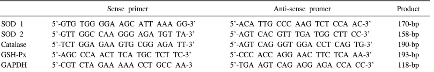
Table 1. PCR primer
Sense primer Anti-sense promer Product
SOD 1 5'-GTG TGG GGA AGC ATT AAA GG-3' 5'-ACA TTG CCC AAG TCT CCA AC-3' 170-bp SOD 2 5'-GTT GGC CAA GGG AGA TGT TA-3' 5'-AGT CAC GTT TGA TGG CTT CC-3' 158-bp Catalase 5'-TCT GGA GAA GTG CGG AGA TT-3' 5'-AGT CAG GGT GGA CCT CAG TG-3' 190-bp GSH-Px 5'-AGC CCA ACT TCA TGC TCT TC-3' 5'-CCC ACC AGG AAC TTC TCA AA-3' 193-bp
GAPDH 5'-CGT CTA GAA AAA CCT GCC AA-3 5'-TGA AGT CAG AGG AGA CCA CC-3' 118-bp
PCR: polymerase chain reaction, SOD: superoxide dismutase, GSH-Px: glutathione peroxidase, GAPDH: glyceraldehyde 3-phosphate dehydrogenase
using AccuPower RT Premix (Bioneer, Korea) with 19-mer oligo dT (Bioneer, Korea) according to the manufacturer's instructions.
Synthesized cDNA was stored at 20oC before use.
5. Design of PCR primers
Primer selection was based on previously published human SOD-1 (CuZn SOD), SOD-2 (MnSOD), catalase, GSH-Px, and glyceraldehyde-3-phosphate dehydrogenase (GAPDH) sequences found through a GenBank database search. GAPDH was used as a control. The primers used are shown in Table 1.
6. Real-time quantitative PCR
Real-time quantitative PCR was carried out in an IQ5 Real- Time PCR Detection System (Bio-Rad Laboratories, USA) using SYBR Green I. The PCR reactions were performed in micro- capillary tubes (Bio-Rad Laboratories, USA) with a final volume of 20μl. The reaction mixture consisted of 1μl of each sense and anti-sense 10μmol/l primer, 10μl of SYBR Green I (Bio-Rad Laboratories, USA), 7μl H2O, and 1μl of the cDNA template. The mRNA levels were quantified using a standard curve method. Standard curves were constructed using a serially diluted standard template. The mRNA levels were normalized to GAPDH to account for differences in reverse transcription efficiencies and the amount of cDNA in the reaction mixtures.
7. Statistical analysis
The statistical analysis of the obtained data was performed by SPSS (version 11.5) software for windows. Data were sta- tistically analyzed using one-way ANOVA and Turkey’s multiple comparison tests. The results are presented as the means±standard error (SE) from at least three experiments. A p-value of <0.05 was considered statistically significant.
RESULTS
1. Cell viability assay for IC50 in LNCaP cells
Treatment of LNCaP cells with increasing concentrations of ALA (0, 0.125, 0.5, 1, 10, 125, 250, 500μM, 1 mM, and 2 mM) resulted in a dose-dependent decrease in cell viability compared to the control (Fig. 1). In particular, a significant induction of cell loss was observed after treatment with ALA at 250μM.
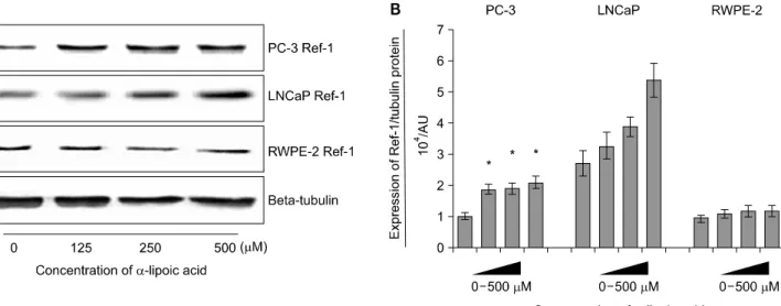
2. Effects of ALA on the expression of Ref-1 protein in PC-3 and LNCaP and RWPE-2 cells
Ref-1 was expressed in all cell lines. The expression of Ref-1 protein was increased with 125, 250, and 500μM of ALA in PC-3 (p<0.05) compared to RWPE-2 cells at 48 hours (Fig. 2).
The expression of Ref-1 protein in LNCaP tended to increase dose-dependently but showed no statistical significance (p>
0.05).
Fig. 2. Effects of α-lipoic acid on the expression of redox factor (Ref)-1 protein in PC-3 and LNCaP prostatic cancer cells and RWPE-2 cells. (A) Ref-1 protein was isolated and Western blot analysis was performed. The bar diagrams (B) show means±SE of Ref-1 expression.
*p<0.05, versus untreated control as analyzed by one-way ANOVA and Turkey’s multiple-comparison tests.
Fig. 3. Effects of α-lipoic acid on the expression of superoxide dismutase (SOD)-1 mRNA in PC-3 (A) and LNCaP (B) prostatic cancer cells and RWPE-2 cells (C). The bar diagrams show means±SE of mRNA expression. *p<0.05, versus untreated control as analyzed by one-way ANOVA and Turkey’s mul- tiple-comparison tests.
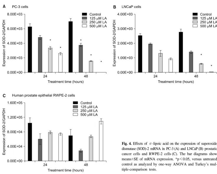
Fig. 4. Effects of α-lipoic acid on the expression of superoxide dismutase (SOD)-2 mRNA in PC-3 (A) and LNCaP (B) prostatic cancer cells and RWPE-2 cells (C). The bar diagrams show means±SE of mRNA expression. *p<0.05, versus untreated control as analyzed by one-way ANOVA and Turkey’s mul- tiple-comparison tests.
3. The effects of ALA on the expression of SOD-1, SOD-2 mRNA in PC-3, LNCaP, and RWPE-2 cells
mRNA expressions of SOD-1, SOD-2 were gradually decreased dose-dependently of ALA in the PC-3 and LNCaP human prostate carcinoma cell lines at 24 hours and 48 hours (Fig. 3, 4). In the expression of SOD-1, PC-3 cells were more affected by ALA at 24 hours and 48 hours, but LNCaP cells had the minimal changes compared to RWPE-2 (p>0.05). Both PC-3 and LNCaP cells, mRNA expressions of SOD-2 decreased at 24 and 48 hours dose-dependently compared to RWPE-2 cells.
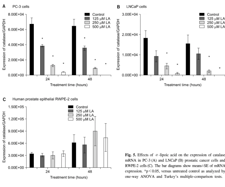
4. The effects of ALA on the expression of catalase, and GSH-Px mRNA in PC-3, LNCaP and RWPE-2 cells
mRNA expression of catalase and GSH-Px was gradually decreased in proportion to the dose of ALA in the PC-3 and
LNCaP human prostate carcinoma cell lines (Fig. 5, 6). In both PC-3 and LNCaP cells, the expressions of catalase were markedly decreased at 250 and 500μM ALA compared to untreated cells at 24 and 48 hours while there were no statistical change in RWPE-2 cells (Fig. 5).
DISCUSSION
The important finding of this study is that ALA affected the expression of Ref-1 protein and antioxidant enzyme mRNA in prostate cancer cells. PC-3, LNCaP, and RWPE-2 cells responded to ALA.
ALA has antioxidative effects and since it was synthesized by Reed,11 has been widely used as a therapeutic agent in the treatment of several conditions such as diabetic neuropathy and hypertension.12,13 Because of its potent antioxidant and redox- regulating properties, ALA was originally proposed for the
Fig. 5. Effects of α-lipoic acid on the expression of catalase mRNA in PC-3 (A) and LNCaP (B) prostatic cancer cells and RWPE-2 cells (C). The bar diagrams show means±SE of mRNA expression. *p<0.05, versus untreated control as analyzed by one-way ANOVA and Turkey’s multiple-comparison tests.
treatment of diseases mediated by free radicals such as heavy metal poisoning, liver disease, radiation poisoning, diabetes, and lung, liver, and skin tumors.7,8,12-14
Over the last decade, oxidative stress has increasingly been considered an important factor in the development and pro- gression of prostate cancer.2,15 Advanced prostate cancer is associated with increased oxidative stress. An increment of malonyldialdehyde, the end product of lipid metabolism, has been observed in hormone-resistant prostate cancer.16 p53 is a tumor suppressor that has been considered to be the oxidative stress response transcription factor. p53 is mutated in advanced stages of prostate cancer as well as in metastatic disease. ALA induces apoptosis and p-53 activation in hepatoma cells.7 Therefore, ALA is suggested to affect the antioxidant system in prostate cancer.
To identify an adequate experimental concentration of ALA, an MTT cell viability assay was performed in LNCaP cells. A
significant reduction in cell viability was observed after treatment with ALA at 250μM. Various test dosages (0, 125, 250, and 500μM) were used.
Ref-1 is a ubiquitously expressed multifunctional protein that plays an important role in the transcriptional response to oxidative stress and in DNA base excision repair. Ref-1 also facilitates the DNA binding activity of transcription factors p53, NF-kB, and AP-1.17,18 An increase in Ref-1 nuclear and cytoplasmic staining is seen in prostate cancer compared to benign prostatic hypertrophy.19 In this study, Ref-1 protein was expressed in all prostate cell lines, PC-3, LNCaP, and RWPE-2, and the ex- pression of Ref-1 protein increased in the prostate cancer cells compared to the RWPE-2 cells. With ALA treatment, the ex- pression of Ref-1 showed a dose-dependent response in prostate cancer cells but not in RWPE-2 cells. The Ref-1 gene is activated at sublethal levels of ROS.20 Elevation of the expression of Ref-1 means a reduction of DNA damage.21 ALA promotes
Fig. 6. Effects of α-lipoic acid on the expression of glutathione peroxidase mRNA in PC-3 (A) and LNCaP (B) prostatic cancer cells and RWPE-2 cells (C). The bar diagrams show means±SE of mRNA expression. *p<0.05, versus untreated control as analyzed by one-way ANOVA and Turkey’s multiple-comparison tests.
the expression of Ref-1 protein in prostate cancer cells compared with RWPE-2, suggesting that ALA may increase DNA damage of cancer cells under pathologic condition.
Cells continuously produce free radicals and ROS as part of metabolic processes. These free radicals are neutralized by an elaborate antioxidant defense system. Cellular antioxidants are composed of enzymatic and nonenzymatic antioxidants. Enzy- matic antioxidants include SOD, catalase, and GSH-Px, while nonenzymatic antioxidants include vitamin E, glutathione, vitamin C, and catechins. Under normal conditions, cells are capable of balancing the production of oxidative stress with antioxidants.22 SOD functions in the cell as one of the primary enzymatic antioxidant defenses against superoxide radicals. SOD exists in three types, cytosolic (CuZnSOD, SOD-1), mitochondrial (Mn- SOD, SOD-2), and extracellular SOD. SOD-2 is considered to be the most important antioxidant enzyme that has antitumor effects. In this study,?mRNA expressions of SOD-1, SOD-2
gradually tend to decrease dose-dependently of ALA in the PC-3 and LNCaP human prostate carcinoma cell lines. Catalase is one of the most potent catalysts known. Catalase catalyses the conversion of hydrogen peroxide, a powerful and potentially harmful oxidizing agent, to water and molecular oxygen. GSH-Px is another antioxidant enzyme with a much greater affinity for hydrogen peroxide than catalase. Elevated expression of catalase and GSH-Px mRNA in PC-3 compared to LNCaP and RWPE-2 cells was observed. In prostate cancer cells, ALA treatment reduced the mRNA expression of catalase and GSH-Px. Several studies show decrease of antioxidant enzyme activity or anti- oxidant enzyme mRNA expression by potent antioxidant.23-25 Further experiments are needed to attested the mechanism for potent antioxidant-induced changes in expression of antioxidant enzymes.
The oxidative stress was high in the prostate cancer cells.
The expression of antioxidant enzymes is elevated in prostate
cancer cell lines compared to normal prostate cells.26 ALA acts as a free radical scavenger in the prostate cell lines. The level of oxidative stress was lower in the prostate cancer cells that were treated with ALA than in the untreated cancer cells. In this study, SOD-1, SOD-2, catalase, and GSH-Px levels were decreased in the prostate cancer cells after ALA treatment.
Also, PC-3 responded well to ALA compared to LNCaP. The changes of expression of antioxidant enzymes by ALA differed according to cell type and were more active in PC-3 than LNCaP cells.
CONCLUSIONS
Ref-1 protein, which has multifunctional roles involved in oxidative DNA damage repair, was increased after ALA exposure in prostate cancer cells. The mRNA expressions of SOD-1, SOD-2, catalase, and GSH-Px were decreased by ALA compared to RWPE-2 in PC-3 and LNCaP cells. ALA affects the anti- oxidant system of prostate cancer cells and may be related to the compensatory changes in their antioxidant defense system.
REFERENCES
1. Ministry of Health and Welfare. 2002 Annual Report of the Korea central cancer registry 2003:11-24
2. Heinonen OP, Albanes D, Virtamo J, Taylor PR, Huttunen JK, Hartman AM, et al. Prostate cancer and supplementation with alpha-tocopherol and beta-carotene: incidence and mortality in a controlled trial. J Natl Cancer Inst 1998;90:440-6 3. Clark LC, Dalkin B, Krongrad A, Combs GF Jr, Turnbull BW,
Slate EH, et al. Decreased incidence of prostate cancer with selenium supplementation: results of a double-blind cancer prevention trial. Br J Urol 1998;81:730-4
4. Chen JZ, Gokden N, Greene GF, Green B, Kadlubar FF.
Simultaneous generation of multiple mitochondrial DNA mutations in human prostate tumors suggests mitochondrial hyper-mutagenesis. Carcinogenesis 2003;24:1481-7
5. Moghadasian MH, Freeman HJ, Godin DV. Endogenous antioxidant status in neoplastic and adjacent tissues in 1,2- dimethylhydrazine-induced colon cancer in rats: effects of olsalazine. Carcinogenesis 1996;17:983-7
6. Shi DY, Liu HL, Stern JS, Yu PZ, Liu SL. Alpha-lipoic acid induces apoptosis in hepatoma cells via the PTEN/Akt pathway.
FEBS Lett 2008;582:1667-71
7. Simbula G, Columbano A, Ledda-Columbano GM, Sanna L, Deidda M, Diana A, et al. Increased ROS generation and p53 activation in alpha-lipoic acid-induced apoptosis of hepatoma cells. Apoptosis 2007;12:113-23
8. Moungjaroen J, Nimmannit U, Callery PS, Wang L, Azad N, Lipipun V, et al. Reactive oxygen species mediate caspase activation and apoptosis induced by lipoic acid in human lung epithelial cancer cells through Bcl-2 down-regulation. J Pha- rmacol Exp Ther 2006;319:1062-9
9. Surapaneni KM, Venkata GR. Lipid peroxidation and antioxidant status in patients with carcinoma of prostate. Indian J Physiol Pharmacol 2006;50:350-4
10. Jung K, Seidel B, Rudolph B, Lein M, Cronauer MV, Henke W, et al. Antioxidant enzymes in malignant prostate cell lines and in primary cultured prostatic cells. Free Radic Biol Med 1997;23:127-33
11. Reed LJ, DeBusk BG, Gunsalus IC, Hornberger CS Jr.
Crystalline alpha-lipoic acid: a catalytic agent associated with pyruvate dehydrogenase. Science 1951;114:93-4
12. Ziegler D. Thioctic acid for patients with symptomatic diabetic polyneuropathy: a critical review. Treat Endocrinol 2004;3:173-89 13. Wollin SD, Jones PJ. Alpha-lipoic acid and cardiovascular
disease. J Nutr 2003;133:3327-30
14. Ho YS, Lai CS, Liu HI, Ho SY, Tai C, Pan MH, et al.
Dihydrolipoic acid inhibits skin tumor promotion through anti-inflammation and anti-oxidation. Biochem Pharmacol 2007;
73:1786-95
15. Pathak SK, Sharma RA, Steward WP, Mellon JK, Griffiths TR, Gescher AJ. Oxidative stress and cyclooxygenase activity in prostate carcinogenesis: targets for chemopreventive stra- tegies. Eur J Cancer 2005;41:61-70
16. Yossepowitch O, Pinchuk I, Gur U, Neumann A, Lichtenberg D, Baniel J. Advanced but not localized prostate cancer is associated with increased oxidative stress. J Urol 2007;178:
1238-43
17. Evans AR, Limp-Foster M, Kelley MR. Going APE over ref-1.
Mutat Res 2000;461:83-108
18. Gaiddon C, Moorthy NC, Prives C. Ref-1 regulates the transactivation and pro-apoptotic functions of p53 in vivo.
EMBO J 1999;18:5609-21
19. Kelley MR, Cheng L, Foster R, Tritt R, Jiang J, Broshears J, et al. Elevated and altered expression of the multifunctional DNA base excision repair and redox enzyme Ape1/ref-1 in prostate cancer. Clin Cancer Res 2001;7:824-30
20. Ramana CV, Boldogh I, Izumi T, Mitra S. Activation of apurinic/apyrimidinic endonuclease in human cells by reactive oxygen species and its correlation with their adaptive response to genotoxicity of free radicals. Proc Natl Acad Sci USA 1998;95:5061-6
21. Lau JP, Weatherdon KL, Skalski V, Hedley DW. Effects of gemcitabine on APE/ref-1 endonuclease activity in pancreatic cancer cells, and the therapeutic potential of antisense oli- gonucleotides. Br J Cancer 2004;91:1166-73
22. Karihtala P, Soini Y. Reactive oxygen species and antioxidant mechanisms in human tissues and their relation to malig- nancies. APMIS 2007;115:81-103
23. Powolny AA, Singh SV. Plumbagin-induced apoptosis in human prostate cancer cells is associated with modulation of cellular redox status and generation of reactive oxygen species.
Pharm Res 2008;25:2171-80
24. Asaumi H, Watanabe S, Taguchi M, Tashiro M, Nagashio Y, Nomiyama Y, et al. Green tea polyphenol (-)-epigallocatech- in-3-gallate inhibits ethanol-induced activation of pancreatic stellate cells. Eur J Clin Invest 2006;36:113-22
25. Siddiqui IA, Raisuddin S, Shukla Y. Protective effects of black tea extract on testosterone induced oxidative damage in prostate. Cancer Lett 2005;227:125-32
26. Chowdhury SK, Raha S, Tarnopolsky MA, Singh G. Increased expression of mitochondrial glycerophosphate dehydrogenase and antioxidant enzymes in prostate cancer cell lines/cancer.
Free Radic Res 2007;41:1116-24