122
A Protocol for Transrectal, Ultrasonography-guided, 41-core Prostate Needle Biopsy
Sang Bong Jeon1, Chen Zhao1, Young Bum Jung1, Young Kyung Park1, Jong Kwan Park1,2
From the 1Department of Urology, Chonbuk National University Medical School,
2Institute for Medical Sciences, and 2Research Institute of Clinical Medicine of Chonbuk National University Hospital, 2Clinical Trial Center of Chonbuk National University Hospital, Jeonju, Korea
Purpose: We evaluated the rate of prostate cancer detection using the transrectal ultrasound (TRUS) guided, 41-core prostate needle biopsy protocol.
Materials and Methods: Between May 2006 and May 2007, 110 patients received a 12-core prostate needle biopsy (Group I) and 62 patients underwent a 41-core prostate needle biopsy (Group II) under local anesthesia. In addition to sextant biopsy, 41-core prostate needle biopsies were obtained from the each lateral peripheral zone (base, mid, and apex glands), midline peripheral zone (base, mid, and apex glands) and each transition zone.
Results: There were no significant differences between the two groups for age, prostate-specific antigen (PSA), or prostate volume. The cancer detec- tion rate in the Group I and Group II was 19.1% and 24.2%, respectively.
We found a significantly higher detection rate in the 41-core biopsy with prostate volumes of more than 40cc (28.1% vs 11.3%). The intraoperative pain score was lower in Group II than Group I (1.57±1.45 vs 3.17±1.73, p<0.01).
Conclusions: The 41-core prostate needle biopsy protocol is safe and intraoperative pain is low. The cancer detection rate of the 41-core prostate needle biopsy was higher than the 12-core prostate needle biopsy in patients with a prostate volume of more than 40cc. (Korean J Urol 2008;
49:122-126)
Key Words: Prostate cancer, Biopsy
대한비뇨기과학회지 제 49 권 제 2 호 2008
1전북대학교 의과대학 비뇨기과학교실,
2전북대학교병원 임상연구소,
2전북대학교 의과학연구소,
2전북대학교병원 지역임상시험센터
전상봉1ㆍ조 신1ㆍ정영범1 박영경1ㆍ박종관1,2
접수일자:2007년 8월 24일 채택일자:2007년 12월 24일
교신저자: 박종관
전북대학교 의과대학 비뇨기과학교실
전북 전주시 덕진구 금암동 634-18
561-712
TEL: 063-250-1510 FAX: 063-250-1564 E-mail: rain@chonbuk.
ac.kr
서 론
혈청전립선특이항원 (prostate specific antigen; PSA)이 증 가하거나 직장수지검사 (digital rectal examination; DRE)상 이상소견을 보이는 환자에서 경직장초음파 유도하 6부위 전립선 생검법 (sextant biopsy)은 1989년 Hodge 등1이 소개 한 후 전립선암을 진단하는 표준검사방법으로 이용되고 있 다. 그러나 6부위 전립선 생검법은 위음성률이 20-30%에 달하고 있고, 전립선 용적이 클수록 전립선암의 진단율이 감소하기 때문에 다양한 전립선 생검법이 개발되고 있다.2-4 또한 생검수를 증가시킨 방법들을 이용할 경우 6부위 전립
선 생검법에 비해 전립선암 발견의 위음성률이 감소한다고 보고되고 있다.5,6 이에 저자들은 본원에서 시행하고 있는 경직장 초음파 유도 41부위 전립선 생검법을 이용한 연구 결과를 보고한다.
대상 및 방법
2006년 5월부터 2007년 5월까지 혈청전립선특이항원치 가 4ng/ml 이상이거나 직장수지검사에서 결절이 만져지는 환자 172명을 대상으로 경직장 초음파 (model 2102, B&K medical, Denmark)유도 전립선 생검을 시행하였다. 172명의 환자 중 110명의 환자에서 12부위 전립선 생검법 (Group I)
Fig. 1. Location of biopsies in the 41-core procedure. A dark circle (sextant and far lateral peripheral zones) means three biopsies performed from the same site. A gray circle means biopsies at the midline peripheral zones and an open circle means biopsies performed in both transition zones.
Table 2. Cancer detection rate according to prostate volume No. of Group I Group II
p-value
patients (%) (%)
Overall 172 21/110 (19.1) 15/62 (24.2) 0.43 Prostate volume (cc)
≤40 87 15/57 (26.3) 6/30 (20.0) 0.513 >40 85 6/53 (11.3) 9/32 (28.1) 0.049 p-values calculated with Pearson chi-square test.
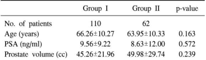
Table 1. Patient characteristics (mean±SD)
Group I Group II p-value
No. of patients 110 62
Age (years) 66.26±10.27 63.95±10.33 0.163
PSA (ng/ml) 9.56±9.22 8.63±12.00 0.572
Prostate volume (cc) 45.26±21.96 49.98±29.74 0.239 PSA: prostate-specific antigen. p-values calculated with Student's t-test.
을 시행하였고, 62명의 환자에서 생검의 개수를 증가시킬 수록 합병증의 빈도가 증가할 수 있다는 설명을 하고 동의 를 얻은 후에 41부위 전립선 생검법 (Group II)을 시행하였 다. 12부위 전립선 생검법은 환자를 쇄석위 또는 측와위를 취한 상태에서 직장 내로 2% 농도의 리도카인 젤리 10cc와 베타딘을 주입하여 국소 마취를 시행한 후 경직장 초음파 유 도 하에 18 gauge 생검침과 자동 전립선 조직검사총 (PRO- MAGTM ULTRA, M&K medical, Gainesville, USA)을 이용하 여 전립선 좌, 우측엽 중엽의 첨부, 중간부, 기저부와 양측 말초대의 첨부, 중간부, 기저부에서 각각 1부위의 생검을 시행하였다. 41부위 전립선 생검법 또한 환자를 쇄석위 또 는 측와위를 취한 상태에서 직장 내로 베타딘을 주입한 후 경직장 초음파 유도 하에 양측 전립선과 정낭 사이의 신경 혈관다발에 1% 리도카인 5cc를 각각 주입하여 국소 마취를 시행한 후 조직생검을 시행하였다. 조직생검은 12부위 생 검과 동일한 기구를 이용하여 전립선 좌, 우측엽 중엽의 첨 부, 중간부, 기저부와 양측 말초대의 첨부, 중간부, 기저부 에서 각각 3부위 생검을 시행하였고, 전립선 중엽의 첨부, 중간부, 기저부와 좌우 이행대에서 각각 1부위 생검을 시행 하여 총 41부위의 조직 생검을 시행하였다 (Fig. 1).
생검 후 지혈을 위해 직장을 통해 생검을 실시한 부위를 2분간 압박하였고, 직장에 베타딘을 주입한 후 gauze pack- ing을 시행하였다. 또한 감염의 예방을 위해 1주일간 fluo- roquinolone 계열의 경구용 항생제와 지혈제를 투여하였다.
통증의 정도는 시술 직후 통증의 정도를 0-10까지 표시한 아날로그 통증지표 (visual analog scale)를 이용하여 평가하 였고, 합병증에 대한 평가는 1주일 후 환자가 외래 방문 시 설문을 통해 조직 검사 후 혈뇨, 혈변, 요로계 감염, 성행위 를 가졌던 환자에서 혈정액 여부 등을 평가하였다.
통계분석은 Student's t-test와 Pearson chi-square test를 이 용하였고 p값이 0.05 미만이면 통계적으로 의미 있는 것으 로 판정하였다. 평균값은 mean±standard deviation (mean±
SD), 통계프로그램은 SPSS (ver 10.0)를 사용하였다.
결 과
Group I과 Group II 간에 연령, 혈청 PSA치와 전립선 용적 은 유의한 차이를 보이지 않았다 (Table 1). 전체 172명 중 36명 (20.9%)이 암으로 진단되었고, Group I이 21명 (19.1%), Group II가 15명 (24.2%)으로 두 군 간의 암진단율은 통계학 적으로 유의한 차이는 없었다.
전립선용적을 40cc를 기준으로 평가하였을 때 전립선용 적이 40cc 이하인 경우, Group I 중 15명 (26.3%), Group II 중 6명 (20.0%)이 암으로 진단되었으나 암진단율의 차이는 통계학적으로 유의하지 않았다. 전립선용적이 40cc를 초과 한 경우 Group I 중 6명 (11.3%), Group II 중 9명 (28.1%)이 암으로 진단되어 Group II에서 통계학적으로 유의하게 암 진단율이 높았다 (Table 2).
생검 직후 시행한 통증관련 설문조사에서 조직검사 동안
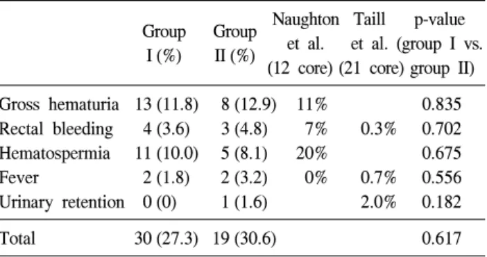
Table 3. Comparison of complications
Naughton Taill p-value Group Group
et al. et al. (group I vs.
I (%) II (%)
(12 core) (21 core) group II) Gross hematuria 13 (11.8) 8 (12.9) 11% 0.835 Rectal bleeding 4 (3.6) 3 (4.8) 7% 0.3% 0.702 Hematospermia 11 (10.0) 5 (8.1) 20% 0.675
Fever 2 (1.8) 2 (3.2) 0% 0.7% 0.556
Urinary retention 0 (0) 1 (1.6) 2.0% 0.182
Total 30 (27.3) 19 (30.6) 0.617
p-values calculated with Pearson chi-square test.
의 통증의 정도는 Group I이 평균 3.17±1.73점, Group II 평 균 1.57±1.45점으로 두 군 간에 유의한 차이를 보였다 (p<
0.001).
전립선 생검 1주일 후에 실시한 합병증에 관한 설문에서 Group I의 27.3%, Group II의 30.6%에서 육안적 혈뇨, 직장 출혈, 혈정액, 발열과 요폐색 중 1개 이상의 합병증이 발생 하였으나 대부분의 환자에서 특별한 처치 없이 자연적으로 소실되었고 발열이 발생한 환자 중 41부위 전립선 생검을 시 행 받은 환자 1명에서 입원치료를 하였다. 합병증의 발생은 두 군 간에 유의한 차이를 보이지 않았다 (p=0.617, Table 3).
고 찰
Hodge 등1이 제시한 6부위 전립선 생검법은 지난 수십 년 동안 전립선암의 진단에 표준적인 방법으로 사용되어 왔 다. Hammerer와 Huland7은 6부위 전립선 생검법을 이용한 전립선암 진단율은 45% 정도였다고 보고하였지만, 이후의 연구에서는 위음성률을 20-30% 정도로 보고하였다. 특히 전립선용적이 큰 경우 전립선암 진단율은 낮고 위음성률은 높아 전립선암 진단율을 높이기 위한 새로운 전립선 생검 법이 시도되었다.2-4
전립선암의 진단율을 높이기 위한 방법으로 전립선 생검 시 sector를 달리하는 방법과 core 수를 늘리는 방법이 있다.
Chen 등8은 6부위 전립선 생검법은 이행대의 전방부 (an- terior transition zone), 말초대의 중간부 (midline peripheral zone), 말초대 전혼의 하방부 (lower portion of the anterior horn of the peripheral zone)에서 조직의 생검이 어려워 진단 이 정확하지 않다고 주장하였다. Eskew 등9은 6부위 전립선 생검법에 말초대의 중간부와 외측을 추가한 방법을 시행하 여 전립선암 진단율의 상승을 보고하였다. 또한 Levine 등10 은 6부위 생검법을 방향을 바꾸지 않은 채 단순히 2번 연속 시행하였을 때 진단율이 10% 향상되었으며, 10부위 생검
시 Presti 등5은 14%, Yeo 등11은 3.1%의 진단율 상승을 보고 하였다. 이 외에도 11부위 생검,12 12부위 생검,13 21부위 생 검14 등이 통상적인 6부위 전립선 생검보다 전립선암 진단 율이 높다고 보고되었다. 그러나 Ashley 등15은 24부위 이상 의 생검이 12-18부위 생검에 비해 전립선암 진단율에 차이 가 없다고 하였다. 저자들이 시행한 41부위 전립선 생검은 12부위 생검에 비해 전립선암 진단율이 5.1% 상승하였으나 통계학적으로 유의한 차이는 없었다. 전립선의 크기가 큰 경우 전립선암 진단율은 낮아지는데, Uzzo 등16은 6부위 전 립선 생검의 전립선암 진단율이 전립선용적이 50cc 미만인 경우 38%, 50cc 이상인 경우 23%로 보고하였고, Park 등4은 전립선용적이 30cc 이상인 경우 전립선암 진단율이 급격히 감소한다고 보고하였다. 전립선용적이 40cc를 초과한 경우 저자들이 시행한 41부위 전립선 생검은 12부위 생검에 비 해 전립선암 진단율이 16.8% 상승하였고 이는 통계학적으 로 유의한 차이를 보여 전립선 용적이 큰 경우 41부위 전립 선 생검법은 전립선암 진단에 효과적일 것으로 생각한다.
생검 숫자가 증가하면 출혈, 감염 등의 합병증의 빈도가 증가할 가능성이 있지만 Naughton 등17은 6부위 생검과 12 부위 생검을 비교하여 두 군 간의 통증이나 질병의 이환율 에 차이가 없다고 보고하였고, de la Taille 등14 또한 6부위 생검과 21부위 생검에서 합병증의 이환율에 차이가 없다고 보고하였다. 일반적으로 육안적 혈뇨는 14-58%에서 발생하 고, 직장출혈은 2-37.1%, 혈정액은 3-45%에서 발생한다. 전 립선 생검 후 감염은 예방적 항생제를 사용한 경우 0.2- 4.2%로 비교적 드물게 발생한다.18,19 41부위 생검은 기존의 생검보다 침을 찌르는 횟수가 더 많아 시술 자체와 연관된 합병증이 증가할 수 있다. 그러나 41부위 생검과 연관된 합 병증 중 육안적 혈뇨는 12.9%, 직장출혈은 4.8%, 혈정액은 8.1%에서 발생하여 기존의 생검 후 발생하는 합병증의 빈 도와 큰 차이를 보이지 않았다. 다만 혈정액은 본원의 경우 조직검사 1주일 후 내원을 하게 되는데, 많은 수의 환자들 이 그 기간 내에 성관계를 갖기가 어려울 수 있어 장기적인 추적관찰이 필요할 것으로 생각한다. 또한 Rietbergen 등18 은 전립선 생검 2시간 전과 생검 후 4시간 후에 항생제를 투여한 경우 4.2%에서 38.5oC 이상의 발열이 발생하였다고 보고하였고, Enlund와 Varenhorst19는 예방적 항생제를 사용 하지 않아도 발열은 2.9%에서만 발생하였다고 보고하였다.
전립선 생검 시 통증은 생검침이 전립선 피막을 관통할 때 주로 발생하며 직장에 탐촉자를 삽입할 때에도 발생한 다.20 전립선 피막이나 간질에는 자율신경 섬유들이 풍부하 게 분포되어 있다. 전립선의 신경지배는 천수 S2-5의 미측 근과 하복 신경총을 경유하는 교감 사슬로부터 파생되고, 이들 섬유들은 전립선 신경총에서 가지를 내어 전립선 기
저부의 후외측면에 위치하는 전립선 혈관경과 함께 주행한 다. 이들 자율신경들이 척수로 내장감각을 전달하여 전립 선 생검 시 통증을 유발한다.21 저자들은 전립선 생검 시 국 소마취 방법으로 12부위 생검은 리도카인 젤을 사용하였고 41부위 생검은 전립선 주위 신경차단술을 시행하였다. 조 직검사 동안의 통증 점수는 전립선 주위 신경차단술을 시 행한 41부위 군이 리도카인 젤을 사용한 12부위 군에 비해 조직 생검의 수가 많았지만 통증 점수가 유의하게 감소하 여 두 가지 방법을 비교할 때 생검 수보다는 정확한 마취방 법이 통증의 감소에 중요한 것으로 생각된다. Issa 등22은 리 도카인 젤을 직장 내에 투여한 후 전립선 생검을 했을 때 통증 점수가 유의하게 감소하였다고 보고하였지만 Des- grandchamps 등23은 리도카인 젤의 직장 내 투여가 전립선 생검 시 환자들의 순응도를 증가시키지 못했다고 보고하고 있어 리도카인 젤을 사용한 국소마취 방법의 효과는 일관 적으로 보고되지 않고 있다. 그러나 전립선 주위 신경차단 술은 대부분 효과가 좋다고 보고되고 있다. Lee 등24은 전립 선 주위 신경차단술을 시행한 군이 리도카인 젤을 사용한 군보다 통증 점수의 유의한 감소를 보고하였고, Byun 등25 또한 전립선 주위 신경차단술을 시행한 경우가 어떠한 마 취도 시행하지 않은 경우에 비해 통증 점수의 유의한 감소 를 보고하였다. 따라서 전립선 주위 신경차단술은 전립선 생검시 먼저 선택해야 할 마취방법으로 생각한다.
결 론
혈관신경총다발의 마취 후 경직장 초음파 유도하의 41부 위 전립선 생검법은 전립선용적이 40cc를 초과한 환자에서 12부위 생검에 비해 전립선암 진단율을 높일 수 있으며, 통 증과 합병증이 적은 비교적 안전한 방법으로 생각한다.
REFERENCES
1. Hodge KK, McNeal JE, Terris MK, Stamey TA. Random systematic versus directed ultrasound guided transrectal core biopsies of the prostate. J Urol 1989;142:71-4
2. Epstein JI, Walsh PC, Sauvageot J, Carter HB. Use of repeat sextant and transition zone biopsies for assessing extent of prostate cancer. J Urol 1997;158:1886-90
3. Stroumbakis N, Cookson MS, Reuter VE, Fair WR. Clinical significance of repeat sextant biopsies in prostate cancer patients. Urology 1997;49(Suppl 3A):113-8
4. Park YS, Kwak C, Shim HB. Effect of prostate size on cancer detection rate of traditional sextant biopsy. Korean J Urol 2003;44:1098-102
5. Presti JC Jr, Chang JJ, Bhargava V, Shinohara K. The optimal
systematic prostate biopsy scheme should include 8 rather than 6 biopsies: results of a prospective clinical trial. J Urol 2000;163:163-6
6. Eskew LA, Bare RL, McCullough DL. Systematic 5 region prostate biopsy is superior to sextant method for diagnosing carcinoma of the prostate. J Urol 1997;157:199-202 7. Hammerer P, Huland H. Systematic sextant biopsies in 651
patients referred for prostate evaluation. J Urol 1994;151:
99-102
8. Chen ME, Troncoso P, Johnston DA, Tang K, Babaian RJ.
Optimization of prostate biopsy strategy using computer based analysis. J Urol 1997;158:2168-75
9. Eskew LA, Woodruff RD, Bare RL, McCullough DL. Prostate cancer diagnosed by the 5 region biopsy method is significant disease. J Urol 1998;160:794-6
10. Levine MA, Ittman M, Melamed J, Lepor H. Two consecutive sets of transrectal ultrasound guided sextant biopsies of the prostate for the detection of prostate cancer. J Urol 1998;
159:471-5
11. Yeo BG, Lee ES, Byun SS. Peripheral 10 site prostate biopsy:
is it really effective? Korean J Urol 2003;44:851-4
12. Babaian RJ, Toi A, Kamoi K, Troncoso P, Sweet J, Evans R, et al. A comparative analysis of sextant and an extended 11- core multisite directed biopsy strategy. J Urol 2000;163:152-7 13. Cho SH, Kim SI, Park HY. The efficacy of routine sextant
prostate biopsy plus selective lesion-directed prostate biopsy.
Korean J Urol 2005;46:700-3
14. de la Taille A, Antiphon P, Salomon L, Cherfan M, Porcher R, Hoznek A, et al. Prospective evaluation of a 21-sample needle biopsy procedure designed to improve the prostate cancer detection rate. Urology 2003;61:1181-6
15. Ashley RA, Inman BA, Routh JC, Mynderse LA, Gettman MT, Blute ML. Reassessing the diagnostic yield of saturation biopsy of the prostate. Eur Urol 2007 [Epub ahead of print]
16. Uzzo RG, Wei JT, Waldbaum RS, Perlmutter AP, Byene JC, Vaughan ED Jr. The influence of prostate size on cancer detection. Urology 1995;46:831-6
17. Naughton CK, Ornstein DK, Smith DS, Catalona WJ. Pain and morbidity of transrectal ultrasound guided prostate biopsy: a prospective randomized trial of 6 versus 12 cores. J Urol 2000;163:168-71
18. Rietbergen JB, Kruger AE, Kranse R, Schroder FH. Com- plications of transrectal ultrasound-guided systematic sextant biopsies of the prostate: evaluation of complication rates and risk factors within a population-based screening program.
Urology 1997;49:875-80
19. Enlund AL, Varenhorst E. Morbidity of ultrasound-guided transrectal core biopsy of the prostate without prophylactic antibiotic therapy. A prospective study in 415 cases. Br J Urol 1997;79:777-80
20. Nash PA, Bruce JE, Indudhara R, Shinohara K. Transrectal ultrasound guided prostatic nerve blockade eases systematic
needle biopsy of the prostate. J Urol 1996;155:607-9 21. Hollabaugh RS Jr, Dmochowski RR, Steiner MS. Neuro-
anatomy of the male rhabdosphincter. Urology 1997;49:426-34 22. Issa MM, Bux S, Chun T, Petros JA, Labadia AJ, Anastasia
K, et al. A randomized prospective trial of intrarectal lidocaine for pain control during transrectal prostate biopsy: the Emory University experience. J Urol 2000;164:397-9
23. Desgrandchamps F, Meria P, Irani J, Desgrippes A, Teillac P, Le Duc A. The rectal administration of lidocaine gel and tolerance of transrectal ultrasonography-guided biopsy of the
prostate: a prospective randomized placebo-controlled study.
BJU Int 1999;83:1007-9
24. Lee YI, Seo IY, Jeong HJ, Rim JS. Comparison of two local anesthetic methods for transrectal ultrasound guided prostate needle biopsy: periprostatic injection of lidocaine and rectal instillation of lidocaine gel. Korean J Urol 2004;45:423-7 25. Byun SS, Lee HJ, Ku JH, Park KJ, Lim DJ, Lee SE, et al.
Effect of periprostatic nerve blockade for transrectal ultrasound guided biopsy of the prostate. Korean J Urol 2004;45:663-6