HIGHLIGHTS
• Focused ultrasound (FUS) has emerged as a potential non-invasive brain stimulation technique.
• Transcranial FUS has exquisite spatial specificity with deep tissue penetration.
• Lasting neuromodulatory effects after FUS may have potential for neurorehabilitation.
• No adverse effects have been reported from large animals/non-human primates/humans.
• Establishment of new safety guideline is warranted for clinical translation.
Special Review
Received: Dec 27, 2020 Revised: Feb 17, 2021 Accepted: Mar 4, 2021 Correspondence to Seung-Schik Yoo
Department of Radiology, Brigham and Women's Hospital, Harvard Medical School, 75 Francis Street, Boston, MA 02115, USA.
E-mail: [email protected]
Wonhye Lee, Daniel S. Weisholtz, Gary E. Strangman,Seung-Schik Yoo
Safety Review and Perspectives of
Transcranial Focused Ultrasound
Brain Stimulation
ABSTRACT
Ultrasound is an important theragnostic modality in modern medicine. Technical
advancement of both acoustic focusing and transcranial delivery have enabled administration of ultrasound waves to localized brain areas with few millimeters of spatial specificity and penetration depth sufficient to reach the thalamus. Transcranial focused ultrasound (tFUS) given at a low acoustic intensity has been shown to increase or suppress the excitability of region-specific brain areas. The neuromodulatory effects can outlast the sonication, suggesting the possibility of inducing neural plasticity needed for neurorehabilitation.
Increasing numbers of studies have shown the efficacy and excellent safety profile of the technique, yet comparisons among the safety-related parameters have not been compiled.
This review aims to provide safety information and perspectives of tFUS brain stimulation.
First, the acoustic parameters most relevant to thermal/mechanical tissue damage are discussed along with regulated parameters for existing ultrasound therapies/diagnostic imaging. Subsequently, the parameters used in studies of large animals, non-human primates, and humans are surveyed and summarized in terms of the acoustic intensity and the mechanical index. The pulse-mode operation and the use of low ultrasound frequency for tFUS-mediated brain stimulation warrant the establishment of new safety guidelines/
recommendations for the use of the technique among healthy volunteers, with additional cautionary requirements for its clinical translation.
Keywords: Low Intensity Pulsed Ultrasound; Neuromodulators; Acoustic Stimulation; Safety
INTRODUCTION
Discovery of piezoelectric phenomena and materials by Curie brothers (Paul-Jacques and Pierre) in the late 19th century catalyzed the development of a piezoelectric transducer that converts electrical signals into mechanical vibration, thereby generating pressure waves in the inaudible frequency domain (> 20 kHz). Ultrasound sonication into an object and subsequent detection of reflected/refracted soundwaves reveal internal structures of an object in a non-invasive fashion, and lead to development of various ultrasound techniques for echo location and imaging. In addition to industrial applications such as assessment of cracks in welded components through ultrasonic non-destructive testing [1], immense contributions
Special Review
Received: Dec 27, 2020 Revised: Feb 17, 2021 Accepted: Mar 4, 2021 Correspondence to Seung-Schik Yoo
Department of Radiology, Brigham and Women's Hospital, Harvard Medical School, 75 Francis Street, Boston, MA 02115, USA.
E-mail: [email protected] Copyright © 2021. Korean Society for Neurorehabilitation
This is an Open Access article distributed under the terms of the Creative Commons Attribution Non-Commercial License (https://
creativecommons.org/licenses/by-nc/4.0) which permits unrestricted non-commercial use, distribution, and reproduction in any medium, provided the original work is properly cited.
ORCID iDs Wonhye Lee
https://orcid.org/0000-0002-4203-2209 Daniel S. Weisholtz
https://orcid.org/0000-0001-6277-4894 Gary E. Strangman
https://orcid.org/0000-0001-8896-2164 Seung-Schik Yoo
https://orcid.org/0000-0002-5150-9857 Conflict of Interest
The authors have no potential conflicts of interest to disclose.
Wonhye Lee ,1 Daniel S. Weisholtz ,2 Gary E. Strangman ,3,4,5 Seung-Schik Yoo 1
1Department of Radiology, Brigham and Women's Hospital, Harvard Medical School, Boston, MA, USA
2Department of Neurology, Brigham and Women's Hospital, Harvard Medical School, Boston, MA, USA
3Neural Systems Group, Massachusetts General Hospital, Harvard Medical School, Charlestown, MA, USA
4Center for Space Medicine, Baylor College of Medicine, Houston, TX, USA
5Translational Research Institute, Houston, TX, USA
Safety Review and Perspectives of
Transcranial Focused Ultrasound
Brain Stimulation
of ultrasound have been seen in medical imaging whereby the ultrasound waves are used to capture and characterize spatiotemporal features of biological organs and physiology [2-4]. Ultrasound has also been deployed as therapeutic methods to promote bone healing [5,6] or alleviate muscle pain [7,8]. Consequently, ultrasound has become an indispensable theragnostic modality in modern medicine.
Ultrasound waves can be focused to a specific region of an object, at a set distance from the transducer, with amplification of mechanical energy at the focus compared to its surroundings.
Efforts through the 1980s and 90s were made in developing novel therapeutic applications of focused ultrasound (FUS), whereby mechanical acoustic energy is transposed to either thermal energy, for example, to ablate uterine fibroid or to high pressure field to break away kidney stones (e.g., extracorporeal shock wave lithotripsy) [9,10]. The acoustic intensities used in these applications (which we will address later) are high enough to induce temperature elevation or to create mechanical shock waves (intensity range is reviewed in Tables 1 and 2).
For transcranial application of FUS, the skull absorbs much of the acoustic energy and, therefore, a different set of technical considerations are important, as compared to conventional ultrasound imaging (see previous review [11]). For example, brain stimulation
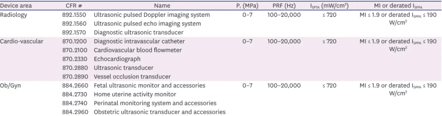
Table 1. Ultrasound-based medical devices and interrogation parameters for regulation (diagnostic ultrasound imaging; ultrasound frequency 1–20 MHz)
Device area CFR # Name Pr (MPa) PRF (Hz) ISPTA (mW/cm2) MI or derated ISPPA
Radiology 892.1550 Ultrasonic pulsed Doppler imaging system 0–7 100–20,000 ≤ 720 MI ≤ 1.9 or derated ISPPA ≤ 190 W/cm2
892.1560 Ultrasonic pulsed echo imaging system 892.1570 Diagnostic ultrasonic transducer
Cardio-vascular 870.1200 Diagnostic intravascular catheter 0–7 100–20,000 ≤ 720 MI ≤ 1.9 or derated ISPPA ≤ 190 W/cm2
870.2100 Cardiovascular blood flowmeter 870.2330 Echocardiograph
870.2880 Ultrasonic transducer 870.2890 Vessel occlusion transducer
Ob/Gyn 884.2660 Fetal ultrasonic monitor and accessories 0–7 100–20,000 ≤ 720 MI ≤ 1.9 or derated ISPPA ≤ 190 W/cm2
884.2730 Home uterine activity monitor
884.2740 Perinatal monitoring system and accessories 884.2960 Obstetric ultrasonic transducer and accessories
For cardiac use: ISPTA ≤ 430 mW/cm2 and MI ≤ 1.9 or derated ISPPA ≤ 190 W/cm2. For continuous-wave fetal imaging and others: ISPTA ≤ 94 mW/cm2 and MI ≤ 1.9 or derated ISPPA ≤ 190 W/cm2 (others category includes abdominal, intraoperative, pediatric, small organ [breast, thyroid, testes, etc.]). For ophthalmic use: ISPTA ≤ 50 mW/cm2 and MI ≤ 0.23. For fetal heart rate monitors with low-power unfocused continuous-wave Doppler transducers: ISATA or ISAPA ≤ 20 mW/cm2.
These exceptions are based on the typical clinical operational conditions that require (1) prolonged application of (2) continuous-wave ultrasound that is more prone to impart thermal energy to the tissue. For ophthalmic use, high sensitivity of the retinal cells toward mechanical pressure reduces the upper limit of the intensity.
CFR, Code of Federal Regulation; Pr, peak negative pressure; PRF, pulse repetition frequency; MI, mechanical index; ISPTA, spatial-peak temporal-average intensity; ISPPA, spatial-peak pulse-average intensity; ISATA, spatial-average temporal-average intensity; ISAPA, spatial-average pulse-average intensity (ISATA and ISAPA
are used in non-focal applications).
Table 2. Ultrasound-based medical devices and interrogation parameters for regulation (therapeutic focused ultrasound; ultrasound frequency 0.1–10 MHz)
Device area CFR # Name Pr (MPa) PRF ISPTA (W/cm2) MI
Gastroenterology/urology 876.4340 High intensity ultrasound system for
prostate tissue ablation 10 N/A continuous wave 1,000–10,000 10–20
876.5990 ESWL 20–110 Variable; 5–20 µs single pulse < 1 200 (≥ 20)
General and plastic surgery 878.4590 Focused ultrasound for tissue heat or
mechanical cellular disruption 12–25 N/A continuous wave 1,000 10–20 MI and ISPTA are approved by case-evaluation.
For cardiac use: ISPTA ≤ 430 mW/cm2 and MI ≤ 1.9 or derated ISPPA ≤ 190 W/cm2. For continuous-wave fetal imaging and others: ISPTA ≤ 94 mW/cm2 and MI ≤ 1.9 or derated ISPPA ≤ 190 W/cm2 (others category includes abdominal, intraoperative, pediatric, small organ [breast, thyroid, testes, etc.]). For ophthalmic use: ISPTA ≤ 50 mW/cm2 and MI ≤ 0.23. For fetal heart rate monitors with low-power unfocused continuous-wave Doppler transducers: ISATA or ISAPA ≤ 20 mW/cm2.
These exceptions are based on the typical clinical operational conditions that require (1) prolonged application of (2) continuous-wave ultrasound that is more prone to impart thermal energy to the tissue. For ophthalmic use, high sensitivity of the retinal cells toward mechanical pressure reduces the upper limit of the intensity.
CFR, Code of Federal Regulation; Pr, peak negative pressure; PRF, pulse repetition frequency; MI, mechanical index; ISPTA, spatial-peak temporal-average intensity; ISPPA, spatial-peak pulse-average intensity; ISATA, spatial-average temporal-average intensity; ISAPA, spatial-average pulse-average intensity (ISATA and ISAPA
are used in non-focal applications); ESWL, extracorporeal shock wave lithotripters.
typically requires frequencies in the 200–700 kHz range. This is much lower compared to the ones used in imaging (on the order of 3–4 MHz), but is necessary to enhance the transmission for the transcranial application. The skull also introduces phase aberrations in ultrasound waves propagation, and additional phase correction schemes are used for focusing, for
example, multi-array ultrasound transducer configuration [12,13] or the use of phase-correcting acoustic lenses [14-16]. The multi-array transducer can electronically steer the depth and location of the focus via adjustment of the wave phase of each transducer element in the array [12,13] while a phase-correcting lens or transducer geometry (e.g., curvature and/or diameter of the piezoelectric material) determines the depth and spatial pattern of the acoustic focus in the case of single-element transducer configuration [14-16]. The intensity of stimulation is controlled by changing the pressure level at an acoustic focus, achieved by controlling the input voltage and power to the piezoelectric material of the transducer. To account for the intensity attenuation by the skull, a derating factor is applied, which is estimated either from direct measurement of transmitted acoustic pressure through ex vivo skull samples or from numerical simulation of acoustic propagation through the skull [17]. Image-guidance has also become a crucial part of the procedure to navigate the focus, avoiding non-therapeutic areas [18]. With these technical advances, the transcranial FUS (tFUS) technique is now used for ablation of brain tissues. For example, high-intensity FUS (HIFU) has been used in functional neurosurgery for essential tremor [19-22] and obsessive-compulsive disorder [23].
The biological effects of ultrasound have been studied over decades by many investigators [24-30], including the Fry brothers (William and Francis) whose early pioneering works demonstrated the ability to modulate of neural excitability using low-intensity FUS on cat thalamus [24]. Rekindled by several studies in the late 2000, ultrasound sonication was shown to reversibly alter the excitability of both peripheral and central nervous tissues [31-39].
With a unique ability to reach deep brain areas with excellent spatial specificity compared to other brain stimulation approaches, FUS has positioned itself as a unique non-invasive brain stimulation modality. Recent studies have shown that the effects of acoustic stimulation can last significantly after the sonication [40,41], which suggests the possibility for inducing therapeutic neuroplasticity. This potential for neurorehabilitation has given FUS significant momentum in its translation into clinical trials, including treatment of major depressive disorders, disorder of consciousness of traumatic brain injury and epilepsy (ClinicalTrials.gov identifiers NCT04405791, NCT04306601, and NCT03868293, respectively).
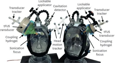
Although abbreviated physical principles of operation as well as hardware schematics for the tFUS-mediated neuromodulation are discussed elsewhere [11], an example of single-element tFUS configuration and its headgear are shown in Fig. 1. The size of the FUS transducer varies depending on the sonication depth. In general, a deeper target requires a larger transducer dimension. A lockable applicator mounted to the headgear is used to hold the transducer in place to achieve the desired orientation. The location and orientation of the head and the transducer are optically tracked for sonication targeting. For uninterrupted delivery of acoustic energy to the targeted area, a compressible hydrogel block is inserted between the transducer surface and the scalp. A cavitation detector, also shown in Fig. 1, can be used for real-time monitoring of potential ‘cavitation events’ (as described in the paragraphs below —
‘Important acoustic parameters relevant to tissue damage’).
Despite growing evidence from animal models, including non-human primates, revealing the effectiveness of the technique, the detailed safety profile of the FUS-mediated brain stimulation has not been established. In this review, we intend to provide: (1) a brief overview
of important acoustic parameters relevant to tissue damage, especially targeting potential thermal and mechanical damages to biological tissue, (2) the United States (U.S.) Food and Drug Administration (FDA)-regulated parameters used for existing ultrasound therapies and diagnostic imaging, and (3) safety information and acoustic parameters regarding the use of FUS for brain neuromodulation of large animals, non-human primates, and humans. In this review, we do not intend to provide the fundamental mechanism behind the neuromodulatory potential of ultrasound, which may involve multi-faceted routes and are still under investigation at this time. The parameters and safety pertaining to studies among small animals (i.e., rodents and rabbits) can be found elsewhere [33,38], and hence is not discussed herein.
IMPORTANT ACOUSTIC PARAMETERS RELEVANT TO TISSUE DAMAGE
There are 2 important mechanisms by which ultrasound can harm biological tissues: (1) heat- related damage by the absorption of ultrasound that yields excessive temperature increase of the tissue and (2) mechanical damage, mainly through cavitation phenomenon (the expansion/contraction or the collapse of bubbles inside biological tissue due to the applied acoustic pressure [42]). Both of these factors must be carefully addressed to avoid damage to the brain tissue.
The absorption of ultrasound by the biological tissue and its conversion to thermal energy is dependent on many factors, mainly the absorption coefficient, heat capacity, and perfusion of the tissue. Osseous structures have high sound absorption rates with lower perfusion compared to other tissues and hence are more susceptible to temperature elevation. In modern FUS systems, energy of incident acoustic waves is distributed over the large area of the skull, and when used in low incident acoustic energy, heat generation at the skull does not pose significant issues in the context of brain stimulation. For generation of heat in the brain, the acoustic intensity, represented as the spatial-peak temporal-average intensity (ISPTA; units of W/
cm2), is considered an important variable. ISPTA indicates the averaged fraction of the acoustic Lockable
applicator Lockable
applicator Cavitation
detector
motionHead tracker Transducer
tracker Transducer
tracker
transducertFUS tFUS
transducer Coupling
hydrogel Coupling
hydrogel Sonication
focus Sonication
focus
Fig. 1. Example of single-element tFUS transducer setup on a mannequin head. Left: a tFUS headgear for targeting deep brain areas (8 cm depth). Right: a tFUS headgear for targeting cortical areas (3 cm depth). Acoustic beam paths are illustrated in green. For the illustration of image-guidance for the tFUS targeting and numerical simulation of acoustic propagation, please refer to the previous article [11].
tFUS, transcranial focused ultrasound.
intensity per second and is derived by spatial-peak pulse-average intensity (ISPPA) multiplied by duty cycle (indicating the fraction of the sonication duration per second). ISPPA is calculated by measuring the pressure of the sound waves (in pascals) using a hydrophone. When operating in pulsed mode, the duty cycle is determined by pulse duration multiplied by pulse repetition frequency. When operating in continuous wave (CW) mode, the duty cycle is 1 (or 100%).
A measure of the likelihood for non-thermal, mechanical bioeffects of ultrasound, including cavitation is expressed in terms of the mechanical index (MI; unitless value). Peak negative pressure (Pr; also called as peak rarefactional pressure), the half of peak-to-peak amplitude of ultrasound pressure wave, is important variable to determine the MI. The MI is defined as Pr
(in MPa) divided by the square root of the fundamental frequency (in MHz) of the ultrasound wave (therefore, higher the MI, the greater the risk of mechanical damage). For example, 250 kHz acoustic pressure waves, which are delivered at a Pr of 450 kPa (0.45 MPa), have a MI of 0.9. The cavitation events are more prone to occur in the media that contains air/
gas, and hence the most cavitation-sensitive tissues are gas-filled organs such as the lungs and intestine. Most of the reported FUS-mediated brain stimulation techniques utilized ultrasound pressures under the FDA limit of the MI for ultrasound imaging (MI = 1.9; except for ophthalmic imaging, Tables 1 and 2); however, the detailed mechanical effects in the lower frequency band used on the skull (in the range of 200–900 kHz compared to the frequency band used in the imaging, i.e., 2–4 MHz) are unknown and warrant further investigation.
Possible adverse effects of tFUS in animals and humans may stem from thermal (from tissue/
skull heating) and mechanical origins (from cavitation or mechanical stretching of the neural tissue). Due to the use of low-intensity ultrasound, which is below or close to the level that are compatible with the ultrasound imaging, studies involving healthy humans and large animals have shown excellent safety record to date. In humans, minor symptoms (e.g., headache) that were not directly related to the sonication have been reported [43]. Only one study on sheep, which utilized excessive repetition of tFUS at an intensity higher than the level for ultrasound imaging (but still much lower than those for HIFU applications), identified the isolated presence of small, non-edema micro-hemorrhage [44]. Albeit excellent records to date, further studies are needed to thoroughly evaluate the short/long-term effects of the tFUS neuromodulation. Recently, efforts are made to form an international consortium (named International Transcranial Ultrasonic Stimulation Safety and Standards), which aims to establish recommendations and guidelines, including contraindications and reporting of adverse events, for safe use of tFUS neuromodulation in humans.
FDA-REGULATED PARAMETERS FOR EXISTING
ULTRASOUND THERAPIES AND DIAGNOSTIC IMAGING
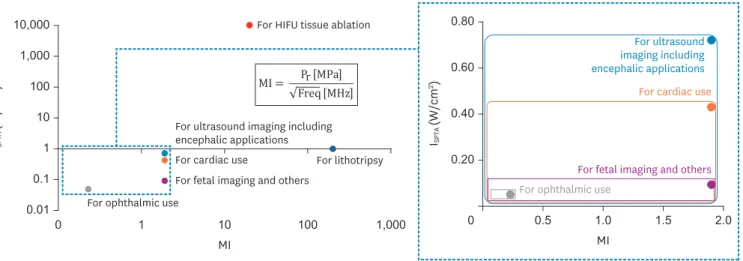
We surveyed the U.S. FDA-approved marketed devices (i.e., ones with the Code of Federal Regulations: CFR) and their regulated operational parameters by the FDA (Tables 1 and 2). In Fig. 2, we illustrated the ranges of parameters (ISPTA and MI) used in clinical practice.
In terms of diagnostic ultrasound procedures, “Guidance for Industry and Food and Drug Administration Staff: Clearance of Diagnostic Ultrasound Systems and Transducers”
(version June 27, 2019) was used to inform the device operation specifications. In terms of therapeutic FUS procedures, the devices are currently identified by the FDA in the field of gastroenterology/urology and general and plastic surgery.
Common FUS therapeutic devices involve tissue ablation or lithotripsy, for which very high intensities are required. For high pressure applications (e.g., shock wave lithotripters), a short burst of focused (or unfocused) ultrasound waves are delivered to the target tissue.
The applied pressure is on the order of 20–110 MPa (i.e., 20,000–110,000 kPa). All these therapeutic applications operate at much stronger acoustic intensity (> 1,000 times higher) or higher pressure level (> 100 times higher; thus MI > 10) than those of the FUS-mediated brain stimulation studies.
One procedure that is comparable, although different, to FUS-mediated brain stimulation would be transcranial Doppler imaging with adult/pediatric encephalic application (CFR 892.1550) to characterize cerebral blood flow. For transcranial Doppler ultrasound, the FDA requires the acoustic output to be ISPTA ≤ 720 mW/cm2, and either MI ≤ 1.9 or ISPPA ≤ 190 W/
cm2. ISPTA ≤ 720 mW/cm2 does not increase the temperature of biological tissue and an MI = 1.9 is the pressure level below which no mechanical damage has been observed. For clinical ultrasound imaging of organs, in the absence of gas-bodies, an MI up to 1.9 is allowed [45], which corresponds to Pr of 3.8 MPa at 250 kHz.
SAFETY INFORMATION AND ACOUSTIC PARAMETERS FOR BRAIN NEUROMODULATION OF LARGE ANIMALS, NON-HUMAN PRIMATES, AND HUMANS
Acoustic parameters used in brain stimulation of animals and humans are reviewed. The
sonication target, the type of FUS transducer, and fundamental frequency of the experiments were listed along with the maximum Pr (and the corresponding MI) and ISPTA. The derating factor (i.e., the amount of attenuation due to the presence of skull) at a specific frequency were estimated based on the ex vivo measurement of the skull samples or through numerical simulation on acoustic propagation reflecting the actual skull anatomy (based on computed tomography [CT]
data) if available. The detraining factor is used to estimate the in situ Pr and derivation of ISPPA. 10,000
1,000 100 10 1 0.1
0.010 1 10 100 1,000
ISPTA (W/cm2)
For HIFU tissue ablation
For lithotripsy For cardiac use
For fetal imaging and others For ophthalmic use
For ultrasound imaging including encephalic applications
MI
ISPTA (W/cm2) 0.80
0.60
0.40
0.20
0 0.5 1.0 1.5 2.0
For cardiac use
For fetal imaging and others For ophthalmic use
For ultrasound imaging including encephalic applications
MI MI = Pr [MPa]
√Freq [MHz]
Fig. 2. Graphical illustration of the ISPTA and MI (in log-scale) used in clinical practice involving ultrasound. The panels with blue dotted line indicate range of the parameters used in diagnostic ultrasound imaging (the magnified panel on the right side shown in linear-scale). The use-specific exceptions for cardiac (in orange), ophthalmic (in gray) and fetal imaging and ‘others’ (in pink) are also noted (‘others category’ includes abdominal, intraoperative, pediatric, small organs such as breast, thyroid, and testes).
ISPTA, spatial-peak temporal-average intensity; MI, mechanical index; HIFU, high-intensity focused ultrasound.
Review of ovine/porcine studies
In studies in sheep (Table 3), in situ Pr of up to 900 kPa and in situ ISPTA of up to 13.8 W/cm2 were applied across multiple FUS sessions to stimulate visual, sensorimotor, and thalamic areas of sheep [44,46,47]. None of these studies reported any negative signs at behavioral, neuroradiological or histological levels. Similarly, based on a study with FUS administration to the sensory thalamic area in a porcine model [48], there was no observed FUS-mediated tissue heating during magnetic resonance (MR) thermometry and no histological finding of tissue damages after the procedure.
Review of non-human primate studies
There are increasing number of tFUS investigations on non-human primates (Table 4)
[40,46,49-55], most of which were done using a single-element FUS transducer with ultrasound frequencies of 250–320 kHz and pulsing schemes of 30%–50% duty cycle. These studies employed in situ ISPTA of up to 25.8 W/cm2, in situ Pr of up to 2.4 MPa, and sonication duration of up to 40 seconds. Even when using much higher intensity, pressure level, and sonication durations (with some higher than typical transcranial Doppler imaging parameters), none of these approaches have shown any negative behavioral or histological impacts.
Table 3. FUS parameters used in large animal models of ovine and porcine
References Target Type of FUS transducer FUS
frequency (kHz)
Maximum in situ Pr
(kPa)
Maximum in situ ISPTA (W/
cm2)
Maximum duty
cycle (%) Sonication duration
(ms)
Maximum in situ MI Lee et al. [44] Sensorimotor cortex,
visual cortex (ovine) Single-element 250 700 7.15 50.0 300 1.40
Yoon et al. [47] Sensorimotor cortex,
thalamus (ovine) Single-element 250 735 12.70 70.0 200 1.47
Gaur et al. [46] Subcortical locations including the LGN and 0–20 mm rostral or caudal to the LGN (ovine)
1,024-element (ExAblate 2100; InSightec,
Tirat Carmel, Israel) 550 900 13.80 50.0 200–300 1.21
Dallapiazza et al.
[48] Thalamus (porcine) Single-element 1,145 567 NR
(ISA of 25–30 W/cm2)
43.7* 40,000* 0.53 1,024-element (ExAblate Neuro 4000;
InSightec) 710 447
990-element (InSightec) 220 249
FUS, focused ultrasound; Pr, peak negative pressure; ISPTA, spatial-peak temporal-average intensity; MI, mechanical index; NR, not reported; LGN, lateral geniculate nucleus; ISA, spatial average intensity; PRF, pulse repetition frequency.
*These parameters in reference [48] was estimated from pulse duration = 43.7 ms with PRF = 10 Hz, for 40 seconds sonication duration (i.e., a total of 400 times of 43.7 ms-long FUS stimulations).
Table 4. FUS parameters used in non-human primates
References Target Type of FUS
transducer FUS frequency
(kHz) Maximum in
situ Pr (kPa) Maximum in situ
ISPTA (W/cm2) Maximum duty
cycle (%) Sonication
duration (ms)Maximum in situ MI
Deffieux et al. [49] Frontal eye field Single-element 320 350 < 0.014* NR 100 0.60
Folloni et al. [50] Amygdala Single-element 250 1,440 19.50 30† 40,000† 2.88
Anterior cingulate cortex 780 5.63 30† 40,000† 1.56
Fouragnan et al. [51] Anterior cingulate cortex Single-element 250 850 NR 30† 40,000† 1.70
Gaur et al. [46] Primary visual cortex Single-element 270 2,400 25.80 50 300 4.62
Khalighinejad et al. [52] Basal forebrain Single-element 250 NR 6.40 30† 40,000† NR
Kubanek et al. [53] Frontal eye field Single-element 270 460 NR (Incident ISPTA
= 0.58) 50 300 0.89
Verhagen et al. [40] Supplementary motor area Single-element 250 880 7.20 30† 40,000† 1.76
Frontal polar cortex Single-element 1,010 9.50 30† 40,000† 2.02
Wattiez et al. [54] Frontal eye field Single-element 320 410 NR NR 100 0.72
Yang et al. [55] Sensory cortex Single-element 250 543 0.45‡ 50 300 1.08
FUS, focused ultrasound; Pr, peak negative pressure; ISPTA, spatial-peak temporal-average intensity; MI, mechanical index; NR, not reported; ISPPA, spatial-peak pulse-average intensity.
*ISPPA 4 W/cm2 × Sonication duration 0.1 seconds/Inter-stimulus interval 30 seconds = 0.013 W/cm2 ISPTA; †These parameters were estimated from pulse duration = 30 ms with PRF = 10 Hz, for 40 seconds sonication duration (i.e., a total of 400 times of 30 ms-long FUS stimulation trials); ‡ISPPA 9.9 W/cm2 × Sonication duration 0.3 seconds × Duty cycle 0.5/Inter-stimulus interval 3 seconds = 0.45 W/cm2 ISPTA.
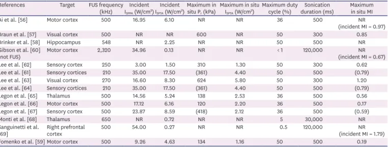
Review of human studies
To date, independent studies have been conducted on healthy human volunteers, an epilepsy patient, and a minimally conscious state patient (Table 5) [56-69]. We added columns showing the incident ISPPA and ISPTA (i.e., acoustic intensity in the absence of skull) in addition to in situ pressure and intensity. Most of these studies have utilized in situ ISPTA of 1.2–5.8 W/
cm2 and in situ Pr of 120–624 kPa, with duty cycles of 36% and 50% and sonication durations of 300 ms and 500 ms, to stimulate the sensory/motor/visual cortices, the hippocampus or the thalamus. Three studies that delivered 30 second- or 120 second-long sonication to the human brain (motor cortex, thalamus, right prefrontal cortex) used duty cycles of 5%
or lower, which yielded a low incident ISPTA of 0.13–0.72 W/cm2. Neither adverse events nor abnormal radiological findings were reported from any of these human studies. Histological examination has never been reported among healthy volunteers.
In terms of MI, the safety of the tFUS brain stimulation techniques is supported by previous investigations among healthy individuals—for example, stimulations of the primary visual cortex (at 270 kHz, maximum in situ MI of 1.2) [63], the motor cortex (at 500 kHz, maximum incident MI of 0.9, in situ MI of 0.17) [66], and the thalamus (at 500 kHz, in situ MI of 0.56) [65]. Most of the ultrasound stimulations are administered with the acoustic intensities and pressures significantly below those used for transcranial Doppler imaging (e.g., ISPTA ≤ 720 mW/
cm2, MI ≤ 0.9 and ISPPA ≤ 7.2 W/cm2). Recently, in the U.S., the first in-human applications of repetitive tFUS were conducted on the thalamus of a minimally-conscious-state patient [68]
and on the hippocampal ictal areas of a patient with drug-resistant temporal lobe epilepsy [58].
The tFUS treatments were successfully delivered without any adverse events in both studies.
Fig. 3 is an illustration showing the ranges of parameters (ISPTA and Pr) used in the context of FUS brain stimulation using large animal models, non-human primates, and in humans.
Some studies in Tables 3-5 are not shown in the graph because the information of in situ ISPTA
or Pr was not reported in the articles.
Table 5. FUS parameters used in humans
References Target FUS frequency
(kHz) Incident
ISPPA (W/cm2) Incident
ISPTA (W/cm2)Maximum in
situ Pr (kPa) Maximum in situ
ISPTA (W/cm2) Maximum duty
cycle (%) Sonication
duration (ms) Maximum in situ MI
Ai et al. [56] Motor cortex 500 16.95 6.10 NR NR 36 500 NR
(incident MI = 0.97)
Braun et al. [57] Visual cortex 500 NR NR 600 NR 50 300 0.85
Brinker et al. [58] Hippocampus 548 NR 2.25 NR NR 50 500 NR
Gibson et al. [60]
(not FUS) Motor cortex 2,320 34.96 0.13 NR NR < 1 120,000 NR
(incident MI = 0.67)
Lee et al. [62] Sensory cortex 250 3.00 1.50 310 1.30 50 300 0.62
Lee et al. [61] Sensory cortices 210 35.00 17.50 (361) 4.40 50 500 (0.79)
Lee et al. [63] Visual cortex 270 16.60 8.30 624 5.80 50 300 1.20
Lee et al. [64] Sensory cortices 210 35.00 17.50 (361) 4.40 50 500 (0.79)
Legon et al. [65] Thalamus 500 14.56 5.24 138 2.53 36 500 0.56
Legon et al. [66] Motor cortex 500 17.12 6.16 120 2.20 36 500 0.17
Legon et al. [67] Sensory cortex 500 23.87 8.59 (418) 2.12 36 500 (0.59)
Monti et al. [68] Thalamus 650 NR 0.72 NR NR 5 30,000 NR
Sanguinetti et al.
[69] Right prefrontal
cortex 500 54.00 0.27 NR NR 0.5 120,000 NR
(incident MI = 1.79)
Fomenko et al. [59] Motor cortex 500 9.26 4.63 134 1.16 50 500 0.19
Numbers within parenthesis are either calculated estimates or relevant values based on information reported in the references.
NR, not reported; FUS, focused ultrasound; Pr, peak negative pressure; MI, mechanical index; ISPPA, spatial-peak pulse-average intensity; ISPTA, spatial-peak temporal-average intensity.
DISCUSSION
Two main acoustic parameters affecting thermal (ISPTA) and mechanical safety (MI) were discussed in the context of tFUS-mediated brain stimulation. Studies to date have revealed the presence of threshold effects in stimulation (i.e., a certain level of minimum acoustic intensity is needed for stimulation), and higher acoustic intensities may yield higher responses to the stimulation [33,34,44]. However, the use of excessively high acoustic intensities risks damaging the brain tissue. Based on our survey, in situ ISPTA of up to 5.8 and 4.4 W/cm2 has been used to stimulate visual and somatosensory areas of the brain without causing any adverse effects among healthy individuals. Much higher in situ ISPTA, for example 25.8 W/cm2, has also been used to stimulate the visual cortical areas in non-human primates while 13.8 W/cm2 was used safely to stimulate subcortical areas including the lateral geniculate nucleus in large animals (sheep) without any observable behavioral or histological anomalies [46].
Although in situ ISPTA is an important parameter for estimating potential temperature elevation, the same mathematical convention to define the ISPTA in ultrasound imaging (i.e., ISPPA × duty cycle) may not be applicable. We note that sonication stimulations are given for the duration typically much shorter than one second with sufficient intervals in-between (> 1 second), whereas no ultrasound imager operates in the same way (i.e., the sonication is always ‘on’ while the image data is acquired). This unique circumstance leads to over- estimation of the ISPTA, which would not reflect its ‘true’ potential for tissue heating. For example, application of 200 ms-long sonication given at 10 W/cm2 ISPPA every one second (1,000 ms) with a duty cycle of 50% yields an ISPTA of 5 W/cm2 (i.e., 10 W/cm2 ISPPA × 0.5 duty cycle) according to the current convention whereas, in reality, 1 W/cm2 (i.e., 10 W/cm2 ISPPA × 200 ms/1,000 ms × 0.5 duty cycle) is given per second. For these reasons, several reports have used different convention defining the ISPTA (Tables 3 and 4).
The pulsing parameters, such as duty cycle and sonication duration (used in each
stimulation), should be conjunctionally designed so as not to raise tissue temperature. The 30
25
20
15
10
5
0 500 1,000 2,000 2,500
In situ ISPTA (W/cm2)
1,500 In situ Pr (kPa) a
b c
d
e
f
g
i h j l k
nm o
p
a b c d e f gh
i kj l m
no p Sheep
Monkey
Human
Lee et al. [44]
Yoon et al. [47]
Gaur et al. [46]
Deffieux et al. [49]
Gaur et al. [46]
Verhagen et al. [40]
Folloni et al. [50]
Yang et al. [55]
Lee et al. [62]
Lee et al. [61]
Lee et al. [63]
Legon et al. [67]
Lee et al. [64]
Legon et al. [65]
Legon et al. [66]
Fomenko et al. [59]
MI = 1.40 @ 250 kHz MI = 1.47 @ 250 kHz MI = 1.21 @ 550 kHz MI = 0.60 @ 320 kHz MI = 2.88 @ 250 kHz MI = 4.62 @ 270 kHz MI = 2.02 @ 250 kHz MI = 1.08 @ 250 kHz MI = 0.62 @ 250 kHz MI = 0.79 @ 210 kHz MI = 1.20 @ 270 kHz MI = 0.79 @ 210 kHz MI = 0.56 @ 500 kHz MI = 0.17 @ 500 kHz MI = 0.59 @ 500 kHz MI = 0.19 @ 500 kHz
Fig. 3. Graphical illustration of the ISPTA and Pr used in the representative FUS-mediated brain stimulation studies. (a-c) are for large animal studies using ovine, (d-h) are for non-human primate studies, and (i-p) are for human studies.
ISPTA, spatial-peak temporal-average intensity; Pr, peak negative pressure; FUS, focused ultrasound; MI, mechanical index.
upper limit of acoustic intensity for non-thermal effects then can be determined for specific sonication parameters, as the threshold for temperature-induced effects has been estimated as 1.5°C–2.5°C above normal body temperature which is held for longer than an hour [70].
For the estimation of the spatiotemporal changes in tissue temperature exposed to the acoustic field, computer-based numerical simulation and non-invasive MR thermometry techniques are now available [17,71,72] and allow for more realistic assessment of thermal effects compared to the convention of ISPTA.
While ISPTA is a time-dependent parameter, the MI is independent from the sonication duration or specific pulsing scheme and should be carefully evaluated. For example, when applied with short sonication duration (e.g., in microseconds) along with sufficient time intervals to allow heat dissipation from the tissue, the sonication can be given at much higher intensities (thus higher pressure level) without increasing the tissue temperature; however, one should consider the limit imposed by the MI. To further provide an example of sonication that operates under the regulatory guideline for imaging applications (i.e., MI = 1.9), Pr of up to 0.95 MPa may be given at 250 kHz fundamental frequency whereby it translates into 30.5 W/cm2 ISPPA. Although cavitation events are not likely under the MI of 1.9, real-time cavitation detection by broad-band hydrophone around the skull and subsequent spectral analysis [73,74] may be used for added-safety (Fig. 1).
Due to the different mode of operation (i.e., pulsed mode with time intervals between the sonication), the current safety FDA-guidelines on ultrasound imaging devices and HIFU devices—which typically operate in CW modes at much higher frequency—warrant establishment of a separate guideline/recommendations for tFUS-mediated brain
stimulation, especially regarding the conventions for the definition of acoustic intensity. In addition, data reporting formats (including the nomenclature for the parameters) should be standardized (as an example, our review found that several key parameter values were not reported or derivable for some studies). The procedures to characterize the acoustic parameters need revision to include a more advanced approach (i.e., hydrophone-based robotic mapping) than existing acoustic force balance measurement (measurement of acoustic absorption by a brush target that is mounted to a balance to measure the force applied to the target, which is good for the characterization of high intensity field). We believe that accrual of safety data from the scientific/medical community may eventually lead to a consensus on using higher acoustic power and pressure waves than those used in ultrasound imaging.
Beyond the need for revision of regulatory parameters on tFUS devices for brain stimulation, several additional safety-related requirements should be considered by the research
community for safe design and conduct in studies involving healthy individuals. First, to avoid stimulation of unintended brain areas, image-guidance and navigation should be used to place and hold the acoustic focus to the desired brain region. The location of the transducer should also be compensated/fixed against potential head movement. For example, a wearable headgear that can hold the transducer in place with respect to the head would be useful. Secondly, neuroanatomical imaging in the form of MR imaging is advised before/
during/after the procedure to detect any structural changes that may be associated with sonication. CT of the head prior to sonication, albeit with a burden of radiation exposure, can be helpful to determine the presence of any abnormal calcification that can distort or absorb the acoustic waves inside the cranium. CT information can also be used to estimate the location and intensity of the acoustic focus via numerical simulations. Finally, neurological
assessment of the subjects before and after the sonication session can help identify any changes in neurological signs that may not be characterized by neuroimaging protocols. Of course, these requirements should be accompanied by the establishment and execution of appropriate inclusion/exclusion criteria of the human volunteers to adequately perform the risk/benefit assessment according to the local regulations.
For the use of the technique with patient groups, (1) the effects from the stimulation should last significantly longer than the sonication, ideally for a duration that can induce neuroplasticity while (2) repeated FUS sessions should be well-tolerated by patients.
Although the long-term effects of FUS-mediated brain stimulation are unknown in humans, emerging evidence based on animal models showed that FUS applied to the sensory areas in rats may induce differential somatosensory evoked potentials persisting more than 35 minutes after the sonication [41], which suggests FUS has potential for inducing neuroplasticity. Another study conducted in non-human primates showed that tFUS applied to the supplementary motor area and the frontal polar cortex resulted in modulatory effects lasting more than 1 hour after the sonication [40], which is long enough to induce long- term potentiation [75,76]. Recently, 2-week long, 3 sessions/week tFUS applications to the left dorsolateral prefrontal cortex in patients with major depressive disorder have shown effectiveness in reducing depressive symptoms with excellent tolerability [77]. These studies suggest the promising translational potential of FUS-mediated brain stimulation.
When considering this technique for neurorehabilitation in patients, additional cautions are needed. For example, stroke-related or tumor-related brain damage may compromise the mechanical integrity of the macro- and microscopic tissue environment (e.g., brain edema, necrotic/liquefaction changes) [78-80], which may increase the risk of mechanical damage by tFUS. Age-dependent, unknown risk factors toward ultrasonic stimulation may also exist as stroke is more prevalent in elderly adults. Excessive calcification within the brain, especially near the acoustic focus, may absorb or scatter the acoustic waves and subsequently confound the stimulatory outcomes or impose additional safety risks to the individual. For potential applications among patients, careful safety evaluation should be conducted considering the changes in brain tissue properties. In addition, patients may have implanted devices (such as brain shunts or aneurysm clips), which may distort the intracranial acoustic propagation, thus deviating from the intended sonication target, or absorb the acoustic energy, thereby elevating the risks for potential tissue heating. These undesirable effects may depend on the material, size, geometry, and orientation of the implanted device. Further investigation is urgently needed to characterize the safety profile of various implanted devices. The potential presence of increased risk to the patient population requires further research in animal models, and may ultimately warrant additional countermeasures (such as the additional use of non-invasive cavitation detector or thermal monitoring) [72,73,81] to offset the risks.
Although clinical applications of low-intensity tFUS remain a ways off, it has demonstrated its safety in animals and heathy humans with emerging efficacy data for therapeutic uses. The spatial specificity, deep brain penetration, and potential for both activating and deactivating brain circuits make tFUS a particularly promising technology for brain stimulation. Conducting thorough assessments of thermal dose and cavitation events will allow researchers and clinicians to administer tFUS safely to both healthy volunteers and patients, providing unprecedented ultrasound-based theragnostic opportunities.
ACKNOWLEDGMENT
This study was supported by NASA TRISH through Baylor College of Medicine (NNX16A069A, to SS Yoo). Authors thank Jennifer Kunes and Kavin Kowsari for their editorial help.
REFERENCES
1. Thornton M, Han L, Shergold M. Progress in NDT of resistance spot welding of aluminium using ultrasonic C-scan. NDT Int 2012;48:30-38.
CROSSREF
2. Jensen JA. Medical ultrasound imaging. Prog Biophys Mol Biol 2007;93:153-165.
PUBMED | CROSSREF
3. Leighton TG. What is ultrasound? Prog Biophys Mol Biol 2007;93:3-83.
PUBMED | CROSSREF
4. Wells PN, Liang HD. Medical ultrasound: imaging of soft tissue strain and elasticity. J R Soc Interface 2011;8:1521-1549.
PUBMED | CROSSREF
5. Claes L, Willie B. The enhancement of bone regeneration by ultrasound. Prog Biophys Mol Biol 2007;93:384-398.
PUBMED | CROSSREF
6. Jung YJ, Kim R, Ham HJ, Park SI, Lee MY, Kim J, Hwang J, Park MS, Yoo SS, Maeng LS, Chang W, Chung YA. Focused low-intensity pulsed ultrasound enhances bone regeneration in rat calvarial bone defect through enhancement of cell proliferation. Ultrasound Med Biol 2015;41:999-1007.
PUBMED | CROSSREF
7. Ebadi S, Henschke N, Nakhostin Ansari N, Fallah E, van Tulder MW. Therapeutic ultrasound for chronic low-back pain. Cochrane Database Syst Rev 2014;CD009169.
PUBMED | CROSSREF
8. Noori SA, Rasheed A, Aiyer R, Jung B, Bansal N, Chang KV, Ottestad E, Gulati A. Therapeutic ultrasound for pain management in chronic low back pain and chronic neck pain: a systematic review. Pain Med 2020;21:1482-1493.
PUBMED | CROSSREF
9. Pishchalnikov YA, Sapozhnikov OA, Bailey MR, Williams JC Jr, Cleveland RO, Colonius T, Crum LA, Evan AP, McAteer JA. Cavitation bubble cluster activity in the breakage of kidney stones by lithotripter shockwaves. J Endourol 2003;17:435-446.
PUBMED | CROSSREF
10. Rassweiler JJ, Knoll T, Köhrmann KU, McAteer JA, Lingeman JE, Cleveland RO, Bailey MR, Chaussy C.
Shock wave technology and application: an update. Eur Urol 2011;59:784-796.
PUBMED | CROSSREF
11. Yoo SS. Technical review and perspectives of transcranial focused ultrasound brain stimulation for neurorehabilitation. Brain Neurorehabil 2018;11:e16.
CROSSREF
12. Hynynen K, Clement GT, McDannold N, Vykhodtseva N, King R, White PJ, Vitek S, Jolesz FA. 500-element ultrasound phased array system for noninvasive focal surgery of the brain: a preliminary rabbit study with ex vivo human skulls. Magn Reson Med 2004;52:100-107.
PUBMED | CROSSREF
13. Marsac L, Chauvet D, Larrat B, Pernot M, Robert B, Fink M, Boch AL, Aubry JF, Tanter M. MR-guided adaptive focusing of therapeutic ultrasound beams in the human head. Med Phys 2012;39:1141-1149.
PUBMED | CROSSREF
14. Maimbourg G, Houdouin A, Deffieux T, Tanter M, Aubry JF. 3D-printed adaptive acoustic lens as a disruptive technology for transcranial ultrasound therapy using single-element transducers. Phys Med Biol 2018;63:025026.
PUBMED | CROSSREF
15. Maimbourg G, Houdouin A, Deffieux T, Tanter M, Aubry JF. Steering capabilities of an acoustic lens for transcranial therapy: numerical and experimental studies. IEEE Trans Biomed Eng 2020;67:27-37.
PUBMED | CROSSREF
16. Xia X, Li Y, Cai F, Zhou H, Ma T, Zheng H. Ultrasonic tunable focusing by a stretchable phase-reversal Fresnel zone plate. Appl Phys Lett 2020;117:021904.
CROSSREF
17. Yoon K, Lee W, Croce P, Cammalleri A, Yoo SS. Multi-resolution simulation of focused ultrasound propagation through ovine skull from a single-element transducer. Phys Med Biol 2018;63:105001.
PUBMED | CROSSREF
18. Jolesz FA. MRI-guided focused ultrasound surgery. Annu Rev Med 2009;60:417-430.
PUBMED | CROSSREF
19. Chang WS, Jung HH, Kweon EJ, Zadicario E, Rachmilevitch I, Chang JW. Unilateral magnetic resonance guided focused ultrasound thalamotomy for essential tremor: practices and clinicoradiological outcomes.
J Neurol Neurosurg Psychiatry 2015;86:257-264.
PUBMED | CROSSREF
20. Elias WJ, Huss D, Voss T, Loomba J, Khaled M, Zadicario E, Frysinger RC, Sperling SA, Wylie S, Monteith SJ, Druzgal J, Shah BB, Harrison M, Wintermark M. A pilot study of focused ultrasound thalamotomy for essential tremor. N Engl J Med 2013;369:640-648.
PUBMED | CROSSREF
21. Elias WJ, Lipsman N, Ondo WG, Ghanouni P, Kim YG, Lee W, Schwartz M, Hynynen K, Lozano AM, Shah BB, Huss D, Dallapiazza RF, Gwinn R, Witt J, Ro S, Eisenberg HM, Fishman PS, Gandhi D, Halpern CH, Chuang R, Butts Pauly K, Tierney TS, Hayes MT, Cosgrove GR, Yamaguchi T, Abe K, Taira T, Chang JW. A randomized trial of focused ultrasound thalamotomy for essential tremor. N Engl J Med 2016;375:730-739.
PUBMED | CROSSREF
22. Lipsman N, Schwartz ML, Huang Y, Lee L, Sankar T, Chapman M, Hynynen K, Lozano AM. MR- guided focused ultrasound thalamotomy for essential tremor: a proof-of-concept study. Lancet Neurol 2013;12:462-468.
PUBMED | CROSSREF
23. Jung HH, Kim SJ, Roh D, Chang JG, Chang WS, Kweon EJ, Kim CH, Chang JW. Bilateral thermal capsulotomy with MR-guided focused ultrasound for patients with treatment-refractory obsessive- compulsive disorder: a proof-of-concept study. Mol Psychiatry 2015;20:1205-1211.
PUBMED | CROSSREF
24. Fry FJ, Ades HW, Fry WJ. Production of reversible changes in the central nervous system by ultrasound.
Science 1958;127:83-84.
PUBMED | CROSSREF
25. Fry FJ, Barger JE. Acoustical properties of the human skull. J Acoust Soc Am 1978;63:1576-1590.
PUBMED | CROSSREF
26. Fry FJ, Goss SA. Further studies of the transkull transmission of an intense focused ultrasonic beam:
lesion production at 500 kHz. Ultrasound Med Biol 1980;6:33-38.
PUBMED | CROSSREF
27. Fry WJ. Intense ultrasound; a new tool for neurological research. J Ment Sci 1954;100:85-96.
PUBMED | CROSSREF
28. Fry WJ. Ultrasound in neurology. Neurology 1956;6:693-704.
PUBMED | CROSSREF
29. Fry WJ, Barnard JW, Fry FJ, Brennan JF. Ultrasonically produced localized selective lesions in the central nervous system. Am J Phys Med 1955;34:413-423.
PUBMED
30. Fry WJ, Fry FJ. Fundamental neurological research and human neurosurgery using intense ultrasound.
IRE Trans Med Electron 1960;ME-7:166-181.
PUBMED | CROSSREF
31. Blackmore J, Shrivastava S, Sallet J, Butler CR, Cleveland RO. Ultrasound neuromodulation: a review of results, mechanisms and safety. Ultrasound Med Biol 2019;45:1509-1536.
PUBMED | CROSSREF
32. Bystritsky A, Korb AS, Douglas PK, Cohen MS, Melega WP, Mulgaonkar AP, DeSalles A, Min BK, Yoo SS.
A review of low-intensity focused ultrasound pulsation. Brain Stimulat 2011;4:125-136.
PUBMED | CROSSREF
33. Kim H, Chiu A, Lee SD, Fischer K, Yoo SS. Focused ultrasound-mediated non-invasive brain stimulation:
examination of sonication parameters. Brain Stimulat 2014;7:748-756.
PUBMED | CROSSREF
34. King RL, Brown JR, Newsome WT, Pauly KB. Effective parameters for ultrasound-induced in vivo neurostimulation. Ultrasound Med Biol 2013;39:312-331.
PUBMED | CROSSREF
35. Lee W, Kim H, Lee S, Yoo SS, Chung YA. Creation of various skin sensations using pulsed focused ultrasound: evidence for functional neuromodulation. Int J Imaging Syst Technol 2014;24:167-174.
CROSSREF
36. Tufail Y, Matyushov A, Baldwin N, Tauchmann ML, Georges J, Yoshihiro A, Tillery SI, Tyler WJ.
Transcranial pulsed ultrasound stimulates intact brain circuits. Neuron 2010;66:681-694.
PUBMED | CROSSREF
37. Tyler WJ, Tufail Y, Finsterwald M, Tauchmann ML, Olson EJ, Majestic C. Remote excitation of neuronal circuits using low-intensity, low-frequency ultrasound. PLoS One 2008;3:e3511.
PUBMED | CROSSREF
38. Yoo SS, Bystritsky A, Lee JH, Zhang Y, Fischer K, Min BK, McDannold NJ, Pascual-Leone A, Jolesz FA.
Focused ultrasound modulates region-specific brain activity. Neuroimage 2011;56:1267-1275.
PUBMED | CROSSREF
39. Yoo SS, Lee W, Kim H. Pulsed application of focused ultrasound to the LI4 elicits deqi sensations: pilot study. Complement Ther Med 2014;22:592-600.
PUBMED | CROSSREF
40. Verhagen L, Gallea C, Folloni D, Constans C, Jensen DE, Ahnine H, Roumazeilles L, Santin M, Ahmed B, Lehericy S, Klein-Flügge MC, Krug K, Mars RB, Rushworth MF, Pouget P, Aubry JF, Sallet J. Offline impact of transcranial focused ultrasound on cortical activation in primates. Elife 2019;8:e40541.
PUBMED | CROSSREF
41. Yoo SS, Yoon K, Croce P, Cammalleri A, Margolin RW, Lee W. Focused ultrasound brain stimulation to anesthetized rats induces long-term changes in somatosensory evoked potentials. Int J Imaging Syst Technol 2018;28:106-112.
PUBMED | CROSSREF
42. Ter Haar G. Ultrasonic imaging: safety considerations. Interface Focus 2011;1:686-697.
PUBMED | CROSSREF
43. Legon W, Adams S, Bansal P, Patel PD, Hobbs L, Ai L, Mueller JK, Meekins G, Gillick BT. A retrospective qualitative report of symptoms and safety from transcranial focused ultrasound for neuromodulation in humans. Sci Rep 2020;10:5573.
PUBMED | CROSSREF
44. Lee W, Lee SD, Park MY, Foley L, Purcell-Estabrook E, Kim H, Fischer K, Maeng LS, Yoo SS. Image-guided focused ultrasound-mediated regional brain stimulation in sheep. Ultrasound Med Biol 2016;42:459-470.
PUBMED | CROSSREF
45. Duck FA. Medical and non-medical protection standards for ultrasound and infrasound. Prog Biophys Mol Biol 2007;93:176-191.
PUBMED | CROSSREF
46. Gaur P, Casey KM, Kubanek J, Li N, Mohammadjavadi M, Saenz Y, Glover GH, Bouley DM, Pauly KB.
Histologic safety of transcranial focused ultrasound neuromodulation and magnetic resonance acoustic radiation force imaging in rhesus macaques and sheep. Brain Stimulat 2020;13:804-814.
PUBMED | CROSSREF
47. Yoon K, Lee W, Lee JE, Xu L, Croce P, Foley L, Yoo SS. Effects of sonication parameters on transcranial focused ultrasound brain stimulation in an ovine model. PLoS One 2019;14:e0224311.
PUBMED | CROSSREF
48. Dallapiazza RF, Timbie KF, Holmberg S, Gatesman J, Lopes MB, Price RJ, Miller GW, Elias WJ.
Noninvasive neuromodulation and thalamic mapping with low-intensity focused ultrasound. J Neurosurg 2018;128:875-884.
PUBMED | CROSSREF
49. Deffieux T, Younan Y, Wattiez N, Tanter M, Pouget P, Aubry JF. Low-intensity focused ultrasound modulates monkey visuomotor behavior. Curr Biol 2013;23:2430-2433.
PUBMED | CROSSREF
50. Folloni D, Verhagen L, Mars RB, Fouragnan E, Constans C, Aubry JF, Rushworth MF, Sallet J.
Manipulation of subcortical and deep cortical activity in the primate brain using transcranial focused ultrasound stimulation. Neuron 2019;101:1109-1116.e5.
PUBMED | CROSSREF
51. Fouragnan EF, Chau BK, Folloni D, Kolling N, Verhagen L, Klein-Flügge M, Tankelevitch L, Papageorgiou GK, Aubry JF, Sallet J, Rushworth MF. The macaque anterior cingulate cortex translates counterfactual choice value into actual behavioral change. Nat Neurosci 2019;22:797-808.
PUBMED | CROSSREF
52. Khalighinejad N, Bongioanni A, Verhagen L, Folloni D, Attali D, Aubry JF, Sallet J, Rushworth MF. A basal forebrain-cingulate circuit in macaques decides it is time to act. Neuron 2020;105:370-384.e8.
PUBMED | CROSSREF