INTRODUCTION
Varicocele is an abnormal physical condition associ- ated with the dilatation of pampiniform plexus that affects 15% of healthy men. It accounts for 35% and 80% of men with primary and secondary infertility, respectively [1]. Infertile men with varicocele have poor semen quality, seminal oxidative stress and sperm DNA damage [2]. Clinically, the main focus is to treat
patients with varicocele having poor semen quality. Al- though varicocele repair could help to improve semen quality and reproductive outcomes, the information about the subcellular changes in the spermatozoa of these patients are limited.
The molecular changes associated with defective spermatozoa are predominantly due to alterations in the expression levels of their proteins. The transcrip- tionally silent spermatozoa depends on translated
Received: Dec 5, 2018 Revised: Jan 23, 2019 Accepted: Feb 5, 2019 Published online Mar 15, 2019 Correspondence to: Ashok Agarwal https://orcid.org/0000-0003-0585-1026
American Center for Reproductive Medicine, Cleveland Clinic, Mail Code X-11, 10681 Carnegie Avenue, Cleveland, OH 44195, USA.
Tel: +1-216-444-9485, Fax: +1-216-445-6049, E-mail: [email protected] Copyright © 2021 Korean Society for Sexual Medicine and Andrology
pISSN: 2287-4208 / eISSN: 2287-4690 World J Mens Health 2021 Jan 39(1): 90-98 https://doi.org/10.5534/wjmh.180118
Proteomic Profiling of Seminal Plasma Proteins in Varicocele Patients
Manesh Kumar Panner Selvam , Ashok Agarwal
American Center for Reproductive Medicine, Cleveland Clinic, Cleveland, OH, USA
Purpose:
Purpose: Seminal plasma provides a nutritive and protective milieu for spermatozoa. It contains factors/proteins required for sperm maturation, hyperactivation, capacitation and acrosome reaction. Alteration in the expression levels of seminal plasma proteins affect the fertilization process. The main objective of this study is to compare the seminal plasma proteome of healthy fertile men (control group) with varicocele patients in order to identify the differentially expressed seminal plasma proteins.
Materials and Methods:
Materials and Methods: Pooled seminal plasma samples from control (n=5) and varicocele (unilateral: n=5 and bilateral: n=5) subjects were used for proteomic profiling and functional bioinformatic analysis. Key differentially expressed proteins (DEPs) associated with binding of zona pellucida (acrosin; ACR), protein folding (heat shock related 70 kDa protein 2; HSPA2), oxidative stress (peroxiredoxin 2; PRDX2), lipid peroxidation and DNA fragmentation (apolipoprotein A2; APOA2) were vali- dated by Western blot. Statistical analysis was conducted using Mann–Whitney test.
Results:
Results: A total of 412 and 486 proteins were detected in seminal plasma of control group and varicocele patients respective- ly. Twenty-eight proteins were identified as DEPs between varicocele and control group. Validation of DEPs revealed down- regulation of HSPA2 (p=0.0037) as well as APOA2 (p=0.0373), and upregulation of PRDX2 (p=0.0474).
Conclusions:
Conclusions: The seminal plasma protein profile of varicocele patients differ from healthy fertile men. Aberrant expression of seminal plasma proteins serve as an indicator of sperm pathology in varicocele patients.
Keywords:
Keywords: Infertility, male; Proteomics; Seminal plasma; Varicocele
This is an Open Access article distributed under the terms of the Creative Commons Attribution Non-Commercial License (http://creativecommons.org/licenses/by-nc/4.0) which permits unrestricted non-commercial use, distribution, and reproduction in any medium, provided the original work is properly cited.
proteins to perform their biological functions [3]. Our previous reports demonstrated an altered expression of key sperm proteins associated with spermatogenesis, sperm motility, capacitation, hyperactivation and zona pellucida binding, and mitochondrial function in vari- cocele patients [4-6]. Although the cause of sperm dys- function in varicocele patients has been explained via a sperm proteomic approach [6,7], it is also crucial to investigate the possible differential expression of semi- nal plasma proteins as they are important for normal sperm function [7]. The seminal plasma provides a pro- tective environment to the spermatozoa and their pro- teins play a major role in the fertilization process by initiating capacitation and acrosome reaction essential for sperm-oocyte interaction [7,8]. It also harbors ap- proximately 30% of the sperm proteins, which reflects the functional state of the spermatozoa [9]. Majority of the secretions are from testis, cauda epididymis and the accessory sexual glands. In varicocele patients, pathophysiological state, compromised testicular func- tion and epididymal dysfunction may change the com- position of the seminal plasma [10,11].
So far, very few studies were focused on the seminal plasma proteomics pertaining to varicocele condition [12-15]. Camargo et al [14], reported that the nitric oxide metabolism was enhanced in the seminal plasma of varicocele patients. Expression of seminal plasma pro- teins related to spermatogenesis, sperm-binding activ- ity, inflammatory response, proliferative activity and apoptosis regulation were altered in adolescents with varicocele [16,17]. However, these studies did not report the changes in the sperm proteome. The present study is an extension of our sperm proteomics [6]. We have compared the seminal plasma proteome profile of vari- cocele patient with healthy fertile donors to identify the differentially expressed proteins (DEPs). The main objective was to evaluate the seminal plasma proteome profile and correlate the DEPs with sperm pathology in varicocele patients.
MATERIALS AND METHODS
1. Ethics statement
The present study involves human subject partici- pation and was approved by the Institutional Review Board (IRB) of Cleveland Clinic (IRB # 17-422), Cleve- land, Ohio, United States. Informed consent was ob- tained by all subjects when they were enrolled.
2. Study subjects
Semen samples were obtained from 10 healthy fertile men without varicocele (control group), and 50 varico- cele patients (33 unilateral and 17 bilateral) of age 20 to 40 years. Patients who attended the clinic for infer- tility treatment from March 2012 to March 2014 were consented and enrolled in this study [6]. Clinical exami- nation, patient history and semen analysis results were considered for sample selection. The clinical diagnosis of varicocele was done by genital examination and scrotal palpation performed by the physician. The con- trol group included men who had initiated pregnancy or fathered a child in the last two years. The sperm proteome of these subjects were analyzed and reported earlier [6]. In the present study, seminal plasma from grade 1 to 3 varicocele patients were used for proteomic analysis. The study was conducted in compliance with the Minimum Information about a Proteomics Experi- ment (MIAPE) guidelines of the Human Proteome Organization’s Proteomics Standards Initiative (HUPO- PSI) for reporting proteomics studies [18].
3. Inclusion and exclusion criteria
Inclusion criteria for control group included healthy men with no varicocele, no chronic diseases, normal genital examination and who had initiated pregnancy or fathered a child in the last two years.
Exclusion criteria for varicocele group included infer- tility of the female partner, genetic defects (Klinefelter syndrome, Y chromosome micro deletion and cystic fibrosis with congenital absence of the vas deferens), chronic prostatitis, and reproductive tract infection.
Additional exclusion criteria for both the study groups included leukocytospermia (Endtz positive), azoospermia and oligozoospermia (<106 sperm/mL), his- tory of systemic illness, inflammation of reproductive tract (orchitis, epididymitis, urethritis, and testicular atrophy), sexually transmitted disease, smoking, and medications.
4. Semen analysis
Following 48 hours of sexual abstinence, semen sam- ples were collected by masturbation at the Andrology Laboratory, Cleveland Clinic. Samples were allowed to liquefy completely for 20 minutes at 37°C and routine semen analysis was performed according to World Health Organization guidelines [19]. Reactive oxygen species (ROS) levels and sperm DNA fragmentation
(SDF) were measured using chemiluminescence assay and terminal deoxynucleotidyl transferase dUTP nick- end labeling (TUNEL) assay, respectively [20]. Semen samples were centrifuged for 7 minutes at 1,000g, and clear seminal plasma was aspirated and stored at -80°C for proteomic analysis.
5. Preparation of samples for proteomic studies
Seminal plasma samples were thawed at room tem- perature and centrifuged at 3,000g for 30 minutes to completely remove contaminating spermatozoa and somatic cells. Protein concentration was determined using a bicinchoninic acid kit (Thermo, Rockford, IL, USA). Pooled samples from varicocele group (unilateral, n=5 & bilateral, n=5) and control group (n=5) were used for proteomic analysis. Equal concentration of protein from each individual sample was used to normalize the protein concentration in each group. In general, pooling of samples is accepted in proteomic analysis and it has been well documented in previous reports [20-22]. All the samples were run in triplicate in one dimensional- Polyacrylamide gel electrophoresis. After electrophore- sis, each gel lane was cut into 6 pieces, digested using 5 μL trypsin (10 ng/μL) and 50 mM ammonium bicar- bonate, and incubated overnight at room temperature.
Prior to in-gel digestion, the samples (cut lanes) were alkylated with iodoacetamine and reduced with dithio- threitol. The peptides from the digested gel were ex- tracted in two aliquots of 30 μL acetonitrile (10%) with formic acid (5%). The two aliquots were pooled together and evaporated to <10 μL and then diluted with 1%
acetic acid to make up a final volume of 30 μL.
6. Liquid chromatography-tandem mass spectrometry analysis
Proteomic profiling of seminal plasma samples were carried out using a Finnigan LTQ linear ion trap mass spectrometer liquid chromatography-tandem mass spectrometry (LC-MS/MS) system. The peptides were fractionated by injecting 5 μL into high performance liquid chromatography column (Phenomenex Jupiter C18 reversed-phase capillary chromatography column).
Fractions containing the peptides were eluted and in- troduced into the source of the mass spectrometer on- line. A full spectral scan was performed by utilizing the data dependent multitask ability of the instrument to determine peptide molecular weights and amino acid
sequence of the peptides [20].
7. Protein identification and quantitative proteomics
Protein identification criteria were established at
>99% probability to achieve false detection rate <1%
as explained in our previous study [6]. Tandem mass spectra were extracted by Proteome Discoverer version 1.4.1.288. All MS/MS results were analyzed using Mas- cot (Matrix Science, London, UK; ver. 2.3.02), Sequest (Thermo Fisher Scientific, San Jose, CA, USA; ver.
1.4.0.288), and X!Tandem (The GPM, thegpm.org; ver.
CYCLONE [2010.12.01.1]). The search was limited to the human protein reference database (http://www.hprd.
org/) and results were uploaded into the Scaffold (ver.
4.0.6.1; Proteome Software Inc., Portland, OR, USA) as previously described [6]. Annotation of proteins was performed using Gene Ontology (GO) terms from Na- tional Center for Biotechnology Information (NCBI).
Relative quantification of the proteins was per- formed by comparing the number of spectra, termed spectral counts in both varicocele and control groups.
The abundance of the proteins was determined by matching the spectra (spectral counts or SpCs), and classified as High (H), Medium (M), Low (L), or Very Low (VL). To overcome the sample-to-sample variation, normalization of spectral counts was done using the normalized spectral abundance factor [6,23].
8. Bioinformatic analysis
DEPs identified in both study groups were subjected to functional annotation and enrichment analysis us- ing both publicly available bioinformatic annotation tools and databases such as UniProt and Reactome.
Proprietary curated database such as Ingenuity path- way analysis (IPA) and MetacoreTM (GeneGo Inc., St.
Joseph, MI, USA) were used to analyze the involve- ment of DEPs in biological and cellular processes, path- ways, cellular distribution, regulatory networks, and protein-protein interactions.
9. Protein selection criteria and validation by Western blot
From the list of 28 proteins identified as DEPs, 4 proteins (heat shock related 70 kDa protein 2: HSPA2, peroxiredoxin 2: PRDX2, apolipoprotein A2: APOA2 and acrosin: ACR) were selected for validation by Western Blot (WB) (n=6, fertile healthy men group
and n=12, varicocele group). Criteria for selection of proteins include: 1) involvement in the top canonical pathways, 2) role of the proteins in reproductive sys- tem development and functions, 3) proteins associated with oxidative stress and sperm DNA damage, 4) pro- teins with a well-described function in the literature. A total of 50 µg of protein per sample was loaded into a 4% to 15% sodium dodecyl sulfate–polyacrylamide gel and electrophoresed for 2 hours at 90 V. WB using spe- cific primary and secondary antibodies (Supplementary Table 1) was performed under standardized conditions [24]. Total protein staining was done with colloidal gold protein stain (Bio-Rad, Hercules, CA, USA) for 2 hours at room temperature by gentle shaking. Stained mem- branes were washed twice with distilled water for 10 minutes and the densitometry image was captured us- ing colorimetric mode on Chemi-Doc (ChemiDocTM MP Imaging System; Bio-Rad).
10. Statistical analysis
Data analysis was performed using MedCalc Statisti- cal Software (V. 17.8; MedCalc Software, Ostend, Bel- gium). Mann–Whitney test was carried out to compare the semen parameters of the fertile donor group and the varicocele group, and the results were considered significant for p<0.05. The same test was used to com- pare the expression levels of the proteins validated us- ing WB technique in both the groups.
RESULTS
1. Semen Parameters
Sperm concentration, motility and normal morphol- ogy were significantly lower in varicocele patients compared to healthy fertile men (Supplementary Table 2). Moreover, ROS and SDF levels were significantly higher in varicocele group when compared to control group (Supplementary Table 2).
2. Proteomic profile of seminal plasma
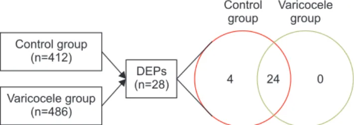
Proteomic analysis of the pooled samples from vari- cocele (unilateral, n=5 & bilateral, n=5) and control group (n=5) resulted in a total of 486 and 412 proteins, respectively. Based on the comparative proteomic analysis 28 proteins were identified to be differentially expressed between varicocele and control group (Fig.
1), with 4 DEPs (T-complex protein 1 subunit delta iso- form a: CCT4, complement factor I preproprotein: CF1,
T-complex protein 1 subunit theta isoform 1: CCT8, T-complex protein 1 subunit alpha isoform a: TCP1) unique to control group (Table 1). The DEPs differed in their abundance in both the varicocele and control groups (Table 1).
3. Pathways regulated by differentially expressed proteins
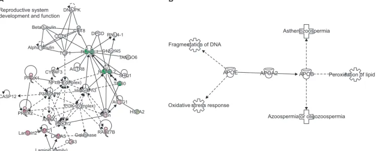
Networks generated using IPA software revealed that reproductive system development and function was the most enriched network with 15 DEPs (Fig. 2A).
Furthermore, functional analysis of DEPs revealed the association of APOA2 protein with DNA fragmenta- tion, oxidative stress response, lipid peroxidation, asthe- nozoospermia, azoospermia or oligozoospermia (Fig. 2B).
4. Transcription factors associated with differentially expressed proteins
Using MetacoreTM, we identified 5 DEPs (elongation factor 1-gamma: eEF1G, heat shock related 70 protein 2: HSPA2, histone H4: HIST-H4, carbonyl reductase [NADPH] 1 isoform 1: CBR1, and acrosin precursor:
ACR) that were predicted to be under the regulation of transcriptional repressor protein yin yang 1 (YY1) (Fig.
3A). Whereas, 3 DEPs (HSPA2, ACR, and CD63 antigen isoform D precursor: CD63) were predicted to be regu- lated by nuclease-sensitive element-binding protein 1 (YB-1) transcriptional factors (Fig. 3B).
5. Selection and validation of differentially expressed proteins by Western blot
Based on the IPA results and metacore analysis we have selected four seminal plasma DEPs (HSPA2, PRDX2, APOA2, and ACR) for validation. HSPA2 and ACR proteins are under the regulation of both the transcription factors YY1 and YB-1. Whereas, the PRDX2 associated with oxidative stress response was
Control group (n=412)
Varicocele group (n=486)
DEPs
(n=28) 4 24 0
Control group
Varicocele group
Fig. 1. The number of proteins identified in the control (n=412) and varicocele groups (n=486) along with the differentially expressed proteins (DEPs) in the control and varicocele groups.
dysregulated in the reproductive system development and function network. Additionally, APOA2 was iden- tified as a multi-functional protein associated to oxida- tive stress response and SDF. WB analysis revealed that HSPA2 and APOA2 proteins were significantly downregulated (0.10-fold variation to control; p=0.0037
and 0.67-fold variation to control; p=0.0373, repectively) (Fig. 4A, 4B), whereas protein PRDX2 was significantly upregulated (1.29-fold variation to control; p=0.0474) in varicocele patients (Fig. 4C). Although, ACR was found to be downregulated (0.81-fold variation to control) in varicocele group, the relative change was not signifi- Table 1. The list of 28 differentially expressed proteins and their abundance and expression in varicocele group compared with the healthy fertile men group
Gene Accession No. Protein name
Healthy fertile
group Varicocele
group NSAF
ratio Expression
SC Abund SC Abund
CCT4 38455427 T-complex protein 1 subunit delta isoform a
2.7 VL 0 - 0.00 Unique to
fertile group
CF1 119392081 Complement factor I preproprotein 1.3 VL 0 - 0.00 Unique to
fertile group CCT8 48762932 T-complex protein 1 subunit
theta isoform 1
5.7 VL 0 - 0.00 Unique to
fertile group TCP1 57863257 T-complex protein 1 subunit
alpha isoform a
2.0 VL 0 - 0.00 Unique to
fertile group
RUVBL1 4506753 RuvB-like 1 8.3 L 0.3 VL 0.02 UE
APOA2a 4502149 Apolipoprotein A-II preproprotein 1.3 VL 0.2 VL 0.04 UE
RUVBL2 5730023 RuvB-like 2 7.7 VL 0.5 VL 0.09 UE
EEF1G 4503481 Elongation factor 1-gamma 10.3 L 2.8 VL 0.24 UE
PGK2 31543397 Phosphoglycerate kinase 2 25.3 M 8.3 L 0.29 UE
CASP12 300360580 Inactive caspase-12 21.7 M 7.7 VL 0.31 UE
ACRa 148613878 Acrosin precursor 10.7 L 3.2 VL 0.32 UE
SEMG1 4506883 Semenogelin-1 preproprotein 698.3 H 395.7 H 0.46 UE
HSPA2a 13676857 Heat shock related 70 protein 2 46.3 M 37.5 M 0.48 UE
SEMG2 4506885 Semenogelin-2 precursor 1321.7 H 765.0 H 0.48 UE
ACTG1 4501887 Actin, cytoplasmic 2 52.3 M 91.5 H 1.73 OE
PRDX1 4505591 Peroxiredoxin-1 13.3 L 35.7 M 2.09 OE
PRDX2a 32189392 Peroxiredoxin-2 11.3 L 32.2 M 2.46 OE
ANXA1 4502101 Annexin A1 7.7 VL 24.7 M 3.05 OE
CD63 383872538 CD63 antigen isoform D precursor 3.0 VL 8.3 L 3.15 OE
TGM4 156627577 Protein-glutamine
gamma-glutamyltransferase 4
31.3 M 181.8 H 3.46 OE
RAB27B 530414276 Ras-related protein Rab-27B isoform X1
2.0 VL 9.5 L 4.03 OE
HIST1H4A 4504301 Histone H4 1.7 VL 9.8 L 5.67 OE
CBR1 4502599 Carbonyl reductase [NADPH] 1 isoform 1
0.7 VL 4.7 VL 6.27 OE
LAMA5 145309326 Laminin subunit gamma-1 precursor 2.0 VL 25.7 M 7.69 OE
LCN15 578817458 Lipocalin-15 isoform X1 0.7 VL 6.2 VL 8.21 OE
GLO1 118402586 Lactoylglutathione lyase 1.3 VL 11.5 L 9.22 OE
LAMC1 578835999 Laminin subunit alpha-5 isoform X1 1.7 VL 36.5 M 12.90 OE
PIGR 530366266 Polymeric immunoglobulin receptor isoform X1
0.3 VL 34.0 M 70.67 OE
SC: spectral count, Abund: abundance, NSAF: normalized spectral abundance factor, VL: very low, L: low, M: medium, H: high, UE: underexpressed, OE: overexpressed.
aThe proteins HSPA2, APOA2, PRDX2 and ACR were selected for validation by Western blotting.
Reproductive system development and function
Asthenozoospermia Asthenozoospermia
Azoospermia or oligozoospermia Azoospermia or oligozoospermia APOA2
APOA2 APOE
APOE Fragmentation of DNA Fragmentation of DNA
Oxidative stress response Oxidative stress response
Peroxidation of lipid Peroxidation of lipid APOB
APOB DNA-PK
DNA-PK
Beta tubulin Beta tubulin
Alpha tubulin Alpha tubulin
CCT4 CCT4
TCP1 TCP1
CCT8
CCT8 DPCDDPCD RNU4-1RNU4-1
TANGO6 TANGO6 C12orf45 C12orf45 RUVBL1 RUVBL1
ACTR8 ACTR8 CYP4F3 CYP4F3 PRDX1 PRDX1
CASP12 CASP12
PRDX2 PRDX2
ANXA1 ANXA1 P38MAPK P38MAPK
NF B (complex) NF B (complex)
Histone h3 Histone h3
TIP60 TIP60 RUVBL2 RUVBL2
SHQ1 SHQ1
ACTG1 ACTG1
HSPA2 HSPA2 Actin
Actin
RAB27B RAB27B Gelatinase Gelatinase LAMA5
LAMA5 CD63 CD63 Laminin (family) Laminin (family) Laminin2
Laminin2LAMC1LAMC1 ERK1/2 ERK1/2
PI3K (complex) PI3K (complex)
A B
Fig. 2. (A) Top most functional enriched network, reproductive system development and function pathway with differentially expressed proteins in seminal plasma of infertile varicocele patient. (B) Functional analysis of the apolipoprotein (APO) A2 protein that was downregulated in the seminal plasma of infertile patients with varicocele.
EFFECTS
TR Transcription Positive/activation Up-regulated (+) Regulation Negative/inhibition Down-regulated (-)
Unspecified YY1
HSPA2
HIST-H4 family
CBR1 ACR
HSPA2
ACR
CD63 YB-1 TR
TR TR
TR
TR TR
TR TR
TR B
B
A B
eEF1G Fig. 3. Upstream transcriptional factors
involved in the regulation of differen- tially expressed proteins identified in the seminal plasma of varicocele patients relative to that of fertile men (control).
(A) Transcriptional repressor protein yin yang 1 (YY1). (B) Nuclease-sensitive el- ement-binding protein 1 (YB-1). HSPA2:
heat shock related 70 kDa protein 2, ACR:
acrosin.
Fig. 4. Protein expression levels of the differentially expressed proteins selected for validation by Western blot in varicocele group relative to fer- tile men. (A) Heat shock related 70 kDa protein 2 (HSPA2), (B) apolipoprotein A2 (APOA2), (C) peroxiredoxin 2 (PRDX2), (D) acrosin (ACR).
cant (p=0.3861) when compared to the control group (Fig. 4D).
DISCUSSION
Seminal plasma, the primary nutritive medium for spermatozoa, plays a crucial role in sperm function and fertilizing potential of male gamate. Previous study from our laboratory have demostrated altered pro- teome profile of sperm in varicocele subjects, specifi- cally underexpression of proteins involved in major en- ergy metabolism pathways, transport, protein folding and proton pumps [6]. Subsequently, we were interested to explore the seminal plasma protein profile of these subjects and eluciate its possible implications on sperm pathology associated with varicocele. Therefore, the present study is an extenstion of the report on sperm proeteomics.
In the present study, the IPA analysis identified re- productive system development and function as one of the top protein-protein interaction network enriched with 15 DEPs (Fig. 2A). PRDX1 and PRDX2 were up- regulated in the varicocele patients. In response to oxidative stress PRDX1 exhibits an increased expres- sion in seminal plasma [25]. In our previous reports, we have demonstrated an increased state of oxidative stress at the subcellular level in the spermatozoa of varicocele patients [6]. Validation results were in agree- ment with proteomic data, suggesting that PRDX2 may serve as potential seminal plasma biomarker in diagnosis of oxidative stress-mediated reproductive dysfunction in varicocele patients. The proteins ruvB- like 1 (RUVBL1) and ruvB-like 2 (RUVBL2), which are involved in DNA damage, were downregulated in the varicocele patients. In our previous study, we observed a significant change in the expression of these proteins in infertile men with high ROS [26]. Hence the altered expression of the DEPs in the network suggests a state of oxidative stress. This results in DNA damage in varicocele patients, which was evident with the sig- nificantly increased ROS levels and SDF compared to control group (Supplementary Table 2). The aberrant expression of seminal plasma proteins could be a major contributing factor for dysfunction of spermatozoa in the varicocele patients.
Spermatozoa of varicocele patients are under the continuous influence of oxidative stress leading to in- creased lipid peroxidation and DNA fragmentation [2].
In the current study, we observed downregulation of APOA2 in the seminal plasma of varicocele patients.
Functional enrichment analysis using IPA revealed that APOA2 was involved in pathways such as oxi- dative stress response, lipid peroxidation and DNA fragmentation (Fig. 2B). Furthermore, our WB results corroborate with the proteomic results (Fig. 4B). The expression of APOA2 in body fluids is noticed during a state of oxidative stress [27]. Hence, the abnormal expression of APOA2 in varicocele subjects indicates seminal oxidative stress and its possible implication on poor semen quality.
Seminal plasma proteome revealed the complete ab- sence of TCP1 and molecular chaperones containing TCP1 subunits such as CCT4 and CCT8 in varicocele group (Table 1). All these three proteins facilitate sperm-oocyte interaction and are essentially required for the sperm-zona pellucida binding [28]. In our previ- ous report we have demonstrated the underexpression of these proteins in spermatozoa of varicocele patients [6]. Absence of these proteins in seminal plasma of varicocele patients reflects the defective function of the spermatozoa. Moreover, the proteomic data as well as WB analysis revealed downregulation of HSPA2, in varicocele patients. HSPA2 is under the regulation of transcription factors YB-1 and YY1 (Fig. 3) associated with sperm-zona binding and fertilization. These find- ings suggest that fertilizing capacity of varicocele pa- tients is affected due to aberrant expression of seminal plasma proteins.
Post-ejaculatory changes associated with spermato- zoa such as hyperactivation, capacitation and acrosome reaction play a vital role in preparing the sperm for fertilization. These events are initiated by the cascade of molecular changes at subcellular level. In the semi- nal plasma of varicocele group, proteins (EEF1G, phos- phoglycerate kinase 2: PGK2 and ACR) associated with normal physiological sperm function were downregu- lated. In general, EEF1G linked to the tyrosine kinase protein is upregulated in the spermatozoa during the sperm capacitation process [26]. PGK2 involved in sper- matogenesis and sperm motility [26] was also down- regulated in varicocele patients and this may be one of the reasons for the poor sperm motility and sperm con- centration in varicocele group (Supplementary Table 1). Earlier reports have suggested PGK2 as a seminal plasma biomarker to differentiate between fertile and infertile men [29,30]. Besides, we observed downregula-
tion of the protein associated with acrosome reaction of spermatozoa, ACR, in the seminal plasma of varicocele patients. ACR is the major protease of mammalian spermatozoa and stored as proacrosin in acrosome. In varicocele patients ACR was reported to be underex- pressed in sperm [26]. Presence of ACR in seminal plas- ma confirms that acrosome integrity is compromised in varicocele patients. Our WB data showed a decreased, but not a significant, expression of ACR in varicocele patients (Fig. 4D). ACR was identified to be regulated by the transcription factors YB-1 and YY1, which play a key role in the regulation of the acrosomal matrix dispersion mechanism (Fig. 3).
The proteomic data and WB analysis revealed an altered seminal plasma proteome in varicocele subjects, which in turn elucidates the pathology associated with spermatozoa. In the present study, we validated only key DEPs associated primarily with oxidative stress and its mediators. However, validation of additional seminal plasma DEPs are warranted to explore the in- volvement of other relevant pathways associated with sperm pathology in varicocele patients.
CONCLUSIONS
The seminal plasma proteome is altered in varico- cele subjects when compared to control group. Altered expression of PRDX2, HSPA2 and APOA2, and its association with oxidative stress and sperm function confirms the dysregulation of physiological processes in the seminal plasma of varicocele subjects.
ACKNOWLEDGEMENTS
The authors thank Tania Dias, M.Sc., Damayanthi Durairajanayagam, PhD, Banu Gopalan, PhD, Rakesh Sharma, PhD, Ana Martins, M.Sc. for their critical reading of the manuscript and helpful suggestions. Be- linda Willard, PhD, Director of Proteomic Core Labora- tory, Lerner Research Institute assisted with proteomic analysis while Bioinformatics data analysis was con- ducted by Banu Gopalan, PhD. Research support was provided by the American Center for Reproductive Medicine at Cleveland Clinic. The authors are grateful to Saradha Baskaran, PhD, for her critical review and constructive comments.
Conflict of Interest
The authors have nothing to disclose.
Author Contribution
Conceptualization: Agarwal A. Data curation: Panner Selvam MK. Formal analysis: Agarwal A, Panner Selvam MK. Funding acquisition: NA. Investigation: Agarwal A. Methodology: Pan- ner Selvam MK. Project administration: Agarwal A. Software:
Panner Selvam MK. Supervision: Agarwal A. Validation: Pan- ner Selvam MK. Writing–original draft: Panner Selvam MK.
Writing–review & editing: Agarwal A, Panner Selvam MK. Ap- proval of the final manuscript: Agarwal A, Panner Selvam MK.
Supplementary Materials
Supplementary materials can be found via https://doi.
org/10.5534/wjmh.180118.
Data Sharing Statement
The data analyzed for this study have been deposited in HARVARD Dataverse and are available at https://doi.
org/10.7910/DVN/Y1ED9R.
REFERENCES
1. Alsaikhan B, Alrabeeah K, Delouya G, Zini A. Epidemiology of varicocele. Asian J Androl 2016;18:179-81.
2. Cho CL, Esteves SC, Agarwal A. Novel insights into the pathophysiology of varicocele and its association with reac- tive oxygen species and sperm DNA fragmentation. Asian J Androl 2016;18:186-93.
3. Panner Selvam MK, Agarwal A. Update on the proteomics of male infertility: a systematic review. Arab J Urol 2018;16:103- 12.
4. Agarwal A, Sharma R, Durairajanayagam D, Ayaz A, Cui Z, Willard B, et al. Major protein alterations in spermatozoa from infertile men with unilateral varicocele. Reprod Biol En- docrinol 2015;13:8.
5. Agarwal A, Sharma R, Durairajanayagam D, Cui Z, Ayaz A, Gupta S, et al. Differential proteomic profiling of spermato- zoal proteins of infertile men with unilateral or bilateral vari- cocele. Urology 2015;85:580-8.
6. Agarwal A, Sharma R, Samanta L, Durairajanayagam D, Sa- banegh E. Proteomic signatures of infertile men with clinical varicocele and their validation studies reveal mitochondrial
dysfunction leading to infertility. Asian J Androl 2016;18:282- 91.
7. Samanta L, Parida R, Dias TR, Agarwal A. The enigmatic seminal plasma: a proteomics insight from ejaculation to fer- tilization. Reprod Biol Endocrinol 2018;16:41.
8. Milardi D, Grande G, Vincenzoni F, Messana I, Pontecorvi A, De Marinis L, et al. Proteomic approach in the identifica- tion of fertility pattern in seminal plasma of fertile men. Fertil Steril 2012;97:67-73.e1.
9. Jodar M, Soler-Ventura A, Oliva R. Semen proteomics and male infertility. J Proteomics 2017;162:125-34.
10. Del Giudice PT, Belardin LB, Camargo M, Zylbersztejn DS, Carvalho VM, Cardozo KH, et al. Determination of testicular function in adolescents with varicocoele: a proteomics ap- proach. Andrology 2016;4:447-55.
11. Vivas-Acevedo G, Lozano-Hernández R, Camejo MI. Varico- cele decreases epididymal neutral α-glucosidase and is associ- ated with alteration of nuclear DNA and plasma membrane in spermatozoa. BJU Int 2014;113:642-9.
12. Zylbersztejn DS, Andreoni C, Del Giudice PT, Spaine DM, Borsari L, Souza GH, et al. Proteomic analysis of seminal plasma in adolescents with and without varicocele. Fertil Steril 2013;99:92-8.
13. Fariello RM, Pariz JR, Spaine DM, Gozzo FC, Pilau EJ, Frai- etta R, et al. Effect of smoking on the functional aspects of sperm and seminal plasma protein profiles in patients with varicocele. Hum Reprod 2012;27:3140-9.
14. Camargo M, Intasqui Lopes P, Del Giudice PT, Carvalho VM, Cardozo KH, Andreoni C, et al. Unbiased label-free quantita- tive proteomic profiling and enriched proteomic pathways in seminal plasma of adult men before and after varicocelec- tomy. Hum Reprod 2013;28:33-46.
15. Del Giudice PT, da Silva BF, Lo Turco EG, Fraietta R, Spaine DM, Santos LF, et al. Changes in the seminal plasma pro- teome of adolescents before and after varicocelectomy. Fertil Steril 2013;100:667-72.
16. Belardin LB, Del Giudice PT, Camargo M, Intasqui P, Anto- niassi MP, Bertolla RP, et al. Alterations in the proliferative/
apoptotic equilibrium in semen of adolescents with varico- cele. J Assist Reprod Genet 2016;33:1657-64.
17. Camargo M, Intasqui P, Bertolla RP. Proteomic profile of seminal plasma in adolescents and adults with treated and untreated varicocele. Asian J Androl 2016;18:194-201.
18. Martínez-Bartolomé S, Deutsch EW, Binz PA, Jones AR, Eise- nacher M, Mayer G, et al. Guidelines for reporting quantita- tive mass spectrometry based experiments in proteomics. J Proteomics 2013;95:84-8.
19. World Health Organization (WHO). WHO laboratory manual for the examination and processing of human semen.
Geneva: WHO; 2010.
20. Sharma R, Agarwal A, Mohanty G, Du Plessis SS, Gopalan B, Willard B, et al. Proteomic analysis of seminal fluid from men exhibiting oxidative stress. Reprod Biol Endocrinol 2013;11:85.
21. Diz AP, Truebano M, Skibinski DO. The consequences of sample pooling in proteomics: an empirical study. Electro- phoresis 2009;30:2967-75.
22. Bogle OA, Kumar K, Attardo-Parrinello C, Lewis SE, Estanyol JM, Ballescà JL, et al. Identification of protein changes in hu- man spermatozoa throughout the cryopreservation process.
Andrology 2017;5:10-22.
23. Zybailov BL, Florens L, Washburn MP. Quantitative shotgun proteomics using a protease with broad specificity and nor- malized spectral abundance factors. Mol Biosyst 2007;3:354- 60.
24. Panner Selvam MK, Agarwal A, Sharma R, Samanta L. Treat- ment of semen samples with α-chymotrypsin alters the ex- pression pattern of sperm functional proteins-a pilot study.
Andrology 2018;6:345-50.
25. Intasqui P, Antoniassi MP, Camargo M, Nichi M, Carvalho VM, Cardozo KH, et al. Differences in the seminal plasma proteome are associated with oxidative stress levels in men with normal semen parameters. Fertil Steril 2015;104:292- 301.
26. Ayaz A, Agarwal A, Sharma R, Arafa M, Elbardisi H, Cui Z.
Impact of precise modulation of reactive oxygen species levels on spermatozoa proteins in infertile men. Clin Proteomics 2015;12:4.
27. Ma C, Li J, Bao Z, Ruan Q, Yu Z. Serum levels of ApoA1 and ApoA2 are associated with cognitive status in older men.
Biomed Res Int 2015;2015:481621.
28. Dun MD, Smith ND, Baker MA, Lin M, Aitken RJ, Nixon B.
The chaperonin containing TCP1 complex (CCT/TRiC) is involved in mediating sperm-oocyte interaction. J Biol Chem 2011;286:36875-87.
29. Rolland AD, Lavigne R, Dauly C, Calvel P, Kervarrec C, Fre- our T, et al. Identification of genital tract markers in the hu- man seminal plasma using an integrative genomics approach.
Hum Reprod 2013;28:199-209.
30. Cui Z, Agarwal A, da Silva BF, Sharma R, Sabanegh E. Evalu- ation of seminal plasma proteomics and relevance of FSH in identification of nonobstructive azoospermia: a preliminary study. Andrologia 2018;50:e12999.