MRI has been widely applied in the evaluation of vari- ous abdominal diseases (1, 2), but its use for the evalua- tion of gastrointestinal disease has been slow to develop because of long acquisition times (resulting in increased motion artifacts, mostly from either respiration or peri- stalsis) and the unavailability of good MR contrast
agents for the gastrointestinal tube (3, 4). As a result, on- ly a small number of published studies have focused on the appearance of intestinal disease processes evaluated by MRI and there have been few reports concerning bowel obstruction (5-8). Several recent reports have de- scribed the use of fast MRI in small bowel disease, in- cluding small bowel obstruction (9-11). In this study, we investigated bowel obstruction using HASTE and true FISP sequences.
Our purpose was to evaluate the efficacy of MRI using HASTE and true FISP sequences in demonstrating bow- el obstruction, in identifying the sites involved, and in determining the cause of obstruction.
The Utility of MR Imaging Using the HASTE and True FISP Sequences in Diagnosing Bowel Obstruction
1Eun Joo Yun, M.D.1, 2, Tae Kyoung Kim, M.D.1, 3, Byung Ihn Choi, M.D.
Purpose: To determine the value of magnetic resonance imaging (MRI) using HASTE (half- Fourier single-shot turbo spin-echo) and true FISP (fast imaging with steady-state precession and heavy T2-weighting) sequences in diagnosing bowel obstruction.
Materials and Methods: Thirty-one consecutive patients in whom suspected bowel ob- struction was revealed at plain abdominal radiography, barium study, and computed tomography (CT) were admitted during an eight-month period. Eighteen of 27 in whom bowel obstruction was confirmed underwent MRI using HASTE and true FISP sequences.
Results: At MRI, bowel obstruction was diagnosed in all patients. Using the HASTE sequence, the site of obstruction was correctly identified in 16 patients(89%) and the cause of obstruction was correctly diagnosed in 15(83%). Using the true FISP se- quence, the corresponding figures were 16(89%) and 16(89%). Of the 18 patients with confirmed bowel obstruction, 33%(6/18) showed better lesion conspicuity at true FISP, for 39%(7/18), conspicuity was equal at both sequences, 28%(5/18) showed better con- spicuity at HASTE.
Conclusion: MRI can be useful for evaluation the presence, site and cause of bowel obstruction. On comparing the findings of HASTE and true FISP sequences, no signifi- cant differences were observed.
Index words : Intestines, obstruction Intestines, MR
1Department of Radiology, Seoul National University College of Medicine
2Department of Radiology, Hanllym University College of Medicine
3Department of Radiology, University of Ulsan College of Medicine Received September 6, 2003 ; Accepted January 31, 2003
Address reprint requests to : Byung Ihn Choi, M.D., Department of Radiology, Seoul National University Hospital, 28 Yongon-dong, Chongno-gu, Seoul 110-744, Korea.
Tel. 82-2-760-2515 Fax. 82-2-743-6385 E-mail: [email protected]
Materials and Methods
Thirty-one consecutive patients in whom suspected bowel obstruction was revealed at plain abdominal radi- ography (n=18), barium study (n=7), and computed to- mography (CT) (n=10), were admitted during an eight- month period. In 26 of the 31, bowel obstruction was confirmed at surgery, and one case was confirmed at colonoscopic biopsy. Of these 27 with confirmed bowel obstruction, 18 underwent MRI using the HASTE and true FISP sequences, and these 18, in whom the respec- tive conditions had been pathologically proven, com- prised our series. The remaining nine, whose condition had been pathologically proven but who did not under- go MRI, were excluded from our study. Among the 18 patients [M:F=8:10; age range=25-93 (mean, 53) years], the common clinical symptoms were nausea, vomiting, abdominal pain, and distention. Thirteen pa- tients had a history of prior surgery (gastrointestinal ma- lignancy, n=10; bowel perforation due to trauma or ul- cer, n=3).
For MR examinations, a 1.0 Tesla scanner (Magnetom
Impact Expert, Siemens, Erlangen, Germany) with a 15 mT/m gradient system and a body phased array coil was used. HASTE (repetition time [TR], infinite; echo time [TE], 87 ms; flip angle, 150°; number of excitations, 1;
matrix number, 192×256; field of view [FOV], 350×
350 mm; slice thickness, 6 mm) and true FISP (TR, 7.6 ms; TE, 3.5 ms; flip angle, 80°; number of excitations, 1;
matrix number, 192×256; FOV, 300×400 mm; slice thickness, 6 mm) images were obtained during single breath-holds lasting an average of 19 seconds. Axial and coronal scans were obtained using both MRI sequences;
fat suppression and cardiac or respiratory gating were not used. No medications for suppressing bowel peri- stalsis and contrast agent were given, and enteric con- trast material or water were not ingested. The total ex- amination time did not exceed 10 minutes.
Two radiologists reviewed the images in terms of the presence, site and cause of bowel obstruction, compar- ing the image quality of the two MRI sequences and reaching their decisions consensually. Obstruction was diagnosed if a definite transition zone was observed, with dilated fluid and/or air-filled loops of small bowel proximal to the site of obstruction and collapsed loops of
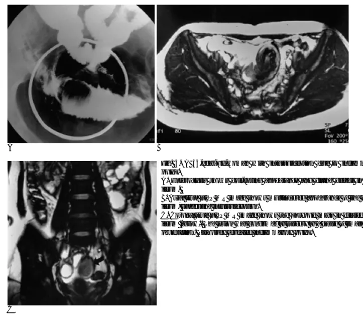
Fig. 1. A 33-year-old man who had total gastrectomy for gastric cancer with recurred tumor in transverse colon.
Coronal true FISP MR image (TR/TE 7.6/3.5, FA 80°) shows wall thickening of distal transverse colon (arrows) and dilated small bowel loops, suggesting malignant obstruction. Surgery revealed recurrence.
Fig. 2. A 47-year-old man who had subtotal gastrectomy for gastric cancer with small bowel obstruction due to postopera- tive adhesion.
Coronal true FISP MR image shows multiple dilated, fluid- filled small bowel loops and an abrupt transition (arrow) of di- lated and collapsed bowel loops. No mass lesions are seen around site of transition. It was confirmed by surgery.
small or large bowel distal to this site, and whether ob- struction involved the proximal or distal small bowel, or the large bowel, was determined. Small and large bowel were differentiated according to their morphology and the central location of small bowel loops versus the pe- ripheral position of the colon, best appreciated at coro- nal imaging (10). The cause of bowel obstruction was determined by evaluating the appearance of the obstruc- tion site and the patient’s past medical history. In these in whom gastrointestinal malignancy had necessitated surgery prior to MRI, post-operative adhesion or tumor recurrence was diagnosed on the basis of the following criteria: beak-like narrowing but the absence of a defi- nite mass for post-operative adhesion, and an incrimi- nating focal intraluminal mass or focal wall thickening of adjacent bowel for tumor recurrence (12). Intussus- ception was diagnosed if an intussuscepted segment and leading point were detected. Inflammatory bowel dis-
ease was diagnosed if mild diffuse bowel wall thicken- ing was present, and colon cancer if a focal intraluminal mass or pronounced focal bowel wall thickening was observed.
Results
In all patients, MRI using the HASTE and true FISP se- quences, demonstrated the existence of bowel obstruc- tion. The obstruction site was correctly identified in 16 of 18 cases; the exception were two cases of distal small bowel obstruction. Obstruction occurred at the distal small bowel in 13 cases, the proximal small bowel in two, the ascending colon in two, and the transverse colon in one; its cause was tumor recurrence after surgery for gastrointestinal malignancy (Fig. 1) in seven cases, postoperative adhesion (Fig. 2) in six, intussuscep- tion (Fig. 3) in two, and colon cancer (Fig. 4), bezoar, and
A
C
B
Fig. 3. A 43-year-old-woman with intussusception due to inflammatory polyp.
A. Enteroclysis shows coil-spring appearance and filling defect in distal ileum.
B. Axial true FISP MR image shows multilayered appearance of the dilated ileum, suggesting intussusception.
C. Coronal true FISP MR image shows the polypoid mass in dilated distal ileum (arrow). The lesion was confirmed at surgery as a cause of small bowel obstruction. Pathology revealed inflammatory polyp.
tuberculosis (Fig. 5) in one case each (Table 1).
At HASTE sequencing, the site of obstruction was cor- rectly identified in 16 patients(89%), and its cause in 15 patients(83%). For the true FISP sequence, the corre- sponding figures were 16(89%) and 16(89%) (Table 2).
For both sequences, the two unidentified cases involved a recurrent tumor and postoperative adhesion. The cause of obstruction diagnosed only at true FISP was a bezoar in the small bowel.
Of the 18 patients with bowel obstruction, 33%(6/18) showed better lesion conspicuity at true FISP. For 39%
(7/18), conspicuity was the same at both sequences, and for 28%(5/18) it was better at HASTE (Table 3).
Discussion
The purpose of radiological imaging in patients with bowel obstruction includes the confirmation or exclu- sion of such obstruction, identification of the site in- volved, and assessment of the severity, and cause of the obstruction and whether strangulation or other compli- cations have arisen. Conventionally, plain radiography,
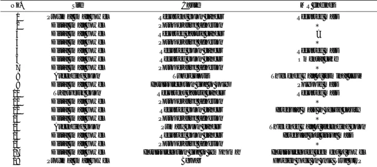
Table 1. Site, Cause and MR Findings of Bowel Obstruction in 18 Patients
No. Site Cause MR findings
01 Proximal small bowel Recurrent colon cancer Recurred mass
02 Distal small bowel Postoperative adhesion *
03 Distal small bowel Recurred gastric cancer ?
04 Distal small bowel Postoperative adhesion *
05 Distal small bowel Recurred colon cancer Recurred mass
06 Distal small bowel Recurred colon cancer Omental cake
07 Distal small bowel Postoperative adhesion *
08 Ascending colon Tuberculosis Thickened wall of terminal ileum
09 Distal small bowel Intussusception due to polyp Polypoid mass
10 Transverse colon Recurrent gastric cancer Recurred mass
11 Distal small bowel Postoperative adhesion *
12 Distal small bowel Recurred colon cancer Irregular mass in pelvic cavity
13 Distal small bowel Postoperative adhesion *
14 Ascending colon Primary colon cancer Thickened wall of ascending colon
15 Distal small bowel Recurred colon cancer Irregular soft tissue mass
16 Distal small bowel Postoperative adhesion *
17 Distal small bowel Intussusception due to lymphoma Intussuscepted segment of bowel
18 Proximal small bowel Bezoar Foreign body on only True FISP
*: abrupt transition of dilated and collapsed bowel loops without any mass
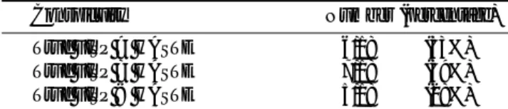
Table 2. Results of Presence, Site and Cause of Bowel Obstruction Using the HASTE and true FISP Sequences in 18 Patients
Presence Site Cause
HASTE 18/18 (100%) 16/18 (89%) 15/18 (83%) True FISP 18/18 (100%) 16/18 (89%) 16/18 (89%)
A B
Fig. 4. A 93-year-old-woman with small bowel obstruction due to primary as- cending colon cancer.
Coronal true FISP (A) and HASTE (B) (TR/TE 10.9/87, FA 150°) MR images show dilated small bowel loops and markedly dilated proximal portion of ascending colon. Another scan shows focal wall thickening of distal portion of ascending colon. She refused surgery. This case was confirmed by colonoscopic biopsy.
barium studies and CT have been widely used (13).
Plain film findings are thought to be diagnostic in about 50-60% of cases and equivocal in about 20-30%
(14). One report showed that enteroclysis correctly pre- dicted the presence of obstruction in 100% of cases and its absence in 88%; the level of obstruction in 89%, and its cause in 86%(15). When small bowel obstruction was classified as high or low grade; CT showed a sensitivity of 81% for high-grade and 48% for low-grade obstruc- tion (16).
Initially, T1-weighted SE imaging along with various types of contrast agent was used, but was limited by
long imaging times, relatively poor image quality, and the expensive nature of the examination. The limitations of MRI in imaging of the bowel include long imaging times and poor spatial and contrast resolution; MR stud- ies suffer from image degradation due to respiration-in- duced ghosting and peristaltic motion artifacts (12). The use of abdominal MRI has increased in recent years, aided by the availability of respiratory triggering and breath-hold sequencing, used to prevent motion arti- facts, and the use of MRI with fast imaging sequences has recently been attempted. An alternative MRI tech- nique is a fast T2-weighted sequence such as HASTE, the advantages of which include its very short imaging time, the fact that a contrast agent is not required, and its possible use in conjunction with barium studies. The value of MRI in patients with inflammatory bowel dis- ease or small bowel tumors is well documented (17- 19), and the use of new scanning sequences is likely to increase its use for the detection of processes involving Table 3. Conspicuity of Lesions in 18 Bowel Obstructions Using
the HASTE and true FISP Sequences
Conspicuity Number (percentage)
True FISP > HASTE 6/18 (33%)
True FISP = HASTE 7/18 (39%)
True FISP < HASTE 5/18 (28%)
A B
C D
Fig. 5. A 50-year-old man with obstruc- tion of the terminal ileum due to tuber- culosis.
MR images (axial true FISP (A), HASTE (B), coronal true FISP (C), HASTE (D)) show concentric bowel wall thickening in the ileocecal area (arrow) and dilated small bowel. The lesion was confirmed at surgery as a cause of small bowel ob- struction. Pathology revealed tubercu- losis.
the gut wall and adjacent mesentery (3). In addition, the introduction of T2-weighted breath-hold sequencing has increased the usefulness of MRI in patients with acute bowel obstruction (10).
In this study, HASTE and true FISP sequences were used as scanning modalities. The recent development of the former, a T2-weighted single-shot fast spin-echo pulse sequence known in full as the half-Fourier acquisi- tion single-shot turbo spin-echo sequence allows the ac- quisition of breathing-independent T2-weighted images in less than 1 second per section. Because of the short acquisition time per image, the HASTE sequence is not sensitive to patient movement or respiratory artifacts, making it suitable for use with uncooperative or pedi- atric patients. It is, furthermore, susceptibility insensi- tive (11, 19).
True FISP displays fluid homogeneously, with a high signal-to-noise ratio, and permits high resolution regard- less of the type of tissue. Motion, however, causes heavy signal loss. Gradient-echo sequences such as true FISP have a number of important advantages over spin- echo sequences: 1, shorter measurement times; 2, in- creased signal-to-noise (SNR) and contrast-to-noise ratio (CNR), with the same imaging time; 3, three-dimension- al imaging; 4, better overall image quality and good anatomic demonstration of the small bowel on T2-like images; 5, fewer motion artifacts. Their disadvantages, however, include artifacts caused by magnetic suscepti- bility and the effects of field inhomogeneity (4, 20-22).
MRI is similar to CT in that the images obtained demonstrate the overall topography of the abdomen. In addition to its lack of ionizing radiation, the inherent ad- vantages of MRI over CT include its multiplanar imag- ing capability and high soft-tissue contrast; its multipla- nar capability is also an important advantage. The lesion itself or the relationship between it and contiguous ali- mentary tract are better demonstrated on coronal or sagittal images (23-26).
In diagnosing bowel obstruction, careful appraisal of MR images may reveal other abnormalities and reduce the number of differential diagnoses. However, the val- ue of MRI findings for specific diagnosis is still to be de- termined. In patients with suspected bowel obstruction who are about to undergo surgery or are acutely ill, MRI may be the imaging technique of choice: because of the short imaging time and the fact that enteric contrast ma- terial is not needed, diagnostic study is possible. The ability of MRI to identify the cause of bowel obstruction could, furthermore, influence clinical management. As
in CT, the major advantage of MRI over contrast studies in evaluating bowel obstruction is its ability to demon- strate extraluminal abnormalities, a fact that is especial- ly important in patients with malignant abdominal le- sions (6, 8).
In this study, MRI was able, in the majority of cases, to discriminate between malignant and benign obstruc- tion in postoperative patients who had undergone surgery to resolve a gastrointestinal malignant tumor.
MRI also correctly diagnosed intussusception, colon cancer, bezoar, and obstruction associated with inflam- matory bowel disease.
Subjective comparison indicated that HASTE and true FISP sequences showed the same diagnostic accuracy, and lesion conspicuity was also similar. True FISP imag- ing, however, more clearly demonstrates a lesion than does HASTE: with HASTE there is blurring in the phase-encoding direction, but on true FISP images, the bowel is surrounded by a sharp hypointense line (though this is a kind of artifact). Thus, the major advan- tage of the true FISP sequence is superior bowel delin- eation. Its disadvantages, however, include the variable signal intensity of intra-abdominal fat and the ferromag- netic artifact in patients with surgical clips. Because they show intra-abdominal fat as hyperintense, HASTE images provide better evaluation of extraluminal struc- tures.
In using MRI with HASTE and true FISP sequences, there are pitfalls and limitations. First, a dilated bowel can show either very high or very low signal intensity, depending on the nature of intraluminal fluid or gas.
The presence of hypointense gas might on occasion be mistaken for a tumor, so careful image analysis is essen- tial. In addition, MRI cannot indicate the possibility of bowel strangulation or intramural gas, or the presence of a small amount of pneumoperitoneum. As mentioned earlier, however, the clear advantages of MRI include its multiplanar capacity and the greater ease with which bowel loops are traced.
This study suffers several limitations. First, the num- ber of patients involved was extremely small; the accu- racy of MRI in the evaluation of bowel obstruction should be tested in a larger population. Second, statisti- cal analysis was not possible, and our findings thus have no application to clinical medicine. Third, although it is absolutely essential that MRI be compared with other widely appreciated and used imaging techniques, we have not correlated our results with these of other imag- ing studies.
In this report, we have shown that MRI using HASTE and true FISP sequences can provide images that permit assessment of the presence, site and cause of bowel ob- struction. These techniques offer the advantages of mul- tiplanar capacity, good spatial resolution, and the ab- sence of ionizing radiation and barium-induced artifact.
The short imaging time combined with the fact that no contrast material is used help reduce the cost of the pro- cedure, which is likely to prove less expensive than pre- vious MR techniques used to evaluate bowel obstruc- tion. Our study has, we believe, highlighted the value of MRI in patients with suspected bowel obstruction and may also be useful in determining guidelines for MRI use in a given institution.
In conclusion, MRI using HASTE and true FISP se- quences can be useful in the evaluation of bowel ob- struction. Further clinical trials and blind comparative study with CT may, however, be needed.
References
1. Low RN, Sigeti JS. MR imaging of peritoneal disease: comparison of contrast-enhanced fast multiplanar spoiled gradient-recalled and spin-echo imaging. AJR Am J Roentgenol 1994;163:1131-1140 2. Brink JA, Borrello JA. MR imaging of the biliary system. Magn
Reson Imaging Clin N Am 1995;3:143-160
3. Goldberg HI, Thoeni RF. MRI of the gastrointestinal tract. Radiol Clin North Am 1989;27:805-812
4. Gourtsoyiannis N, Papanikolaus N, Grammatikakis J, Maris T, Prassopoulos P. MR imaging of the small bowel with a true-FISP sequence after enteroclysis with water solution. Invest Radiol 2000;
35:707-711
5. Vlahos L, Gouliamos A, Athanasopoulou A, et al. A comparative study between Gd-DTPA and oral magnetic particles (OMP) as gastrointestinal (GI) contrast agents for MRI of the abdomen. Magn Reson Imaging 1994;12:719-726
6. Bagley AS, Semelka RC. Investigating bowel disease with ultra- sound and MRI. Abdom Imaging 1994;19:403-404
7. Beall DP, Regan F. MRI of bowel obstruction using the HASTE se- quence. J Comput Assist Tomogr 1996;20:823-825
8. Chou CK, Liu GC, Chen LT, Jaw TS. The use of MRI in bowel ob- struction. Abdom Imaging 1993;18:131-135
9. Regan F, Cavaluzzi J, Nguyen B. Fast MR abdominal imaging us- ing the HASTE sequence. AJR Am J Roentgenol 1998;170:1471- 1476
10. Regan F, Beall DP, Bohlman ME, Khazan R, Sufi A, Schaefer DC.
Fast MR imaging and the detection of small-bowel obstruction.
AJR Am J Roentgenol 1998;170:1465-1469
11. Lee JK, Marcos HB, Semelka RC. MR imaging of the small bowel using the HASTE sequence. AJR Am J Roentgenol 1998;170:1457- 1463
12. Semelka RC, Shoenut JP, Silverman R, Kroeker MA, Yaffe CS, Micflikier AB. Bowel disease: prospective comparison of CT and 1.5T pre- and postcontrast MR imaging with T1-weighted fat-sup- pressed breath-hold FLASH sequences. J Magn Res Imaging 1991;
1:625-632
13. Maglinte DD, Balthazar EJ, Kelvin FM, Megibow AJ. The role of radiology in the diagnosis of small-bowel obstruction. AJR Am J Roentgenol 1997;168:1171-1180
14. Mucha P Jr. Small intestinal obstruction. Surg Clin North Am 1987;
67:597-620
15. Shrake PD, Rex DK, Lappas JC, Maglinte DD. Radiographic evalu- ation of suspected small bowel obstruction. Am J Gastroenterol 1991;86:175-178
16. Maglinte DD, Gage SN, Harmon BH, et al. Obstruction of the small intestine: accuracy and role of CT in diagnosis. Radiology 1993;188:61-64
17. Shoenut JP, Semelka RC, Silverman R, Yaffe CS, Micflikier AB.
Magnetic resonance imaging in inflammatory bowel disease. J Clin Gastroenterol 1993;17:73-78
18. Shoenut JP, Semelka RC, Magro CM, Silverman R, Yaffe CS, Micflikier AB. Comparison of magnetic resonance imaging and en- doscopy in distinguishing the type and severity of inflammatory bowel disease. J Clin Gastroenterol 1994;19:31-35
19. Semelka RC, John G, Kelekis NL, Burdendy DA, Ascher SM.
Small-bowel neoplastic disease: demonstration by MRI. J Magn Reson Imaging 1996;6:855-860
20. Pavlidis J, Trierweiler R, Allis J, Groschel. Fast steady-state imag- ing: DESS, True FISP and CISS. Educational updates, SIEMENS, 1995;1-7
21. Deshpande VS, Shea SM, Laub G, Simonetti OP, Finn JP, Li D. 3D magnetization-prepared true-FISP: a new technique for imaging coronary arteries. Magn Reson Med 2001;46:494-502
22. Gourtsoyiannis N, Papanikolaus N, Grammatikakis J, Maris T, Prassopoulos P. MR enteroclysis protocol optimization: compari- son between 3D FLASH with fat saturation after intravenous gadolinium injection and true FISP sequences. Eur J Radiol 2001;
11:908-913
23. Chou CK, Chen LT, Sheu RS, Wang ML, Jaw TS, Liu GC. MRI manifestations of gastrointestinal wall thickening. Abdom Imaging 1994;19:389-394
24. Chou CK, Liu GC, Chen LT, Jaw TS. Retrograde air insufflation in MRI: a technical note. Abdom Imaging 1993;18:211-214
25. Chou CK, Liu GC, Chen LT, Jaw TS. MRI manifestations of peri- toneal carcinomatosis. Gastrointest Radiol 1992;17:336-338 26. Kim JH, Ha HK, Sohn MJ, et al. Usefulness of MR imaging for dis-
eases of the small intestine: comparison with CT. Korean J Radiol 2000;1:43-50
대한방사선의학회지 2003;48:485-492
장폐쇄의 진단에 있어서 HASTE와 True FISP기법을 이용한 자기공명영상의 유용성1
1서울대학교 의과대학 방사선과학교실
2한림대학교 의과대학 방사선과학교실
3울산대학교 의과대학 방사선과학교실 윤 은 주1, 2・김 태 경1, 3・최 병 인
목적:장폐쇄를 진단하는데 있어 HASTE기법과 true FISP기법을 이용한 자기공명영상의 유용성에 대하여 알아보고자 하였다.
대상과 방법:8개월동안 단순복부촬영, 바륨검사, CT에서 장폐쇄가 의심되는 31명의 환자 중 자기공명영상을 시행하고
원인이 수술이나 생검을 통해 확진된 18명의 환자를 대상으로 하였다. 자기공명영상은 1.0 T를 이용하여 half- Fourier acquisition single-shot turbo spin-echo(HASTE)기법과 fast imaging with steady-state precession and heavy T2-weighting(true FISP)기법으로 영상을 얻었다. 두 기법 각각에서 장폐쇄의 유무, 위치와 원인에 대해 평가 하였고 병변의 선명도를 비교분석하였다.
결과:18명 모두에서 자기공명영상으로 장폐쇄의 유무를 확인하였다. HASTE기법에서 장폐쇄의 위치는 16명(89%) 에서 확인되었고, 원인은 15명(83%)에서 진단되었다. True FISP기법에서 장폐쇄의 위치는 16명(89%)에서 확인되 었고, 원인은 16명(89%)에서 진단되었다. 병변의 선명도는 6명(33%)에서는 true FISP기법이, 5명(28%)에서는 HASTE기법이 우수하였고 7명(39%)에서는 유의한 차이가 없었다.
결론:장폐쇄의 유무와 위치확인, 원인의 진단에 있어 자기공명영상은 우수한 검사법이며 그 기법인 HASTE와 true FISP기법에서의 소견은 큰 차이가 없었다.