간동맥은 정상과 다양한 해부학적 변이(variants)를 가지고 있다. 복강동맥(celiac trunk)에서 기원하는 간분지(hepatic branches)로부터 모든 간엽(hepatic lobes)의 동맥 공급을 받 는 경우는 25 - 75 % 정도이다 (1). 변이를 가지는 경우, 각 각의 간엽은 상장간막동맥(superior mesenteric artery), 좌위 동맥(left gastric artery), 대동맥(aorta), 또는 다른 복강분지 (visceral branches)로부터 동맥공급을 받을 수 있다. 또한 이 런 변이 혈관들은 정상 혈관 분지가 존재하면서 동시에 관찰 되는 부분지(accessory branch)일 수도 있고, 간엽의 일차적 동맥 공급이 없는 부위에 분지하는 전위분지(replaced branch)
일 수도 있다. 따라서 이에 대한 정확한 평가는 화학색전술이 나 혈관을 포함하는 간이식 등의 술기에 필수적인 조건으로 대 부분 고식적 혈관조영술에 의해 이루어지고 있다(2). 그러나 술기 자체의 침습성과 이차원적인 단면상의 단점을 보완하기 위해 횡단면 CT 영상(axial CT image) 혹은 삼차원 재구성 CT 혈관조영술을 얻는다. 횡단면 영상은 정상 간동맥의 주행 과 분지 및 변이 간동맥(variant hepatic artery)의 유무를 평 가하는데 있어서 도움이 되지만 정확한 분지의 여부는 알기 힘 들고, 다양한 변이 혈관들이 비슷하게 나타내므로 고식적 혈관 조영술에 앞서 단순히 보조적인 역할을 하는데 그치고 있다.
이에 반해 삼차원 재구성 CT 혈관조영술은 혈관구조를 입체 적으로 재현할 수 있으므로, 고식적 혈관조영술만큼 정확한 간
Multidetector Helical CT-angiography를 이용한 간동맥의 해부학적 변이의 평가
1이동훈・이준우・전웅배・이석홍
목적 : multidetector helical CT technology를 이용하여 oblique thick-slab maximal inten- sity projection(MIP) 기법으로 hepatic angiography를 얻은 후 간동맥의 해부학과 그 변이를 평가해보고자 하였다.
대상과 방법 :간 질환을 평가하기 위해 multidetector helical CT technology를 이용하여 간 CT 를 시행한 70명의 환자를 대상으로 하였다. 사용된 CT 기기는 multidetector helical CT였으 며, 비이온성 조영제를 4 cc/sec로 총 120 cc를 전완정맥을 통하여 주입한 후, 간 하연에서 간 상연까지 초기 동맥기, 후기 동맥기, 문맥기의 세 영상을 얻었다. 초기 동맥기 영상은 high speed mode, 절편 두께(slice thickness) 2.5 mm, table speed 15 mm/rotation으로, 조영제 주입 25 초 후 밑에서 위로 스캔을 시행하였으며, 1.25 mm 간격으로 영상을 재구성하였다. Computer workstation(Advantage Window Voxtol 3.03, GE system)으로 영상을 전송한 후 MIP 기법 을 이용하여 간동맥의 CT-angiography 영상을 얻었다.
결과 : 모든 예에서 복강동맥, 상장간막동맥, 간동맥, 좌위동맥, 비장동맥 및 위십이지장동맥이 관찰되었다. 70예 중 정상 간동맥 유형은 53예(75.7%)였다. 좌간동맥이 좌위동맥에서 기시하 는 형(LH-LG)이 8예(11.4%), 우간동맥이 상장간막동맥에서 기사하는 형(RH-SM)이 3예 (4.3%), 우간동맥이 위십이지장동맥에서 기시하는 형(RH-GD)이 2예(2.6%)로 나타났다. 그 리고 고유간동맥이 좌위동맥에서 기시하는 예가 1예, 간동맥이 상장간막동맥에서 기시하는 예 (hepatomesenteric trunk) 1예, 간동맥과 좌위동맥이 같이 기시하면서 동시에 비장동맥과 상 장간막동맥이 같이 기시하는 형(hepatogastric/splenomesenteric trunk) 1예, 복강동맥과 상장 간막동맥이 같이 기시하는 형(celiomesenteric trunk) 1예로 나타났다
결론 : Multidetector helical CT를 이용한 삼차원 재구성 CT 혈관조영술은 현재 고식적 혈관 촬영술을 대체할 수 있는 가장 발전된 영상방법이며, 검사 비용이 저렴하고 비침습적이면서 간 동맥의 정상 및 변이를 고식적 혈관조영술만큼 잘 반영하여, 간동맥의 해부학적 평가를 요하는 간의 술전 검사 또는 간암의 치료에 있어 많은 도움을 줄 수 있을 것으로 생각한다.
1부산대학교 의과대학 진단방사선과학교실
이 논문은 2001년 1월 22일 접수하여 2001년 6월 7일에 채택되었음.
동맥의 평가방법이 될 수 있음이 이전의 연구에서 보고되었다 (3). 따라서 이번 연구에서 high pitch와 얇은 절편을 적용할 수 있는 multidetector helical CT를 이용하여 횡단면 영상을 얻은 후 삼차원으로 재구성한 입체 영상을 통해 간동맥의 해 부학과 그 변이를 평가해보고 삼차원 영상이 가지는 유용성에 대해 알아보고자 하였다.
대상과 방법
간 질환을 평가하기 위해 multidetector helical CT를 시행 한 환자 93명 중에서, 호흡조절이 불가능하거나, 초기 동맥기 에서 간동맥의 조영증강이 충분하지 않거나, 수술 후 금속 보 형물(metallic surgical clip) 또는 장관 내 조영제에 의한 인공 물(artifact)이 나타나는 경우, 그리고 기술적으로 부적절한 경 우 등 23명을 제외한, 초기 동맥기 영상에서 간동맥과 복부동 맥이 충분히 조영되었던 70명(남자 48명, 여자 22명, 평균연 령 56세)을 대상으로 하였다.
사용된 CT 기기는 Four Multidetector Helical CT (LightSpeed Qx/i, GE medical system)였다. 조영제는 전완 정맥을 통하여 Ultravist 300주사(Iopromide, Schering, Germay)를 초당 4 ml로 총 120 ml를 자동주입기를 사용하여 주입하였다. 스캔은 간 하연에서 간 상연까지 초기 동맥기, 후 기 동맥기, 문맥기의 세 영상을 얻었다. 초기 동맥기 영상은 조 영제 주입 25초 후 고속 모드(high speed mode)를 이용하여 절편 두께(slice thickness) 2.5 mm, table speed 15 mm/rota- tion, pitch 6으로 밑에서 위로, 후기 동맥기는 같은파라미터 (parameter)를 이용하여 초기 동맥기 영상을 얻은 직후 위에 서 아래로 스캔하였다. 문맥기 영상은 조영제 주입 60-65초 후 고화질모드(high quality mode)를 이용하여 절편 두께 2.5 mm, table speed 7.5 mm/rotation, pitch 3으로 다시 아래에 서 위로 스캔을 실시하였다. 모든 영상은 1.25 mm 간격으로 영상을 재구성하였다. 그 중에서 초기 동맥기 영상을 comput- er workstation(Advantage Window Voxtol 3.03, GE sys- tem)으로 전송한 후 oblique thick-slab maximum intensity projection(MIP) 기법을 이용하여 간동맥의 삼차원 CT 시상 면, 관상면 및 사위관상면(oblique coronal) 혈관조영술 영상 을 얻었다. MIP 영상의 관심영역은 판두께(slab thickness)를 30-40 mm로 하여 간동맥의 주요 분지 혈관과 변이 혈관들 이 나타나게 하였다. 시상면 영상은 복부 대동맥에서 기시하는 복강동맥과 상장간막동맥이 잘 묘출되도록 만들었으며, 관상면 및 사위관상면 영상은 변이 혈관들의 기시부위가 잘 표현되도 록 하였고, 혈관의 주행경로는 특히 정맥관 인대열(fissure for ligamentum venosum) 및 문맥-하대정맥공간(porto-caval space)을 통과하는 혈관의 존재 유무를 주시하면서 혈관을 추 적하여 영상을 만들었다. 모든 간동맥의 삼차원 혈관조영술 영 상은 방사선과 전공의 1인에 의해 만들어졌으며, 이후 간동맥 의 정상 혹은 변이에 대한 분류는 복부를 전공하는 방사선과 전문의 1명과 함께 이루어졌다.
결 과
CT 기기에서 workstation으로 한 환자의 영상을 전송하는 데 5-10분 정도 걸렸으며, 이 영상을 MIP기법을 이용하여 삼 차원 CT 혈관조영술 영상을 얻는데 15-30분(평균 25분) 정 도 소요되었다. 모든 예에서 복강동맥, 상장간막동맥, 간동맥 (hepatic artery), 좌위동맥, 비장동맥(splenic artery) 및 위십 이지장동맥(gastroduodenal artery)이 관찰되었다. 간내 혈관 은 고유간동맥(proper hepatic artery)에서 평균 3-4분지까지 관찰할 수 있었다. 70예 중 정상간동맥 유형은 53예(75.7%) 였다. 좌간동맥(left hepatic artery)이 좌위동맥에서 기시하는 형(LH-LG)이 8예(11.4%), 우간동맥(right hepatic artery)이 상장간막동맥에서 기사하는 형(RH-SM)이 3예(4.3%), 우간 동맥이 위십이지장동맥에서 기시하는 형(RH-GD)이 2예 (2.6%)로 나타났다. 그리고 고유간동맥이 좌위동맥에서 기시 하는 예가 1예, 간동맥이 상장간막동맥에서 기시하는 예 (hepatomesenteric trunk) 1예, 간동맥과 좌위동맥이 같이 기 시하면서 동시에 비장동맥과 상장간막동맥이 같이 기시하는 형 (hepatogastric/splenomesenteric trunk) 1예, 복강동맥과 상 장간막동맥이 같이 기시하는 형(celiomesenteric trunk) 1예로 나타났다 (Table 1).
고 찰
간동맥은 정상과 다양한 해부학적 변이를 가지고 있다. 변이 를 가지는 경우, 각각의 간엽은 상장간막동맥, 좌위동맥, 대동 맥, 또는 다른 복강분지로부터 동맥공급을 받을 수 있다.
또한 이런 변이 혈관들은 정상 혈관 분지가 존재하면서 동 시에 관찰되는 부분지일 수도 있고, 간엽의 일차적 동맥 공급 이 없는 부위에 분지하는 전위분지일 수도 있다. Michels등(4) 은 복강동맥에서 총간동맥(common hepatic artery)으로, 그리 고 우, 중, 좌간동맥으로 나뉘는 것은 단지 55%에 불과하다고 보고하였다. 또한 현재 널리 행해지는 간암에 대한 간동맥조영 술이나 경동맥 화학색전술시에 총간동맥 조영술만을 시행하면
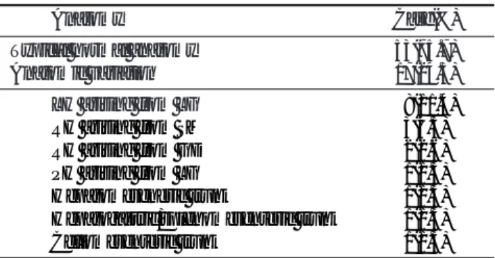
Table 1. Findings on 3D CT Hepatic Angiography
Anatomy Case(%)
Typical normal anatomy 53(75.7)
Anatomic variation 17(24.3)
LH arising from LG 08(11.4)
RH arising from SM 03(4.3)
RH arising from GD 02(2.6)
PH arising from LG 01(1.3)
Hepatomeseneric trunk 01(1.3)
Hepatogastric/splenomesenteric trunk 01(1.3)
Celiomesenteric trunk 01(1.3)
LH : left hepatic artery, LG : left gastric artery
RH : right hepatic artery, SM : superior mesenteric artery GD : gastroduodenal artery, PH : proper hepatic artery
복강동맥, 좌위동맥, 대동맥, 상장간막동맥등에서 기시하는 변 이 간동맥을 발견할 수 없다 (5). 따라서 간동맥의 정상변이에 대한 정확한 지식은 간동맥 조영술 및 경동맥화학색전술과 외 과 의사의 간이식술 등을 포함하는 간수술에 매우 중요하다.
간동맥의 해부학적인 변이를 알고자 시체(cadever)해부와 혈관조영술을 통한 여러 연구가 시행되었다 (4, 6-12). 최초 의 광범위한 연구는 Michels등(4)에 의해 이루어졌고, 이 연구 에서 200예를 분석하여 10가지의 간동맥의 해부학적 변이를 기술하였다. 그 보고에서 일반적인 정상 구조는 200예 중 110 예로 전체의 55%에 불과하였고, 가장 흔한 정상변이로 변이 좌간동맥(replaced left hepatic artery)과 변이 우간동맥 (replaced right hepatic artery)이 각각 20예(10%)와 22예 (11%)로 나타났다. 그외 부좌간동맥(accessory left hepatic artery)이 16예(8%), 부우간동맥(accessory right hepatic artery)이 14예(7%)를 차지했다. Brems등(13)은 172예의 간 이식 수술을 분석하여 Michels의 분류를 6가지로 변형(mod- ified Michels’classification)하여 외과 의사들에게 도움이 되
게 하였다. 또한 정 등(5)은 2000예를 분석하여 Michels의 연 구와는 다른, 혈관조영술자에게 도움이 되는 분류방식을 제시 하였다. 이 연구에 따르면 모든 간동맥이 고유간동맥에서 기시 하는 형태가 전체의 60%를 차지하고, 총간동맥에서 모든 간 동맥이 기시하는 경우가 75%였다. 흔한 변이로는 좌간동맥이 좌위동맥에서 기시하는 형이 15%, 우간동맥이 상장간막동맥 에서 기시하는 형이 11%, 우간동맥이 복강동맥에서 직접 기 시한 형이 3%의 빈도로 나타났다.
임상에서 실제 환자를 대상으로 간동맥의 해부학적 변이에 대해 알고자 할 경우 대부분 고식적 카테터 혈관조영술을 사 용한다. 간암의 경동맥화학색전술이나 간이식 수술 전의 혈관 지도(vascular map)는 일반적으로 카테터 혈관조영술을 이용 하게 되고, 여기서 간동맥과 문맥의 분지와 측부혈관(collat- eral vessel)의 유무를 찾게 된다 (14). 그러나 고식적 혈관조 영술은 여러 단점을 가지고 있다. 이 방법은 침습적인 방법으 로 출혈, 전해질 불균형, 신 기능 장애 등의 위험요소를 가지 며, 환자의 입원과 시술 후 안정을 요구한다. 또한 변이 혈관
A B
Fig. 1. Coronal oblique MIP image shows a replaced right hepatic artery(arrow) from superior mesenteric artery(A). Note the re- placed right hepatic artery(open arrow) at porto-caval space on axial MIP image(B).
A B
Fig. 2. Coronal oblique MIP image shows a replaced left hepatic artery(arrow) arising from left gastric artery(A). Axial MIP image shows a replaced left hepatic artery(open arrow) in the fissure of the ligamentum venosum(B).
의 존재 시 환자와 시술자의 방사선 피폭 시간이 길어진다 (15).
따라서 간동맥의 정상과 변이의 발견에 고식적 혈관조영술을 대신할 방법으로 이차원 횡단면 영상과 삼차원 CT 혈관조영 술이 활발히 연구되고 있다.
횡단면 CT 영상은 여러 가지 정보를 제공한다. CT는 간실 질과 복강내 맥관구조(abdominal vasculature)를 평가할 수 있 을 뿐 아니라, 이전의 알지 못했던 복강내 질병을 찾을 수도 있다. Chambers등(16)은 간동맥의 해부학적 평가에 있어 횡 단면 CT 영상의 유용성을 보여주었다. 이 연구에 따르면 횡단 면 CT 영상에서 변이 간동맥을 발견할 확률은 96%의 민감도 와 87%의 특이도를 가진다. 이 등(17)의 보고에 따르면 문 맥-하대정맥공간과 정맥관 인대열의 혈관을 관찰함으로써 간 동맥 변이 유무를 알 수 있다고 하였다. 저자들은 문맥-하대 정맥 공간을 횡으로 주행하는 혈관을 가질 경우 민감도 94%, 특이도 100%, 양성예측률 100%, 음성예측률 99%로 변이 간 동맥의 예측이 가능하며, 정맥관 인대열내 혈관을 가질 경우
A B
Fig. 3. Coronal(A) & sagittal(B) MIP images show a replaced left hepatic artery(arrow) arising from left gastric artery and a replaced right hepatic artery(open arrow) arising from superior mesenteric artery.
A B
Fig. 4. The common hepatic artery, left gastric artery, and splenic artery have a replaced origin from the superior mesenteric artery on coronal(A) and oblique coronal(B) MIP images.
Fig. 5. Coronal MIP image shows a replaced right hepatic artery(arrow) arising from gastroduodenal artery(open arrow).
민감도 88%, 특이도 100%, 양성예측률 100%, 음성예측률 98%로 변이 간동맥을 발견할 수 있다고 보고하였다. 이 연구 에서 문맥-하대정맥 공간을 횡으로 주행하는 혈관은 16예 중 11예가 우간동맥이 상장간막동맥에서 기시하는 형이고, 2예가 복강동맥에서 직접 우간동맥이 기시하는 형, 그리고 부우간동 맥이 복강동맥에서 기시하는 형, 부우간동맥이 상장간막동맥에 서 기시하는 형, 총간동맥이 복강동맥에서 기시하는 형이 각각 1예 씩 나타났다. 또한 저자들의 연구에서 정맥관 인대열내 혈 관을 가진 14예 중 좌위동맥에서 좌간동맥이 기시하는 형이 11예로 나타났으며 그 외 좌위동맥이 좌간동맥에서 기시하는 형이 2예, 부좌간동맥이 좌위동맥에서 기시하는 형이 1예로 나 타났다. 그러나 횡단면 CT 영상은 간동맥 변이의 유무를 판단 하는 데는 도움을 주지만 변이 혈관이 어느 동맥으로부터 기 시하는지, 즉 어떤 종류의 변이인지의 정확한 판단은 힘들다.
일반적으로 문맥-하대정맥 공간의 혈관은 상장간막동맥에서 기원하는 변이 우간동맥일 경우가 많으나, 많은 다른 변이 혈 관들이 비슷한 주행을 가질 수 있다 (3, 18). 상장간막동맥에 서 기시하는 부우간동맥, 상장간막동맥에서 기원하는 변이 총 간동맥(replacement of the entire hepatic trunk), 간동맥이 조 기에 좌,우간동맥으로 분지하고 우간동맥이 문맥-하대정맥공 간을 주행하는 경우, 복강동맥에서 기원하는 변이 우간동맥등 이 유사한 영상소견을 보일 수 있다 (3, 16, 19). 따라서 횡단 면 CT 영상만으로는 혈관구조의 변이를 정확히 평가하기가 곤 란한 경우가 많다.
최근에는 CT의 기술학이 눈부시게 발전함에 따라 삼차원 CT 혈관조영술이 간동맥의 평가에 점차 사용되고 있다. 삼차 원 CT 혈관조영술은 얇은 절편의 횡단면 영상을 workstation 으로 전송한 후, 실시간으로 여러 방향의 간동맥의 해부학적 위치와 주위와의 관계를 묘사할 수 있다. Winter등(3)은 간이 식 지원자의 술전 검사로 shaded surface display(SSD)와 maximum intensity projection(MIP) techniques를 사용하여 얻은 CT 혈관조영술의 유용성을 증명하였다. 이 연구에서 저 자들은 삼차원 CT 혈관조영술이 고식적 혈관조영술만큼 정확
하게 간동맥의 해부학적 평가를 할 수 있으며, 검사 비용은 25%에 불과하다는 것을 밝혔다. 동맥의 해부학적 평가를 위 해 삼차원 CT 혈관조영술을 사용하는 것은 많은 장점이 있다.
비용의 감소 이외에도, 고식적 혈관조영술만큼 정확한 검사방 법이면서 비침습적인 방법으로써 환자의 불편과 위험요소가 감 소되며, 환자가 입원하지 않고 쉽게 할 수 있을 뿐 아니라, 환 자와 의사의 방사선 피폭을 감소시킬 수 있다 (3, 15).
Workstation상에서 여러 방향으로의 회전이 가능하므로 임상 의사들이 다양한 변이 혈관들을 쉽게 이해할 수 있을 뿐 아니 라, 단면적인 고식적 혈관조영술에서는 잘 묘사하기 힘든 췌 장-십이지장 아케이드(pancreaticoduodenal arcade)를 잘 묘 사하여 수술에 따른 질병률(morbidity)과 사망률(mortality)을 감소시킬 수 있다 (20, 21).
Multidetector helical CT등을 포함하는 CT장비의 발달로 인 한 다양한 pitch와 이에 따른 더 넓어진 포함영역과 얇은 절편 두께, 그리고 자동주입기를 이용한 조영제의 단일일시주사 (single bolus injection)와 함께, 재구성 소프트웨어와 삼차원 재구성 기법의 발달에 힘입어 삼차원 CT 혈관조영술의 영상 의 질은 계속 향상되고 있다 (22-26). Four multidetector- row helical CT의 사용으로 이전의 single 또는 double mul- tidetector row helical CT에 비해 포함영역(volume cover- age)이 넓어졌다. Section sensitivity profile과 영상 잡음 (image noise), 그리고 영상 인공물(image artifact)등의 항목 에 대한 비교를 통해 Hu등(27)은 four multidetector-row helical CT가 single multidetector-row helical CT에 비해 비 슷하거나 더 나은 질의 영상을 얻으면서 volume coverage speed가 2-3배 정도 빠르다고 하였다. Volume coverage speed가 2-3배 증가할 경우 기존의 장비에 비해 포함영역이 2-3배 증가한다는 것 이외에도, 주입된 조영제를 이용하여 더 넓은 범위를 포함할 수 있으므로 조영제의 양을 줄일 수 있고, 다중위상연구(multiphase study)일 경우 초기 동맥기와 문맥 기의 분리를 용이하게 할 수 있는 장점이 있다. 따라서 스캔의 적절한 시간 조절이 중요한 폐색전증이나, 다중위상연구가 필
A B
Fig. 6. The celiac trunk and the superior mesenteric artery have a same origin. And the other vessels are normal in course on coro- nal(A) & sagittal(B) MIP images.
요한 경우, 넓은 포함영역이 요구되는 삼차원 CT 혈관조영술 이나 신경 또는 여러군데의 외상의 평가에 유용하게 사용될 수 있다.
삼차원 CT 혈관조영술은 몇가지 제한점을 가지고 있다. 환 자의 호흡조절이 안되거나 금속 등에 의한 인공물이 나타나 CT 스캔 자체가 힘든 경우와 재구성 기법에 의한 문제가 나 타날 수 있다. MIP기법은 빠르고 가장 흔히 사용하는 방법이 지만, 얻어진 helical CT 자료의 10%정도만 영상의 재구성에 사용되고 나머지는 버려지게 된다. 또한 주위 장기나 다른 구 조물은 영상에 나타나지 않으며, 비스듬히 주행하는 단면에서 혈관의 표면이 매끈하지 않게 나타날 수도 있다. 따라서 이런 단점을 극복하기 위해 SSD기법이나 three-dimensional vol- ume-rendering기법이 같이 사용되고 있다 (28). 미래에는 조 영증강 자기공명영상(contrast-enhanced magnetic reso- nance image)을 이용한 혈관조영술이 간동맥의 해부학적 평 가에 고식적 혈관조영술의 대체 방법이 될 수도 있겠지만, 현 재의 MR 기술로는 작은 동맥혈관을 삼차원 CT 혈관조영술만 큼 잘 나타내지는 못한다. 따라서 삼차원 CT 혈관조영술이 현 재 고식적 혈관조영술을 대체할 가장 발전된 영상기법이 될 수 있다.
이번 연구는 몇가지 제한점을 가지고 있다. 첫째, 대상의 선 택에 있어서 특정 질환을 대상으로 한 것이 아니라, 간 질환의 평가과정 중에 간 CT를 촬영한 70명을 임의로 선정하였다. 따 라서 모든 환자에 대한 간 질환의 진단 또는 조직학적 확진은 이루어지지 않았다. 둘째, 고식적 혈관조영술과의 비교가 이루 어지지 않았다. 그러나 간동맥의 평가에 있어 삼차원 혈관조영 술이 고식적 혈관조영술의 대체방법이 될 수 있다는 연구 결 과들을 참고하여 볼 때 이것은 이번 연구의 결과에 큰 영향을 주지는 않을 것이라 사료된다 (3, 15, 28, 29). 셋째, 대상 인 원의 수가 적어, 다양한 변이에 대한 이전 연구 결과들과의 통 계학적인 비교가 되지 못했다. 이것은 이번 연구가 multide- tector helical CT를 이용한 삼차원 재구성 CT 혈관조영술의 유용성에 초점을 둔 것으로, 앞으로 통계학적으로 유의한 인원 을 대상으로 하는 광범위한 연구가 필요할 것으로 보인다.
결론적으로 multidetector helical CT를 이용한 삼차원 재구 성 CT 혈관조영술은 현재 고식적 혈관조영술을 대체할 수 있 는 가장 발전된 영상방법이며, 검사 비용이 저렴하고 비침습적 이면서 간동맥의 정상 및 변이를 고식적 혈관조영술만큼 잘 반 영하여, 간동맥의 해부학적 평가를 요하는 간의 술전 검사 또 는 간암의 치료에 있어 많은 도움을 줄 수 있을 것으로 생각 한다.
참 고 문 헌
1. Nelson TM, Pollak R, Jonasson O, et al. Anatomic variants of the celiac, superior mesenteric, and inferior mesenteric arteries and their clinical relevance. Clin Anat 1988;1:75-91
2. Makisalo H, Chaib E, Krokos N, Caine R. Hepatic arterial varia- tions and liver-related diseases of 100 consecutive donors. Transpl Int 1993;6:325-329
3. Winter T Ⅲ, Freeny P, Nghiem H, et al. Hepatic arterial anatomy in transplantation candidates : evaluation with three-dimensional CT arteriography. Radiology 1995;195:363-370
4. Michels NA. Blood supply and anatomy of the upper abdominal or- gans. Philadelphia: Lippincott,1995:210-291
5. 정진욱. 간동맥의 해부학과 측부순환. 혈관 및 중재적방사선과학 증례집 1996;2:14-22
6. Michels NA. Newer anatomy of the liver and its variant blood sup- ply and collateral circulation. Am J Surg 1966;112:337-347 7. Rong GH, Sindelar WF. Aberrant peripancreatic arterial anatomy :
considerations in performing pancreatectomy for malignant neo- plasms. Am Surg 1987;53:726-729
8. Kemeny MM, Hogan JM, Goldberg DA, et al. Continuous hepatic artery infusion with an implantable pump : problems with hepatic arterial anomalies. Surgery 1986;99:501-504
9. Rygaard H, Forrest M, Mygind T, et al. Anatomic variants of the hepatic arteries. Acta Radiol Diagn 1986;27:425-427
10. Daly JM, Kemeny N, Botet J. Long-term hepatic arterial infusion chemotherapy. Arch Surg 1984;119:936-941
11. Niederhuber JE, Ensminger WD. Surgical considerations in the management of hepatic neoplasia. Semin Oncol 1983;10:135-147 12. Jonathan RH, Joubin G, Ronald WB. Surgical anatomy of the he-
patic arteries in 1000 cases. Ann Surg 1994;220:50-52
13. Brems JJ, Millis JM, Hiatt JR, et al. Hepatic artery reconstruction during liver transplantation. Transplantation 1989;47:403-406 14. Ferris J, Marsh J, Litle A. Presurgical evaluation of the liver trans-
plantation candidate. Radiol Clin North Am 1995;33:497-520 15. Smith PA, Klein AS, Heath DG, et al. Dual-phase spiral CT angiog-
raphy with volumetric 3D rendering for preoperative liver trans- plant evaluation. J Comput Assist Tomogr 1998;22:868-874 16. Chambers TP, Fishman EK, Bluemke DA, Urban BA, Venbrux
AC. Identification of the aberrant hepatic artery with axial spiral CT. J Vasc Intervent Radiol 1995;6:959-964
17. 이재영, 정진욱, 김태경. 나선식 CT 횡단면 영상에서 간동맥 정상 변이의 진단: 문맥-하대정맥 공간과 정맥과인대열에 있는 혈관의 중요성. 대한방사선의학회지 1997;37:473-478
18. Ito K, Choji T, Fujita T, et al. Imaging of the portocaval space. AJR Am J Roentgenol 1993;161:329-334
19. Winter TC Ⅲ, Ngbiem HV, Freeny PC, Hommeyer SC, Mack LA.
Hepatic arterial anatomy : demonstration of normal supply and vascular variants with three-dimensional CT angiography.
Radiographics 1995;15:771-780
20. Cikrit DF, Dalsing MC, Sawchuk AP, et al. Vascular injuries dur- ing pancreatobiliary surgery. Am Surg 1993;59:692-696
21. Woods MS, Traverso LW. Sparing a replaced common hepatic artery during pancreatico- duodenectomy. Am Surg 1993;59:719- 721
22. Bluemke DA, Fishman EK. Spiral CT of the abdomen: clinical ap- plications. Crit Rev Diagn Imag 1993;34:103-157
23. Ney DR, Fishman EK, Magid D, et al. Three-dimensional volumet- ric display of CT data: effect of scan parameters upon image quali- ty. J Comput Assist Tomogr 1991;15:875-885
24. Kuszyk BS, Heath DG, Ney DR, et al. CT angiography with vol- ume rendering: image findings. AJR Am J Roentgenol 1995;165:445- 448
25. Johnson PT, Heath DG, Bliss DF, Cabral B, Fishman EK. Three-di- mensional CT: real-time interactive volume rendering. AJR Am J Roentgenol 1996;167:581-593
26. Fishman EK, Magid D, Ney DR, et al. Three-dimensional imaging.
Radiology 1991;181:321-337
27. Hui HH, David HW, Dennis F, et al. Four multidetector-row heli- cal CT: Image quality and volume coverage speed. Radiology 2000;
215:55-62
28. Hong KC, Freeny PC. Pancreaticoduodenal arcades and dorsal pancreatic artery : comparison of CT angiography with three-di- mensional volume rendering, maximum intensity projection, and
shaded-surface display. AJR Am J Roentgenol 1999;172:925-931 29. Nghiem HV, Dimas CT, McVicar JP, et al. Impact of double helical
CT and three-dimensional CT arteriography on surgical planning for hepatic transplantation. Abdom Imaging 1999;24:278-284
J Korean Radiol Soc 2001;45:35-41
Address reprint requests to : Jun Woo Lee, M.D., Department of Diagnostic Radiology, Pusan National University Hospital 1-10, Ami-dong, Seo-ku, Pusan 602-739, Korea.
Tel. 82-51-240-7354 Fax. 82-51-244-7534
Assessment of Hepatic Arterial Variation Using Multidetector Helical CT-Angiography
1Dong-Hoon Lee, M.D., Jun Woo Lee, M.D., Woong bae Jun, M.D., Suk Hong Lee, M.D.
1Department of Diagnostic Radiology, Pusan National University
Purpose: To evaluate the anatomy of the hepatic artery and normal variants using oblique thick-slab maximal intensity projection (MIP) 3-D CT angiography and multidetector helical CT technology.
Materials and Methods: In 70 patients, axial three-phase CT together with multidetector helical CT and a non- ionic contrast agent was used to evaluate liver disease. During the early arterial phase, the parameters were as follow: slice thickness, 2.5 mm; table speed, 15 mm/rotation, pitch, 6; contrast material, 4 ml/sec; total 120 ml.
Using the MIP technique and an Advantage window voxtal 3.03 system (GE), the images obtained were recon- structed as 3D angiograms. In each case, the arterial anatomy and its variants were recorded.
Results: A typical anatomy was found in 53 cases (75.7 %). Common variants were a left hepatic artery arising from the left gastric artery(8 cases, 11.4 %) and a right hepatic artery arising from the superior mesenteric artery(3 cases, 4.3 %). Other variant cases were a right hepatic artery arising from the gastroduodenal artery(2 cases, 2.9%), a proper hepatic artery arising from the left gastric artery (1 case, 1.4%), a hepatomesenteric trunk (1 case), a hepatogastric/splenomesenteric trunk(1 case), and a celiomesenteric trunk (1 case).
Conclusion: 3-D hepatic angiography using multidetector helical CT technology is non-invasive and as accu- rate as conventional angiography for the evaluation of hepatic arterial anatomy. It is thus considered that 3-D CT angiography is very helpful for the evaluation of hepatic arterial anatomy prior to liver surgery such as transplantation or the treatment of hepatocellular carcinoma.
Index words :Liver, CT Angiography
Computed tomography (CT), three-dimensional