INTRODUCTION
Lung cancer continues to be one of the most lethal malig- nant tumors. Lung cancer screening using low-dose CT has expanded in clinical medicine due to the presence of high risk populations and the relatively asymptomatic nature of the dis- ease in the early stages. With the increased use of helical CT for lung cancer screening, the detection rate of small lung cancer lesions has also increased (1-4).
Small pulmonary nodules suspicious for a malignancy mandate a pathological diagnosis. CT-guided transthoracic needle aspiration biopsy is a well-established method for the cytological diagnosis of pulmonary nodules (5-8). However,
histological evaluation by tissue-core cutting-needle biopsy is better than cytology for the determination of a specific diag- nosis, especially for benign lesions and subsolid lesions (9, 10). CT fluoroscopy provides real-time imaging and allows radiologists to manipulate a patient in response to respiratory movement. Therefore, this imaging approach may permit rapid localization of lesions that are smaller and/or located in less accessible areas, as well as in patients that have difficulty cooperating with the procedure (11, 12). Kim et al. (13) re- ported that CT fluoroscopy-guided percutaneous biopsy of pulmonary lesions provided high diagnostic accuracy compa- rable to that of conventional CT-guided procedures, and with fewer complications. However, radiation exposure to both pa-
J Korean Soc Radiol 2011;65(4):373-379
Received June 17, 2011; Accepted August 5, 2011 Corresponding author: Yoon Kyung Kim, MD Department of Radiology, Korea University Guro Hospital, Korea University College of Medicine, 97 Gurodong-gil, Guro-gu, Seoul 152-703, Korea.
Tel. 82-2-2626-1338 Fax. 82-2-863-9282 Department of Radiology, Gachon University Gil Hospital, 1198 Guwol-dong, Namdong-gu, Incheon 405-760, Korea.
Tel. 82-32-3060 Fax. 82-32-460-3065 E-mail: [email protected]
Copyrights © 2011 The Korean Society of Radiology
Purpose: To evaluate the efficacy of CT fluoroscopy-guided core biopsy of small pulmonary nodules.
Materials and Methods: This study included 62 patients (35 men, 27 women; age range, 36-85 years) that had a small (≤ 20 mm) pulmonary nodule and underwent CT fluoroscopy-guided core biopsy. The overall diagnostic accuracy and complica- tion rate were calculated. The diagnostic accuracy was compared between two groups according to the nodule size (≤ 10 mm vs. > 10 mm), and nodule density (solid vs. subsolid).
Results: Malignant or premalignant lesions were finally diagnosed in 39 patients;
36 true-positive and three false-negative findings (sensitivity, 92%). A benign lesion was finally diagnosed in 23 patients, with no false-positive results (specificity, 100%). The overall diagnostic accuracy was 95%. The sensitivity and diagnostic ac- curacy were 85% and 91% for nodules ≤ 10 mm, and 96% and 97% for nodules >
10 mm (p > 0.05). The sensitivity and diagnostic accuracy were 93% and 96% in the solid group and 90% and 92% in the subsolid group (p > 0.05). Seventeen (27%) patients had a pneumothorax and two (3%) required a closed thoracostomy.
Conclusion: CT fluoroscopy-guided core biopsy of small pulmonary nodules yields high diagnostic accuracy with acceptable complication rates.
Index terms
Computed Tomography
Computed Tomography Fluoroscopy Biopsy, Needle
Lung Neoplasms Pulmonary Nodules
CT Fluoroscopy-Guided Core Biopsy for Diagnosis of Small (≤ 20 mm) Pulmonary Nodules
1CT 투시촬영 유도하 중심부바늘생검을 이용한 폐 소결절(≤ 20 mm)의 진단1
Hye Larn Lee, MD
1, Yoon Kyung Kim, MD
1,2, Ok Hee Woo, MD
1, Hwan Seok Yong, MD
1, Eun-Young Kang, MD
1, Hyun Koo Kim, MD
3, Bong Kyung Shin, MD
41Department of Radiology, Korea University Guro Hospital, Korea University College of Medicine, Seoul, Korea
2Department of Radiology, Gachon University Gil Hospital, Incheon, Korea
Departments of 3Thoracic Surgery, 4Pathology, Korea University Guro Hospital, Korea University College of Medicine, Seoul, Korea
64-multi-detector CT scanner (Brilliance 64; Philips Medical Systems, Cleveland, OH, USA). At the time of the biopsy, se- lected images were obtained in the area of interest with 5-mm- thick contiguous transverse CT sections. Biopsies were planned such that they crossed the fewest pleural surfaces and avoided fissures or visible bullae. The procedure was performed with the patient in the prone, supine, or lateral decubitus position, depending on the location of the lesion. After local anesthe- sia, a core biopsy specimen was obtained with an 18-gauge automated cutting needle (Bio-gun; M.I.Tech, Seoul, Korea).
The needle was inserted through the pleura and advanced to a position close to a target lesion under CT fluoroscopy guid- ance. To avoid irradiating the radiologists’ fingers, the opera- tor manipulated the needle with a needle holder during CT fluoroscopy-guided biopsy. To minimize the radiation to the patient and radiologist, the real-time scanning was limited to short glimpses to visualize the position of the needle tip. After the position of the needle tip was confirmed, the automated needle biopsy system was fired. Specimen acquisition was re- peated until the radiologist determined that the specimens were adequate. Core specimens were immersed in 10% for- malin for a subsequent pathology examination. A low dose CT was performed over the biopsy site immediately after the biopsy. Follow-up chest radiographs were obtained 1 hour and 24 hours after the biopsy to evaluate for a pneumothorax.
A thoracostomy was considered when a patient had a symp- tomatic, large (> 30%) pneumothorax.
Statistical Analysis
The sensitivity, specificity, and diagnostic accuracy were calculated. The biopsy was considered as truly benign if the results of the biopsy evaluation were confirmed as benign by a surgically resected specimen, and if the lesion was stable in size for at least 12 months or decreased in size. A biopsy was considered to be truly malignant if the results of the biopsy were confirmed by a surgically resected specimen or if the le- sion increased in size. A biopsy was considered non-diagnos- tic if the pathology yielded no specific benign or malignant diagnoses. Non-diagnostic cases were excluded from the cal- culation of sensitivity, specificity, and diagnostic accuracy.
The results were compared between the two groups according to lesion size and lesion density. Fisher’s exact test and the tient and doctor were significantly higher than conventional
CT-guided biopsy.
The present study evaluated the diagnostic accuracy and complications of CT fluoroscopy-guided core biopsy of small (≤ 20 mm) pulmonary nodules.
MATERIALS AND METHODS
Patients and Lesions
A retrospective analysis was performed on the imaging re- cords of all patients that underwent CT fluoroscopy-guided core biopsy of pulmonary nodules at this institution between February 2007 and February 2010. The study included pa- tients that underwent biopsy for small pulmonary nodules with a diameter of less than 20 mm and with a final diagnosis established by surgical resection of the nodule or imaging fol- low-up. Patients diagnosed with benign lesions by biopsy that remained stable in size and were followed for < 12 months, were excluded from the study. Finally, 62 patients (35 men and 27 women; age range, 36-85 years) were enrolled in the study. Among these patients, 35 underwent surgical resection and 27 patients underwent follow-up exams. The medical and imaging records of these 62 patients were reviewed by two ra- diologists. The diameter of all nodules and lesion depth (length of the aerated lung traversed by the needle from the surface of the pleura to the proximal margin of the target le- sion) were measured on lung window settings. The patients were divided into two groups on the basis of nodule size (≤ 10 mm, n = 25 vs. > 10 mm, n = 37) and nodule density (solid, n = 49 vs. subsolid, n = 13) for comparison of the diagnostic accu- racy. When the lesions contained a solid portion less than 50%, the lesion was defined as subsolid. Lesions containing more than 50% of a solid portion were defined as solid lesions. The complications associated with the procedure such as pneumo- thorax and thoracostomy insertion rates were recorded.
Procedures
The biopsies were performed by experienced chest radiolo- gists that had four and 10 years of experience performing tho- racic biopsy procedures, respectively. Written informed consent was obtained from patients before their biopsy. All biopsies were performed under CT fluoroscopy guidance using a
lymph node in 1, and inflammatory pseudotumor in 1), with no false-positive results. Non-diagnostic results were obtained in three out of the 62 lesions (5%). All three non-diagnostic samples were lesions smaller than 10 mm and were benign le- sions (intrapulmonary lymph node in 1, chondroid hamartoma in 1, and no change on follow up images at 24-month follow- up periods in 1); these lesions were excluded from the calcula- tions of sensitivity, specificity and diagnostic accuracy.
The mean lesion diameter was 1.3 ± 0.4 cm (range, 0.5-2.0 cm). The overall sensitivity, specificity and diagnostic accura- cy were 92%, 100%, and 95%, respectively. The sensitivity and diagnostic accuracy were 85% (11/13) and 91% (20/22) for nodules ≤ 10 mm, 96% (25/26) and 97% (36/37) for nodules
> 10 mm. The sensitivity and diagnostic accuracy were 93%
and 96% in the solid lesion group, and 90% and 92% in the subsolid group. The sensitivity and diagnostic accuracy were not significantly different between the two groups of nodule sizes and densities (Tables 1, 2).
Seventeen (27%) patients developed a pneumothorax, with two (3%) requiring placement of a thoracostomy tube. After the thoracostomy, all patients fully recovered without other complications. The frequency of pneumothorax for patients with a nodule size ≤ 10 mm and > 10 mm was similar (24%
Student’s t-test were used to assess the statistical significance of the difference between the two groups. A p-value of less than 0.05 was considered to indicate statistical significance.
RESULTS
A total of 59 cases out of 62 were diagnosed by CT fluoros- copy-guided core biopsy. Malignant or premalignant lesions were finally diagnosed in 39 patients (adenocarcinoma in 20, metastases from an extrathoracic malignancy in 7, squamous cell carcinoma in 4, small cell carcinoma in 3, bronchioloalve- olar carcinoma in 3, and atypical adenomatous hyperplasia in 2) with 36 true-positive (adenocarcinoma in 18, metastases from an extrathoracic malignancy in 7, squamous cell carci- noma in 4, small cell carcinoma in 2, bronchioloalveolar car- cinoma in 3, and atypical adenomatous hyperplasia in 2) and 3 false-negative findings (chronic inflammation in 1, anthra- cofibrosis in 1, type II pneumocyte hyperplasia in 1) (Fig. 1).
A benign lesion was finally diagnosed in 23 patients (chronic granulomatous inflammation in 6, organizing pneumonia in 4, tuberculosis in 4, chondroid hamartoma in 3, chronic inter- stitial inflammation in 1, fungus ball in 1, cryptococcosis in 1, actinomycosis in 1, rheumatoid nodule in 1, intrapulmonary
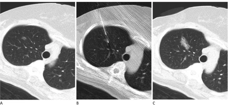
A B C
Fig. 1. CT fluoroscopy-guided core biopsy in a 59-year-old woman.
A. A 1 cm-sized subsolid nodule is seen in the right upper lobe.
B. CT scan obtained during biopsy shows needle targeting of the nodule.
C. Post-biopsy CT scan shows pulmonary hemorrhage along the needle passage. The histopathological diagnosis by biopsy was bronchioloalveo- lar carcinoma confirmed by surgery.
out of 62 lesions (5%), which were all benign lesions measur- ing less than 1 cm.
The diagnostic accuracy for lesions < 1 cm in diameter (91%) was lower than that for lesions > 1 cm in diameter (97%).
However, this difference was not statistically significant. Nod- ule size is an important factor influencing the diagnostic ac- curacy of CT fluoroscopy-guided core biopsy. Montaudon et al.
(15) reported that a lesion size ≤ 1 cm was significantly associ- ated with a higher rate of false negative diagnoses of a malig- nancy. They suggested that by using a coaxial automated de- vice, a high diagnostic yield could be obtained. Li et al. (16) reported that the diagnostic accuracy for lesions < 1.5 cm in diameter (74%) was statistically different from lesions > 1.5 cm in diameter (97%). These differences in the diagnostic ac- curacy of small and large pulmonary nodules might be asso- ciated with the aspiration needle used instead of the core bi- opsy needle. Two prior studies (15, 16) used conventional CT for biopsy guidance. However, the diagnostic accuracy could have been higher if CT fluoroscopy was used for guidance be- cause it might have been easier to target small lesions < 1 cm in diameter.
Special attention to patients and radiologists is needed for CT fluoroscopy-guided biopsies due to the hazard of radia- tion exposure. During CT fluoroscopy-guided biopsy, a pa- tient is exposed to radiation at or near the needle puncture site. The fixed position of the scanning plane in combination with high exposure factors may lead to a high accumulation of radiation exposure to the patient’s skin. Radiologists are also exposed to scattered radiation because of their close prox- imity to the scanning. Kim et al. (13) studied that radiation doses to the patients and doctors and diagnostic performance for CT-guided percutaneous needle aspiration biopsy of pul- monary lesions were performed with or without fluoroscopic guidance. The surface radiation doses to the patient and the doctor as well as the hand measured using a thermolumines- cent dosimeter was significantly higher in the group with flu- oroscopic guidance. However, the procedure time was signifi- cantly shorter, and the number of punctures and complication rate were lower. To minimize radiation exposure we wore lead covered clothing, gloves, and glasses. To avoid irradiating the hands, the needle was manipulated with a needle holder dur- ing CT fluoroscopy-guided biopsy and fluoroscopy time was and 30%, respectively). The pneumothorax frequency for solid
and subsolid nodules was found in 32% and 25% of proce- dures, respectively. Lesion size and depth were not significantly different between the two groups, with or without pneumotho- rax (Table 3). Post procedural pulmonary hemorrhage oc- curred near the pulmonary nodule in 35 patients (56%); how- ever, no significant massive hemoptysis developed.
DISCUSSION
CT fluoroscopy-guided core biopsy is a well-established ac- curate method used for the diagnosis of pulmonary nodules.
According to Hiraki et al. (14), the diagnostic yield of CT flu- oroscopy-guided core biopsies performed in 1,000 lesions was 99.4%, with only six non diagnostic results (0.6%). The overall diagnostic accuracy was 95.2% regardless of lesion size. For lesions measuring less than 1 cm, the diagnostic accuracy was 92.7%. In our study, the diagnostic yield was 95% and the overall diagnostic accuracy was 95%. For lesions measuring less than 1 cm, the diagnostic accuracy was 91%. The results of our study showed a similar diagnostic accuracy compared to previously reported rates in the medical literature (10, 14).
In our study, non diagnostic samples were obtained in three Table 1. Diagnostic Accuracy of Small (≤ 20 mm) Pulmonary Nod- ules Based on Lesion Size
Lesion Diameter p-value
≤ 10 mm > 10 mm
Sensitivity 85% (11/13) 96% (25/26) 0.253 Accuracy 91% (20/22) 97% (36/37) 0.549 Lesion depth (cm) 2.0 ± 1.5 1.6 ± 1.4 0.285 Table 2. Diagnostic Accuracy of Small (≤ 20 mm) Pulmonary Nod- ules Based on Lesion Density
Lesion Density p-value
Subsolid (n = 12) Solid (n = 47)
Sensitivity 90% (9/10) 93% (27/29) 1.000 Accuracy 92% (11/12) 96% (45/47) 0.501 Lesion depth (cm) 2.1 ± 1.3 1.7 ± 1.5 0.411 Lesion size (cm) 1.1 ± 0.4 1.4 ± 0.4 0.030
Table 3. Factors Influencing the Occurrence of Pneumothorax With
Pneumothorax
Without
Pneumothorax p-value Lesion size (cm) 1.34 ± 0.43 1.32 ± 0.42 0.874 Lesion depth (cm) 1.72 ± 1.38 1.80 ± 1.47 0.865
used in an automated gun are important precautions in avoid- ing unnecessary bleeding complications (18).
The limitations of our study include the following. First, our study had a relatively small number of enrolled patients, especially in the subsolid nodule group and the group of nod- ules equal to or less than 10 mm. To achieve statistical signifi- cance, more studies with large data sets will be needed. Second, our study had a follow-up period of 12 months. Pulmonary nodules are generally considered benign if they are stable in size for two years. GGO lesions such as atypical adenomatous hyperplasia or bronchioloalveolar carcinoma may persist with- out size change over two years. Therefore, a 12-month follow up period might have missed a lung malignancy in patients without histological confirmation. However, our study pa- tients were followed with CT that had a specific benign pa- thology such as tuberculosis and chondroid hamartoma. Pa- tients that had a nonspecific pathological diagnosis such as chronic inflammation underwent surgical resection. None of the patients that were followed with CT had subsolid nodules.
Only solid nodules were followed by CT; therefore, a short follow up period does not appear to significantly affect the overall diagnosis. Third, biopsies were performed in patients that had a chest CT that was suspicious for a malignant pul- monary lesion. The diagnostic accuracy for malignant disease was 90%, compared to 80% for benign disease. Our study in- cluded more malignant lesions than benign lesions, which might have contributed to the high diagnostic accuracy.
In conclusion, CT fluoroscopy-guided core biopsy was dem- onstrated to be a safe and accurate modality for the diagnosis of small (≤ 20 mm) pulmonary nodules. This technology could be used with high diagnostic accuracy even in lesions equal to or less than 10 mm and for subsolid nodules.
REFERENCES
1. Kaneko M, Eguchi K, Ohmatsu H, Kakinuma R, Naruke T, Suemasu K, et al. Peripheral lung cancer: screening and detection with low-dose spiral CT versus radiography. Ra- diology 1996;201:798-802
2. Sone S, Takashima S, Li F, Yang Z, Honda T, Maruyama Y, et al. Mass screening for lung cancer with mobile spiral com- puted tomography scanner. Lancet 1998;351:1242-1245 minimized.
Contrary to general assumptions that the diagnostic accu- racy of CT-guided biopsy for ground-glass opacity (GGO) le- sions is less than for solid lesions, Kim et al. (9) reported that CT-guided core biopsy of pulmonary GGO lesions could yield a high diagnostic accuracy approaching that of solid lesions.
In fact, the diagnostic accuracy of GGOs was 91%, with a posi- tive predictive value of 97%, and a negative predictive value of 75%. This result might be related to the use of a cutting needle rather than an aspiration needle. The results of our study showed a 92% diagnostic accuracy for GGO lesions. This re- sult was lower than for solid lesions (96%); however, the dif- ference was not statistically significant. Therefore, subsolid le- sions could be biopsied when needed; if a cutting biopsy needle is used, the diagnostic accuracy for subsolid lesions is as high as for solid lesions. However, there is still debate over whether small pure GGO nodules need to be biopsied.
In several studies, investigators have compared the frequen- cy of pneumothorax after CT fluoroscopy-guided lung biopsy.
Kirchner et al. (17) showed that the frequency of pneumotho- rax was 38% (8/21) after CT fluoroscopy-guided biopsies of lung lesions. In our study, complications such as pneumotho- rax and thoracostomy occurred in 27% of 62 patients, which is in the mid range of reported complication rates (12.5-69%) (13). The prevalence of pulmonary hemorrhage in our study was relatively higher than rates reported (20.2-30%) in the lit- erature (9, 15, 18). Further, pulmonary hemorrhage along the needle path or around the target lesion developed in 35 pa- tients (56%), but none required specific treatment. Our study included a higher proportion of small nodules and GGO nodules, which are risk factors for bleeding, compared to pre- vious studies (18). Laurent et al. (19) found that when using a coaxial 20-gauge automated cutting needle, there was a 43%
alveolar hemorrhage rate and a 6% hemoptysis rate in 67 bi- opsied of lung nodules < 20 mm in size, compared to a 14%
alveolar hemorrhage rate and a 5% hemoptysis rate in 135 bi- opsies of larger lesions. When taking a biopsy from a small le- sion or GGO lesion, the cutting needle often includes a por- tion of aerated lung, which results in a lower tamponading ef- fect than solid tissue. Therefore, embedding the cutting nee- dle into lung lesions without violating the aerated lung and avoiding the trauma incurred by the rapidly firing needle
13. Kim GR, Hur J, Lee SM, Lee HJ, Hong YJ, Nam JE, et al. CT fluoroscopy-guided lung biopsy versus conventional CT- guided lung biopsy: a prospective controlled study to as- sess radiation doses and diagnostic performance. Eur Ra- diol 2011;21:232-239
14. Hiraki T, Mimura H, Gobara H, Iguchi T, Fujiwara H, Sakurai J, et al. CT fluoroscopy-guided biopsy of 1,000 pulmonary lesions performed with 20-gauge coaxial cutting needles:
diagnostic yield and risk factors for diagnostic failure.
Chest 2009;136:1612-1617
15. Montaudon M, Latrabe V, Pariente A, Corneloup O, Be- gueret H, Laurent F. Factors influencing accuracy of CT- guided percutaneous biopsies of pulmonary lesions. Eur Radiol 2004;14:1234-1240
16. Li H, Boiselle PM, Shepard JO, Trotman-Dickenson B, McLoud TC. Diagnostic accuracy and safety of CT-guided percutaneous needle aspiration biopsy of the lung: com- parison of small and large pulmonary nodules. AJR Am J Roentgenol 1996;167:105-109
17. Kirchner J, Kickuth R, Laufer U, Schilling EM, Adams S, Liermann D. CT fluoroscopy-assisted puncture of thoracic and abdominal masses: a randomized trial. Clin Radiol 2002;57:188-192
18. Yeow KM, Su IH, Pan KT, Tsay PK, Lui KW, Cheung YC, et al.
Risk factors of pneumothorax and bleeding: multivariate analysis of 660 CT-guided coaxial cutting needle lung bi- opsies. Chest 2004;126:748-754
19. Laurent F, Latrabe V, Vergier B, Montaudon M, Vernejoux JM, Dubrez J. CT-guided transthoracic needle biopsy of pulmonary nodules smaller than 20 mm: results with an automated 20-gauge coaxial cutting needle. Clin Radiol 2000;55:281-287
3. Henschke CI, McCauley DI, Yankelevitz DF, Naidich DP, McGuinness G, Miettinen OS, et al. Early Lung Cancer Ac- tion Project: overall design and findings from baseline screening. Lancet 1999;354:99-105
4. Sobue T, Moriyama N, Kaneko M, Kusumoto M, Kobayashi T, Tsuchiya R, et al. Screening for lung cancer with low- dose helical computed tomography: anti-lung cancer as- sociation project. J Clin Oncol 2002;20:911-920
5. Westcott JL. Direct percutaneous needle aspiration of lo- calized pulmonary lesions: result in 422 patients. Radiolo- gy 1980;137:31-35
6. Khouri NF, Stitik FP, Erozan YS, Gupta PK, Kim WS, Scott WW Jr, et al. Transthoracic needle aspiration biopsy of be- nign and malignant lung lesions. AJR Am J Roentgenol 1985;144:281-288
7. Stanley JH, Fish GD, Andriole JG, Gobien RP, Betsill WL, Laden SA, et al. Lung lesions: cytologic diagnosis by fine- needle biopsy. Radiology 1987;162:389-391
8. Westcott JL. Percutaneous transthoracic needle biopsy.
Radiology 1988;169:593-601
9. Kim TJ, Lee JH, Lee CT, Jheon SH, Sung SW, Chung JH, et al. Diagnostic accuracy of CT-guided core biopsy of ground-glass opacity pulmonary lesions. AJR Am J Roent- genol 2008;190:234-239
10. Yamagami T, Iida S, Kato T, Tanaka O, Toda S, Kato D, et al.
Usefulness of new automated cutting needle for tissue- core biopsy of lung nodules under CT fluoroscopic guid- ance. Chest 2003;124:147-154
11. Katada K, Kato R, Anno H, Ogura Y, Koga S, Ida Y, et al.
Guidance with real-time CT fluoroscopy: early clinical ex- perience. Radiology 1996;200:851-856
12. Moore EH. Technical aspects of needle aspiration lung bi- opsy: a personal perspective. Radiology 1998;208:303-318
CT 투시촬영 유도하 중심부바늘생검을 이용한 폐 소결절(≤ 20 mm)의 진단1
이혜란
1· 김윤경
1,2· 우옥희
1· 용환석
1· 강은영
1· 김현구
3· 신봉경
4목적: 폐 소결절(≤ 20 mm)의 진단에 있어 CT 투시촬영 유도하 중심부바늘생검의 유용성에 대해 알아보고자 하였다.
대상과 방법: 폐 소결절(≤ 20 mm)에 대하여 CT 투시촬영 유도하 중심부바늘생검을 시행 받은 62명의 환자를 대상으
로 진단적 정확도와 합병증에 대해 알아보았다. 조직검사 결과와 수술(n = 35) 또는 추적검사(n = 27)로 확인된 최종
결과를 비교하여 진단적 정확도를 계산하였으며, 진단적 정확도는 소결절의 크기(≤ 10 mm 대 > 10 mm)와 결절의 밀 도(고형결절 대 아 고형결절)에 따라 차이가 있는지 알아보았다.
결과: 최종적으로 39명의 환자에서 악성 또는 전암 병소가 확인되었다. CT 투시촬영 유도하 중심부바늘생검을 통해 이 중 36명은 진양성, 3명은 가음성으로 나왔다(민감도, 92%). 23명의 환자들은 양성 병소로 밝혀졌으며 가양성 결과는 없 었다(특이도, 100%). CT 투시촬영 유도하 폐 결절 중심부바늘생검의 진단적 정확도는 95%였다. 10 mm 이하의 폐 결절 의 민감도와 진단적 정확도는 각각 85%와 91%였으며, 10 mm보다 큰 폐 결절의 민감도와 진단적 정확도는 각각 96%
와 97%였다(p > 0.05). 고형 폐 결절의 민감도와 진단적 정확도는 각각 93%와 96%였으며, 아 고형 폐 결절의 민감도 와 진단적 정확도는 각각 90%와 92%였다(p > 0.05). 17명의 환자(27%)에서 기흉이 발생하였으며 이 중 2명(3%)은 흉강 배액관 삽입을 시행하였다.
결론: CT 투시촬영 유도하 중심부바늘생검은 폐 소결절(≤ 20 mm) 진단에 있어 비교적 정확하고 안전한 검사이다.
1고려대학교 의과대학 구로병원 영상의학과학교실, 2가천의과학대학교 길병원 영상의학과학교실,
고려대학교 의과대학 구로병원 3흉부외과학교실, 4병리과학교실