https://doi.org/10.14193/jkfas.2018.22.2.62
서 론
부주상골은 후경골근건이 주상골에 붙는 곳에서 발생하는 부골 이다.1) 발생률은 인구에서 4%∼21%까지 확인되는 비교적 흔한 질 환이다.2-4) 부주상골은 대부분은 증상이 없이 지내다가 우연히 발 견되는 경우가 많지만 일부 환자들에게서 주상골 부근의 족부 내 측 통증을 유발하는 직접적인 원인이 된다.5)
부주상골은 대부분 증상이 없지만 과도한 운동에 의한 자극, 외 상성, 연골결합의 퇴행성 변화, 또는 돌출된 뼈 부위의 염증에 의
Original Article
This is an Open Access article distributed under the terms of the Creative Commons Attribution Non-Commercial License (http://creativecommons.org/licenses/CC
by-nc/4.0) which permits unrestricted non-commercial use, distribution, and reproduction in any medium, provided the original work is properly cited.
Copyright 2018 Korean Foot and Ankle Society. All rights reserved.ⓒ
Purpose: To evaluate the clinical significance and usefulness of a bone scan in accessory navicular bone.
Materials and Methods: Eighty-five patients with foot pain and accessory navicular bone on radiography, who underwent bone scan
from 2012 to 2015, were analyzed retrospectively. The subjects was divided into a symptomatic and asymptomatic group according to the presence of navicular bone tenderness. The grade of bone scan uptake was divided into 3 grades. Age, gender, grade of bone scan and size of the accessory navicular bone were analyzed. The symptomatic group were divided into a low (grade 0, 1) and high uptake (grade 2) group to determine the appropriate treatment. The low uptake group was treated conservatively for 3 months. The high uptake group was initially treated conservatively for 3 months and surgery was performed if pain persisted. For the clinical evaluation, the visual analogue scale, American Orthopaedic Foot and Ankle Society midfoot scale were evaluated in the first examination and last follow-up date. The patient’s satisfaction grade was also evaluated at the last follow-up.Results: The asymptomatic group mostly showed no uptake in the bone scan. On the other hand, some patients in the asymptomatic
group showed an increase in uptake. In these patients, the size of accessory navicular bone was related to the grade of bone scan uptake, showing that the bone scan uptake grade can be predicted when applying different cut off values for the bone size. The symptomatic group mostly showed uptake in the bone scan and the grade of uptake had a positive correlation with the size of the accessory navicular bone (p<0.05). Age and gender were not related to the bone scan uptake. In the clinical evaluation, conservative and surgical treatment showed a good outcome.Conclusion: The bone scan uptake grade alone cannot be used to completely predict the symptoms. On the other hand, the size of the
accessory bone can increase the bone scan uptake. Therefore, the size of the accessory bone, and patient symptoms should be considered in patients with a high uptake when deciding treatment.Key Words: Bone scan, Accessory navicular bone
골 주사 검사를 이용한 부주상골의 평가와 예후 및 치료에 대한 임상적 의의
박성해, 이준영, 장현웅
조선대학교 의과대학 정형외과학교실
Evaluation of Accessory Navicular Bone Using a Bone Scan and Its Clinical Significance for the Prognosis and Treatment
Sung Hae Park, Jun Young Lee, Hyun Woong Jang
Department of Orthopaedic Surgery, Chosun University School of Medicine, Gwangju, Korea
Received December 12, 2017 Revised May 17, 2018 Accepted May 21, 2018 Corresponding Author: Jun Young Lee
Department of Orthopaedic Surgery, Chosun University Hospital, 365 Pilmun- daero, Dong-gu, Gwangju 61453, Korea
Tel: 82-62-220-3147, Fax: 82-62-226-3379, E-mail: [email protected] ORCID: https://orcid.org/0000-0002-9764-339X
Financial support: This work was supported by a grant from the Clinical Medicine Research Institute of the Chosun University Hospital (2014).
Conflict of interest: None.
www.jkfas.org 였고 충분한 보존적 처치 치료 후에도 통증이 지속되면 수술적 처 치를 시행하였다.
연구대상의 평균 연령은 18세(16∼34세)였으며 방사선학적 분 류상 분리된 부주상골이 연골 연합에 의해 주상골에 결합된 2형의 부주상골을 대상으로 하였다. 연구대상 중 후족부의 외반이나 편 평족, 후경골근 기능 부전 등이 동반된 환자는 없었다. 평균 크기 는 5.3 mm (2∼12 mm)였으며, 85명 중 남성이 24명(28.2%), 여성 이 61명(71.8%)이었다. 양측으로 발생한 경우는 총 7예(8.2%) 있 었다(Table 1).
소아환자에 있어서는 European recommendation9)을 참고하여 주사하였다. 4시간 경과 후 전신 스캔을 시행하였고, 이후 양측 발 의 족배부 영상을 획득하였다.
모든 스캔 이미지는 후향적으로 분석되었으며 영상 분석자들 은 최종 진단까지 족부에 부골이 있는지 없는지만을 파악한 상태 에서 분석을 시행하였다. 두 명의 핵의학 의사들이 상의하여 족부 내측 부골 주변의 골흡수량 증가 정도에 대하여 평가하였다. 골 흡 수량 정도는 3단계로 구분하였는데, 0단계는 증가가 없는 경우, 1 단계는 경한 증가(mild uptake)를 보이는 경우, 2단계는 큰 증가량 을 보이는 경우(hot uptake)로 구분하였다. 1단계는 인접한 설상골 (cuneiform)에 비하여 약간 증가되어 있는 경우로 정의하였다. 2단 계는 인접한 뼈에 비하여 월등히 흡수량이 증가되어 있는 경우로 정의하였다(Fig. 1).
증상을 호소하는 그룹에서 0단계와 1단계의 증가량을 보인 경우 저흡착군(low uptake)으로 분류하고 2단계의 증가량을 보인 경우 고흡착군(high uptake)으로 분류하였다(Fig. 2).
해서 유발될 수 있다.3,6) 대부분 증상을 나타내지 않으나 통증이 유 발되는 경우 보존적 혹은 수술적 치료를 요한다. 부주상골의 존재 여부는 단순 방사선 촬영을 통하여 쉽게 진단이 가능하나, 부주상 골의 통증 유발 여부 및 수술적 처치에 대한 결정은 어려운 경우가 많다.
족부 통증으로 내원한 환자에 대해서 환자가 호소하는 임상 증 상과 단순 방사선학적 검사를 통해서 부주상골의 유무를 진단한 다. 그리고 이에 대해서 자기공명영상(magnetic resonance imag- ing)에서 부주상골 주변의 골수부종이나 주변 연부조직의 염증 소 견 등이 관찰되고 골주사검사에서 혈액풀기(blood fool phase)와 지연기(deylaed phase)에서 흡수량 증강을 보여 족부 통증의 원인 이 부주상골로 의심되는 경우에 우선 보존적 치료를 시행할 수 있 다. 하지만 3개월 이상의 보존적 치료 후에도 증상이 지속되면 수 술적 치료를 고려할 수 있다.7)
Chiu 등8)은 골주사 검사에서 흡수량 증가를 보이지 않는 경우 증상이 있는 부주상골 증후군을 진단에서 배제할 수 있다고 하였 다. 하지만 증상이 없는 부주상골에서도 흡수량 증가가 발생할 수 있다. 따라서 증상이 없는 환자에서 골주사 검사 시 부주상골의 흡 수량 증가에 대해서는 조심스럽게 해석을 해야 한다. 하지만 증상 이 없는 부주상골에서 흡수량 증가가 발생하였을 때 이러한 원인 등에 대한 연구들이 많지 않다.
따라서 본 연구에서는 부주상골 증후군의 골주사 검사를 시행 시 흡수량의 증가를 보일 경우, 이것이 부주상골 증후군의 증상 발 현과 관련이 있는지를 알아보고자 하였다. 또한 이러한 골주사 검 사상 흡수량 증가에 영향을 끼칠 수 있는 인자들에 대하여 분석하 고, 골주사 검사가 수술적 처치를 결정하는 데 도움을 줄 수 있을 지를 알아보고자 하였다.
대상 및 방법
본 연구는 본원의 기관윤리심의위원회의 승인(IRB no. 2016-12- 024-001)을 받고 후향적 연구로 이루어졌다. 2012년 8월부터 2015 년 9월까지 본원에 족부 통증을 주소로 내원한 환자 중 족부 단순 방사선학적 검사상 부주상골을 확인한 85명을 대상으로 골주사 검 사(Tc-99m hydroxymethylenediphosphonate bone scan)를 시행 하였고 이 환자들에 대하여 후향적으로 분석을 시행하였다. 환자 의 선정 기준으로는 다음과 같이 시행하였다. 환자들은 족부 통증 을 호소하였고, 족부 단순 방사선학적 검사상 부주상골이 확인되 었고, 골주사 검사를 시행하였으며, 골주사 검사 시행 이후 추시되 어 최종적 진단을 하였다. 최종적 진단 이후 실제 부주상골과 증상 의 관련 여부를 판단하여 증상이 있는 부주상골과 증상이 없는 부 주상골 두 그룹으로 분류하였다. 증상이 없는 부주상골 그룹의 경 우 증상 발생 여부를 확인하기 위해 추시되었으며, 증상이 있는 부 주상골 그룹에서 치료는 골주사 검사 이후 보존적 치료를 시행하
Table 1. Dermography
Variable Value
Age (yr) 18 (16∼34)
Type All type 2*
Size (mm) 5.3 (2∼12)
Sex
Male 24
Female 61
Symptom†
Unilateral 78
Bilateral 7
Bone scan uptake
Non-symptomatic accessory navicular
Low uptake 29
High uptake 3
Symptomatic accessory navicular
Low uptake 23
High uptake 30
Values are presented as mean (range) or number only.
*Type 2 accessory navicular: an accessory ossification center may be medial to the navicular.
†Bilateral accessory navicular were 23 cases, but the symptomatic bilateral accessory navicular were 7 cases.
임상적 평가로 수술 전과 최종 추시 시의 American Orthopaedic Foot and Ankle Society (AOFAS) midfoot scale과 통증 정도(visual analogue scale, VAS)를 측정하였다. 최종 추시 시 환자의 주관 적인 만족도는 만족(satisfied), 약간 만족(somewhat satisfied), 불 만족(not satisfied)으로 구분하였다. 증상이 있는 그룹과 없는 그 룹 간의 비교는 Mann-Whitney 검정을, 각 군의 치료 전후 비교는 Wilcoxon 검정을 시행하였다. 또한 증상이 없는 부주상골 증후군 에서도 흡수량이 증가하는 경우가 있기 때문에 증상과 흡수량의 연관성을 찾기 위하여 흡수량 증가에 대하여 영향을 끼칠 수 인자 들에 대하여 분석을 시행하였다. 이에 환자의 나이, 성별, 증상의 지속시간, 부골의 크기를 조사하였는데, 부골의 크기는 족부 단순 전후면 방사선학적 검사에서 부골의 세로 직경과 가로직경의 평균 을 구하여 부골의 직경(mm)을 측정하여 크기를 구분하였다. 통계 적인 방법으로는 t-test, chi-square test, chi-square for trend, simple correlation과 Mann-Whitney U test, Fisher’s exact test, or Wilcox- on’s test 등이 사용되었다. 로지스틱 회귀분석(logistic regression) 과 receiver-operator curve analysis 또한 시행되었다. MedCalc software package (ver. 13.3.3.0; MedCalc, Ostend, Belgium)가 분 석에 사용되었으며, p-value가 0.05 미만일 경우 통계적으로 유의 하다고 판단하였다.
결 과
외래 추시 시 총 85명의 부주상골을 가진 환자들 중 32명의 부주 상골 이외의 원인으로 판정된 그룹(증상이 없는 환자군)과 53명의 부주상골 증후군 그룹(증상이 있는 환자군)으로 분류되었다.
증상이 없는 환자군에서 23명은 골주사 검사상 흡수량 증가를 보이지 않았다(Fig. 1). 그러나 증상이 없는 환자군에서도 1단계 흡수량이 6명, 2단계 흡수량을 보인 경우도 3명이 있었다. 증상이 없는 환자군 32명 모두 추시 마지막까지 증상이 유발되지 않았으 며 최종적으로 증상이 없는 부주상골로 진단되었다. 증상이 없는 환자군에서 족부 통증을 유발하는 요인으로는 족관절 만성 불안 증상을 호소하는 환자에 대해서는 초기 2주간의 부목 고정하에
과잉활동을 삼가고 필요에 따라 비스테로이드성 소염제를 복용하 도록 하는 보존적 치료를 3개월 이상 시행하였다. 보존적 치료를 3 개월간 시행한 후 증상이 다소 호전된 경우에는 경과 관찰하였고, 호전되지 않아 환자가 수술적 치료를 원할 경우에는 수술적 처치 를 시행하였다.
수술적 처치 받은 모든 환자에서 변형된 Kidner 술식을 이용하 여 수술적 처치를 시행하였다.1,10) 앙와위 자세에서 부주상골 위로 내측에 피부 절개를 가하고, 부주상골과 후경골건을 노출시킨 후 연골 결합을 확인하였다. 부주상골을 제거한 후 주상골의 연골 결 합 부위에 대해 소파술을 시행하였다. 후경골건을 suture anchor를 이용하여 주상골에 고정한 후, 골막, 피하조직, 피부 순으로 봉합 하였다. 수술 후 6주간 단 하지 부목 고정하에 비체중부하 보행을 시행하였다.
A B C
Figure 1. Grades on bone scan. Plantar images of bone scans showed grade 0 (A), grade 1 (B; arrow) and grade 2 (C; arrow).
Patients who underwent bone scan for r/o symptomatic AN (n=85)
Grade 0 (no uptake)
Grade 1 (mild uptake)
Grade 2 (hot uptake)
Grade 0 (no uptake)
Grade 1 (mild uptake)
Grade 2 (hot uptake) Enrolled asymptomatic AN(n=32)
Bone scan uptake on AN?
Enrolled symptomatic AN(n=53)
Bone scan uptake on AN?
Figure 2. Flow chart of enrollment and grades on bone scan. r/o: rule out, AN:
www.jkfas.org 약간 만족이었고, 고흡착군 중 보존적 처치를 시행한 9예 모두 만 족 또는 약간 만족이었다. 최종적으로 고흡착군 중 수술을 시행 받 은 21예의 경우에도 20예에서 만족 또는 약간 만족이었으며, 1예 의 불만족이 있었다.
결론적으로 보존적 처치를 시행한 저흡착군 23예, 고흡착군 9예 에서 모두 증상의 호전을 보였고 보존적 처치가 듣지 않아 수술을 시행한 고흡착군의 21예 중 17예는 수술 후 증상 호전, 수술 전후 비슷한 경우가 3예, 악화 1예가 있었다. 검사 및 수술과 관련된 합 병증은 없었다.
골주사 검사 시행 시 흡수량의 정도와 관계 있는 인자들을 파악 하기 위하여 증상이 있는 그룹과 없는 그룹으로 나누어 결과 분석 을 시행하였다.
증상이 없는 환자군에서 부주상골의 크기가 클수록 흡수량 이 높은 것을 확인할 수 있었다(correlation coefficient r=0.6317, p=0.0003 on simple correlation). 2단계 이상의 흡수량을 예측하는 경우 수신자 조작 특성 곡선(receiver operating characteristic [ROC]
curve)에서 판정 기준치(cut off value)가 14.2 mm 초과일 경우 민 감도 100%, 특이도 92.31%를 보이고, 양성예측치 50%, 음성예측 치 100%를 보였다. 1단계 또는 2단계의 흡수량을 예측하는 경우 ROC curve에서 판정 기준치가 6.8 mm 초과일 경우 민감도 100%, 특이도 44.44%를 보이고, 양성예측치 50%, 음성예측치 100%를 보 였다. 따라서 증상이 없는 부주상골에서 크기가 14.2 mm 이하일 경우 2단계 이상의 흡수량을 보일 수 없었고, 6.8 mm 이하일 경우 1단계나 2단계 흡수량을 보일 수 없었다(Table 3). 이는 크기가 작 아질수록 흡수량이 줄어들어 나타나지 않을 수 있고 크기가 클수 록 흡수량이 높음을 확인할 수 있었다.
증상이 있는 군에서도 부주상골의 크기가 클수록 흡수량이 높은 것을 확인할 수 있었다(correlation coefficient r=0.3950, p=0.0088 on simple correlation).
증상이 있는 군에서도 최종 진단 시 로지스틱 회귀분석을 시행 해 본 결과 부주상골의 크기(odds ratio=1.26, p=0.0035)와 흡수량 의 단계(odds ratio=22.94, p<0.0001)가 모두 의미 있는 결과를 보 여 진단을 하는 데 있어 도움을 주는 것을 확인할 수 있었다. 그러 나 다중회귀분석 시행 결과 증상이 있는 부주상골의 진단에 있어 서 흡수량의 단계만이 진단에 의미가 있음을 확인할 수 있었다.
또한 ROC curve 분석상에서 골주사 검사에서 흡수량 증가는 증 정증, 삼각인대의 손상, 경골하 부골 등이 있었으며 원인을 알 수
없는 경우도 7예 있었다. 추시 기간은 6∼35개월로 평균 19.49±
12.31개월이었다.
두 명의 핵의학 의사들 간의 흡착 정도에 대한 일치도는 매우 좋 았다(weighted κ=0.868).
증상이 있는 환자군에서는 1단계 흡수량이 20명, 2단계의 흡수 량이 30명에서 관찰되었으며, 0단계 흡수량도 3명 관찰되었다(Fig.
1). 0단계와 1단계를 포함한 저흡착군 23예 및 2단계의 흡수량을 보인 고흡착군 30예에서 모두 보존적 처치를 먼저 시행하였다. 저 흡착군 23예는 모두 호전을 보였지만, 고흡착군 30예 중 9예는 호 전, 21예는 증상 지속 및 악화를 보였고, 호전되지 않은 21예에 대 해서는 수술적 처치를 시행하였다.
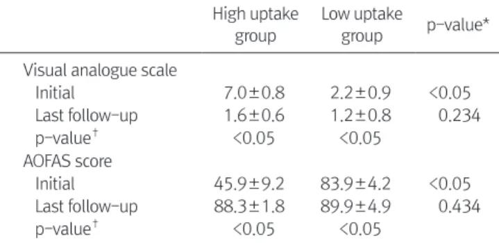
고흡착군은 저흡착군에 비해 초기 VAS 점수가 높았고 AOFAS 점수가 낮았다(p<0.05). 저흡착군은 보존적 치료만으로 VAS (치 료 전 2.2±0.9, 치료 후 1.2±0.8; p<0.05) 및 AOFAS 점수(치료 전 83.9±4.2, 치료 후 89.9±4.9; p<0.05)의 호전을 보였으며 통계적 으로 유의하였다. 또한 저흡착군에서 추시 기간 동안 증상의 악화 를 보인 환자는 없었다. 고흡착군(n=30)에서 수술한 경우(n=21), 술 전에 비해 VAS (술 전 7.0±0.8, 술 후 1.6±0.6; p<0.05) 및 AOFAS 점수(술 전 45.9±9.2, 술 후 88.3±1.8; p<0.05)가 호전되 었으며 통계적으로 유의하였다. 최종 추시 시, 보존적 치료를 받은 저흡착군과 수술적 치료를 받은 고흡착군 간의 VAS (p=0.234) 및 AOFAS 점수(p=0.434)의 유의한 차이는 없었다(Table 2).
환자의 주관적 만족도는 저흡착군의 경우 23예 모두 만족 또는
Table 2. Clinical Assessment between the High and Low Uptake Groups High uptake
group
Low uptake
group p-value*
Visual analogue scale
Initial 7.0±0.8 2.2±0.9 <0.05
Last follow-up 1.6±0.6 1.2±0.8 0.234
p-value† <0.05 <0.05
AOFAS score
Initial 45.9±9.2 83.9±4.2 <0.05
Last follow-up 88.3±1.8 89.9±4.9 0.434
p-value† <0.05 <0.05
AOFAS score: American Orthopaedic Foot and Ankle Society midfoot score.
*Mann-Whitney test, †Wilcoxon test, comparing the initial and last follow-up value.
Table 3. Predicting Uptake Level with Size in Asymptomatic Accessory Navicular Bone Group Cut off value
(size of bone, mm) Sensitivity (%) Specificity (%) Positive predictive value (%) Negative predictive value (%) Predicting grade 2 uptake level
14.2 100 92.31 50 100
Predicting grade 1 & 2 uptake level
6.8 100 44.44 50 100
예측할 수 있음을 시사한다.
하지만 본 연구 결과에서와 같이 부주상골의 크기가 증상이 없 는 부주상골에서나 증상이 있는 부주상골에서의 흡수량 정도와 관련이 있는 것으로 보아 골주사 검사상 흡수량의 단계적 평가 는 추가적 정보를 얻는 데 도움이 될 것으로 보인다. Romanowski 와 Barrington5)은 증상이 있는 부주상골 군 10예에서 골주사 검사 를 시행하였고 부주상골이 양측으로 존재할 확률이 50%∼89%임 을 고려할 때12,13) 통증이 있는 쪽의 부주상골에서만 흡수량 증가를 보였다고 보고하였다. 반면에 Chiu 등8)은 본 연구와 비슷한 연구 를 13명의 증상이 있는 군과 10명의 증상이 없는 군으로 나누어 시 행하였고, 골주사 검사상 흡수량이 증가하는 것은 증상을 나누는 데 도움이 되지 않는다고 하였다. 하지만 증상이 없는 부주상골 군 에 있어 골주사 검사상 흡수량 증가는 크기에 따라 증가할 수 있으 나 증상을 유발시키는 예상인자로 영향이 없음을 알 수 있었다. 따 라서 골주사 검사상 흡수량 정도만 가지고 진단과 분류를 내려서 는 안되며 증상이 없는 환자에게 있어서는 흡수의 단계적 분류는 의미가 없음을 알 수 있었다. 따라서 흡수의 단계적 분류는 증상이 있는 부주상골 환자에게서 임상적인 증상과 연관되어 사용되어야 할 것으로 생각된다.
증상이 있는 부주상골이 의심되는 환자들 중 0단계의 흡수량은 음성 예측치 100%이므로 이 단계의 흡수량을 보인 족부 통증을 호 소 환자는 부주상골 이외의 원인에 의한 통증으로 생각할 수 있다 (Table 4). 또한 족부의 통증이 있는 경우, 증상이 없는 부주상골을 배제하기 위해서는 진단 기준에 있어 흡수량을 1단계 이상으로 하 는 것이 2단계로 하는 것보다 음성예측치가 높아 더 도움이 될 것 으로 보인다.
본 연구는 후향적 연구로 수술적 처치를 시행한 자들에서 모두 2단계의 높은 흡수량을 보였지만 이에 대하여 정확한 상관관계를 얻기 위해서는 전향적인 연구로의 진행이 필요할 것으로 보인다.
또한 본 연구에서는 부주상골의 크기와 증상의 정도가 흡수량 증 가에 영향을 미칠 것이라고 제시하였지만 다른 여러 인자에 대한 추가적 연구 및 보완이 필요할 것으로 보인다.
결 론
본 연구의 결과로 볼 때, 골주사 검사는 족부의 부주상골 진단과 보존적 치료에 대한 기대 정도를 미리 예측하는 데 도움이 되고, 수술적 처치를 결정하는 데 유용하게 활용될 수 있으나 임상적 요 인 및 부주상골의 크기 등을 고려하여 진단 및 처치의 방향을 결정 해야 할 것이다.
REFERENCES
상이 있는 부주상골을 진단하는 데 도움이 됨을 알 수 있었다(area under the curve=0.901, standard error=0.0360, 95% confidence interval, 0.808∼0.958; p<0.0001).
흡수량을 1단계 이상 혹은 2단계 이상으로 나누어 진단 기준으 로 설정했을 때의 결과들을 정리하였고 1단계 이상을 진단 기준으 로 사용할 경우 음성예측치가 100%로, 증상이 있는 흡수량이 0단 계일 경우 부주상골은 배제할 수가 있어 진단에 있어 도움이 됨을 확인할 수 있었다(Table 4).
나이, 성별, BMI 같은 다른 임상적 요인들은 증상이 있는 군과 증상이 없는 군 모두에서 흡수량 증가와 관계가 없었다(p>0.005).
고 찰
본 연구는 족부의 부주상골을 진단하고 치료 방침을 계획하는 데 있어 골주사 검사의 유용성을 보고하고자 하였다.
증상이 없는 부주상골에 있어서 흡수량 증가가 발생하기도 하는 데, 이에 대한 원인은 확실하지 않다. Chisin 등11)은 엄지발가락 종 자골에 있어 흡수량 증가가 증상이 없는 활동적인 일을 많이 하는 대상 중 29%에서 나타났고, 앉아서 활동적인 일을 하지 않는 대상 에서 26%까지 나타나 흡수량이 증가하여도 정상적일 수 있다고 제 시하였다. 또한 Chiu 등8)은 증상이 없는 환자들이 증상이 발현되 지 않는 것으로 미루어보아 증상이 없는 대상에서 골 흡수량 증가 를 보이는 것은 추후 증상을 유발하는 위험요소가 되지 않을 거라 고 주장했다. 본 연구에서는 외래 추시 중 흡수량 증가를 보이는 증상이 없는 부주상골이 증상이 있는 부주상골이 되는 경우는 없 었다. 이는 기존의 연구들과 비슷한 결과로, 골주사 검사상 흡수량 Table 4. Diagnostic Performance of Symptomatic Accessory Navicular Bone by Uptake Grade on Tc-99m Hydroxymethylenediphosphonate Bone Scintigraphy
Diagnostic criteria Value 95% CI
Grade ≥1 (mild uptake)
Sensitivity (%) 100.00 91.78∼100.00
Specificity (%) 66.67 47.19∼82.71
Positive likelihood ratio 3.00 1.81∼4.98
Negative likelihood ratio 0.00 -
Disease prevalence (%) 58.90 46.77∼70.29 Positive predictive value (%) 81.13 68.03∼90.56 Negative predictive value (%) 100.00 83.16∼100.00 Grade ≥2 (hot uptake)
Sensitivity (%) 60.47 44.41∼75.02
Specificity (%) 93.33 77.93∼99.18
Positive likelihood ratio 9.07 2.33∼35.36 Negative likelihood ratio 0.42 0.29∼0.62 Disease prevalence (%) 58.90 46.77∼70.29 Positive predictive value (%) 92.86 76.50∼99.12 Negative predictive value (%) 62.22 46.54∼76.23 CI: confidence interval.
www.jkfas.org calcaneo-cuboid-cuneiform osteotomies and the modified Kid-
ner procedure for severe flatfoot associated with symptomatic accessory navicular in children and adolescents. J Orthop Surg Res. 2014;9:131.
2. Miller TT. Painful accessory bones of the foot. Semin Musculo- skelet Radiol. 2002;6:153-61.
3. Mellado JM, Ramos A, Salvadó E, Camins A, Danús M, Saurí A.
Accessory ossicles and sesamoid bones of the ankle and foot:
imaging findings, clinical significance and differential diagnosis.
Eur Radiol. 2003;13 Suppl 6:L164-77.
4. Keats TE, Anderson MW. Atlas of normal roentgen variants that may simulate disease. 9th ed. Philadelphia: Elsevier Saunders;
2012.
5. Romanowski CA, Barrington NA. The accessory navicular-an important cause of medial foot pain. Clin Radiol. 1992;46:261- 4.
6. Jain S, Karunanithi S, Agarwal KK, Kumar G, Roy SG, Tripathi M.
Incremental value of single photon emission tomography/com- puted tomography in 3-phase bone scintigraphy of an accessory navicular bone. Indian J Nucl Med. 2014;29:191-2.
7. Veitch JM. Evaluation of the Kidner procedure in treatment of symptomatic accessory tarsal scaphoid. Clin Orthop Relat Res.
1978;(131):210-3.
8. Chiu NT, Jou IM, Lee BF, Yao WJ, Tu DG, Wu PS. Symptomatic and asymptomatic accessory navicular bones: findings of Tc- 99m MDP bone scintigraphy. Clin Radiol. 2000;55:353-5.
9. Lassmann M, Biassoni L, Monsieurs M, Franzius C, Jacobs F, EANM Dosimetry and Paediatrics Committees. The new EANM paediatric dosage card. Eur J Nucl Med Mol Imaging.
2008;35:1748.
10. Lee J, Youn H, Choi WJ, Lee JW. Comparison of clinical outcome of excision versus osteosynthesis in type II accessory navicular. J Korean Foot Ankle Soc. 2011;15:72-8.
11. Chisin R, Peyser A, Milgrom C. Bone scintigraphy in the assess- ment of the hallucal sesamoids. Foot Ankle Int. 1995;16:291-4.
12. Geist ES. The accessory scaphoid bone. J Bone Joint Surg.
1925;7:570-4.
13. Mygind HB. The accessory tarsal scaphoid; clinical features and treatment. Acta Orthop Scand. 1953;23:142-51.