INTRODUCTION
Neurofibromatosis type 2 (NF2) is a dominantly inherited tumor-prone disorder characterized by multiple schwan- nomas and meningiomas with associated symptoms of tin- nitus, hearing loss, and balance dysfunction [1]. The NF2 tumor-suppressor gene (MIM#101000) is the only gene known to be associated with NF2. Mutation scanning com-
bined with gene-dosage analysis to identify deletions or duplications in single exons increases the mutation-detec- tion rate to nearly 72% in simplex cases and to more than 90% in familial cases [2, 3]. However, the NF2mutation characteristics in Korean patients have not been elucidat- ed to date. In one study, a pathogenic mutation was iden- tified in 1 of 15 clinically diagnosed Korean patients [4].
However, the inference that the mutation rate of NF2in this population is much lower than that in other popula- tions is questionable, because direct sequencing and gene- dosage tests were not employed in the previous study. Here, we conducted a comprehensive mutational analysis in Kore- an NF2 patients by performing direct sequencing and a gene-dosage assessment.
190 190
Molecular Characterization of the NF2 Gene in Korean Patients with Neurofibromatosis Type 2: A Report of Four Novel Mutations
Moon-Woo Seong, M.D.
1,4, Im Kyung Yeo, B.S.
1, Sung Im Cho, M.S.
1, Chul-Kee Park, M.D.
2, Seung-Ki Kim, M.D.
2, Sun Ha Paek, M.D.
2, Dong Gyu Kim, M.D.
2, Hee-Won Jung, M.D.
2, Hyunwoong Park, M.D.
1, So Yeon Kim, M.D.
1,
Ji Yeon Kim, M.D.
3, and Sung Sup Park, M.D.
1,3Departments of Laboratory Medicine1and Neurosurgery2, Clinical Research Institute3, Seoul National University Hospital, Seoul;
Department of Laboratory Medicine4, National Cancer Center, Goyang, Korea
190 190
Background : Neurofibromatosis type 2 (NF2) is an autosomal dominant syndrome caused by the NF2 tumor suppressor gene. However, the NF2 mutation characteristics in Korean patients are not sufficiently understood. In this study, we conducted a comprehensive mutational analysis in 7 Korean NF2 patients by performing direct sequencing and gene-dosage assessment.
Methods : We analyzed all exons and flanking regions of NF2 by direct sequencing and screened the deletions or duplications involving NF2 by multiplex ligation-dependent probe amplification.
Results : Four novel NF2 mutations, including 2 splice-site mutations (c.364-1G>A and c.886- 3C>G), 1 frameshift mutation (c.524delA), and 1 missense mutation (c.397T>C; p.Cys133Arg), were identified in our patients. No large deletion or duplication was identified in our series. Subsequently, we identified an abnormal splicing product by using reverse transcription-PCR and direct sequenc- ing in 2 patients with a novel splice-site mutation. The missense mutation c.397T>C was predicted to have harmful effects on protein function.
Conclusions : The detection rate of NF2 mutations in Korean patients (57%) is similar to those in other populations. Our results provided a greater insight into the mutational spectrum of the NF2 gene in Korean subjects. (Korean J Lab Med 2010;30:190-4)
Key Words : Hereditary cancer, Neurofibromatosis type 2, NF2
Received :October 29, 2009 Manuscript No :KJLM09-134 Revision received :March 5, 2010
Accepted :March 19, 2010
Corresponding author :Sung Sup Park, M.D.
Department of Laboratory Medicine, Seoul National University Hospital, 28 Yeongeon-dong, Jongno-gu, Seoul 110-744, Korea Tel : +82-2-2072-3206, Fax : +82-2-747-0359
E-mail : [email protected]
MATERIALS AND METHODS 1. Subjects
Seven unrelated patients with clinically diagnosed NF2 were included in this study. All patients provided informed consent for the use of their clinical data and blood sam- ples. All patients fulfilled the Manchester criteria for NF2 (Table 1) [1]. Briefly, the study subjects comprised 2 famil- ial cases and 5 simplex cases; 4 patients had bilateral ves- tibular schwannoma (BVS) and 3 had unilateral vestibular schwannoma (UVS).
2. Mutational analysis of NF2
Total genomic DNA was extracted from peripheral blood by using Gentra PureGene DNA isolation kits (Gentra Sys- tems, Minneapolis, MN, USA), and total RNA was prepared by using RNAzol B (Tel-Test, Friendswood, TX, USA) ac- cording to the manufacturer’s protocol.
PCR was performed using primers designed to flank the splice junctions of all coding exons on the NF2gene (Table 2). Additional primer sets for exons 1 and 6 were designed to enhance the sequencing quality. The amplified products were sequenced on an ABI 3730 analyzer (Applied Biosys- tems, Foster City, CA, USA) using BigDyeTM Terminator v3.1 Cycle sequencing kits (Applied Biosystems, Foster City, CA, USA). Sequences were analyzed using SeqScape soft- ware (Applied Biosystems , Foster City, CA, USA) and Muta-
tion Surveyor (Softgenetics, State College, PA, USA).
Multiplex ligation-dependent probe amplification to detect large deletions or duplications involving the NF2region was performed using the SALSA P044-NF2 kit (MRC-Hol- land, Amsterdam, Netherlands). PCR products were ana- lyzed on an ABI 3130 analyzer by using Genemarker ver.
1.51 (Softgenetics, State College, PA, USA). The peak heights were normalized, and a deletion or duplication was sus- pected when the normalized peak ratio was less than 0.75 or greater than 1.30.
A) Bilateral vestibular schwannomas B) First-degree family relative with NF2 AND
- Unilateral vestibular schwannoma OR
- Any 2 of the following: meningioma, schwannoma, glioma, neurofibroma, posterior subcapsular lenticular opacities C) Unilateral vestibular schwannoma AND any 2 of the following:
meningioma, schwannoma, glioma, neurofibroma, posterior subcapsular lenticular opacities
D) Multiple meningiomas (2 or more) AND - Unilateral vestibular schwannoma OR
- Any 2 of the following: schwannoma, glioma, neurofibroma, cataract
Table 1. Manchester clinical diagnostic criteria for neurofibro- matosis type 2
Exon Name Sequence (5′→ 3′) PCR product size (bp)
1 1-1F GGGAAAGTCCTGCCTACCTT 542
1-1R CCTGCACTCTGAGCCCTTTA
1-2F ACTCCCCTTTCCGCTCAG 540
1-2R CAGGAGCATCCAGCTTCTTC
2 2F TTTCCCACTCATGGGTTTGT 579
2R AGGCATAAAGCCAGAAGCAA
3 3F CTGTGGCCCTGAGAACATTT 670
3R GGACCCATTTTCAAGGAGGT
4 4F CTCTCCACCTGTCTGCATCA 481
4R TCTGCACACCACACACACAC
5 5F AAACATGCCCACATTTCCAT 545
5R CTAGTCCTGGTGACCCCAAA
6 6F GATGGCTTCTGAGCATGTGA 564
6R CCAGCTCTCCCCTTTTCTTT 6-1R GCCCATAAAGGAATGTAAACCA 6-2R CTTTAAGGCAAAAAAAAAAAAAAAAG
7 7F GGATGGGAAATTCTGCTTGA 588
7R GGACGGAGATCTCACAGAGC
8 8F CTTCTACCTGCCCCAATTCA 452
8R AACAACCACACCCTCAAAGC
9 9F TCAAGAATCCCTTCCCACAC 446
9R GCGCCAAGTGAGATACCATT
10 10F TGCATGTTTCCAGAGCTGAC 591
10R GATGCATGCACTCTTGGCTA
11 11F TGTTTTTCAAGTGGCACAGC 582
11R GTAGTGCCCAGGCTGAGAAG
12 12F GGGAATGTGGCTTGTCATTT 585
12R ACTGAGTTCCTGTGCCCAAC
13 13F GCTGCAGAAGGTCTGGTTTC 513
13R GCTCTCTGCACCTCTCATCC
14 14F ATGTGGAGGGAGTGAAGTGG 440
14R CCAGGGTGTAAGAGCAGAGC
15 15F ACCCTAGATCGCACACCAAG 523
15R GGCTCAAAATCCACCCTGTA
16 16F TCACGATTTCAGGCCTATCC 492
16R ATGCCACCAAGACAAAGGAC
17 17F TGTCAAGAGGCAATGCTGAC 462
17R CTCAGCTGGGGAAAGTTCTG Table 2. Primer sequences used in this study
3. Mutational effect of novel mutations
Reverse transcription-PCR (RT-PCR) and sequence anal- ysis of mRNA products were used to identify aberrant spli- cing products of potential splice-site mutations. To deter- mine the significance of the novel missense variant, we determined the allele frequency in control subjects, obtained information from amino acids and proteins and performed
in-silico prediction using the 3 software programs: Polyphen (http://genetics.bwh.harvard.edu/pph/), Pmut (http://mm- b2.pcb.ub.es:8080/PMut/), and SIFT (http://sift.jcvi.org) [5-7].
Sequence variants were described at the cDNA level by using GenBank reference sequences (NM_000268.3) accord- ing to the HUGO-approved systematic nomenclature for description of sequence variations (http://www.hgvs.org/
Marker XIII
Marker VI P4
P2 Control
0.63 kb 0.55
kb
c.364-I G>A
Exon 3
T C T T A C A G
T C T T A C A G
G C C A S A G
T A A G C T G
T A A G C T G A T T C T C C A G
A T T C T C C A G
A T T C T C C
A G
A T T C T C C A G
G T A A A G A A
G T A A A G A A
T A T G G T G A
Exon 4/Exon 5
P4
Control
c.886-3C>G
Intron 9 Exon 10
Exon 9 Exon 10
P2
P2
Control 0.62
kb Control
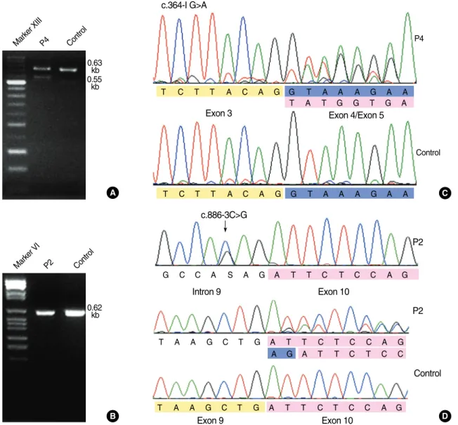
Fig. 1. Results of RT-PCR and sequence analysis for the 2 splice-site mutations c.364-1G>A (patient P4) and c.886-3C>G (patient P2).
(A) The c.364-1G>A mutation yields a 0.55-kb abnormal product as well as 0.63-kb normal product in RT-PCR with the following primers:
F-5′-AAGCAACCCAAGACGTTCAC-3′, R-5′-CCGGATTGCAAAGTAGTTCA-3′. (B) The c.886-3C>G cannot be discriminated from nor- mal products by using following primers: F-5′-CTGACCCCCAAGATCTCCT-3′, R-5′-GCTTCAGCTGATCTGCCTCT-3′. (C) Exon 4 skip- ping is shown in the cDNA sequence of patient 4 with c.364-1G>A. (D) Patient P2 is heterozygous for c.886-3C>G, and 2-bp insertion of AG (blue) between exon 9 (yellow) and exon 10 (pink) is shown in the cDNA sequence. This insertion is caused by the introduction of a new splice acceptor site from c.886-4A to c.886-3C>G and subsequent inclusion of original splice acceptor site (c.886-2_-1AG) in the mature transcript.
C
D A
B
mutnomen/).
RESULTS
Four novel NF2mutations, including 2 splice-site muta- tions (c.364-1G>A and c.886-3C>G), 1 frameshift muta- tion (c.524delA), and 1 missense mutation (c.397T>C), were identified in our patients (Table 3). No large deletion or duplication was identified in our series.
For the 2 splice-site mutations, we performed RT-PCR and analyzed the obtained mRNA using direct sequencing.
The RT-PCR product of c.886-3C>G was not distinct from normal-sized products (Fig. 1). However, subsequent se- quence analysis revealed that a new splice-acceptor site was introduced at intron 9, and the effect of this intro- duction was similar to that of AG insertion between cod- ing nucleotides 885 and 886 (c.885_886insAG, p.Ile296Argf-
sX14). The c.364-1G>A mutation yielded an abnormal RT- PCR product, and direct sequencing confirmed the absence of exon 4 (Fig. 1). The frameshift mutation, c.524delA, in- troduced a premature stop codon in the transcript.
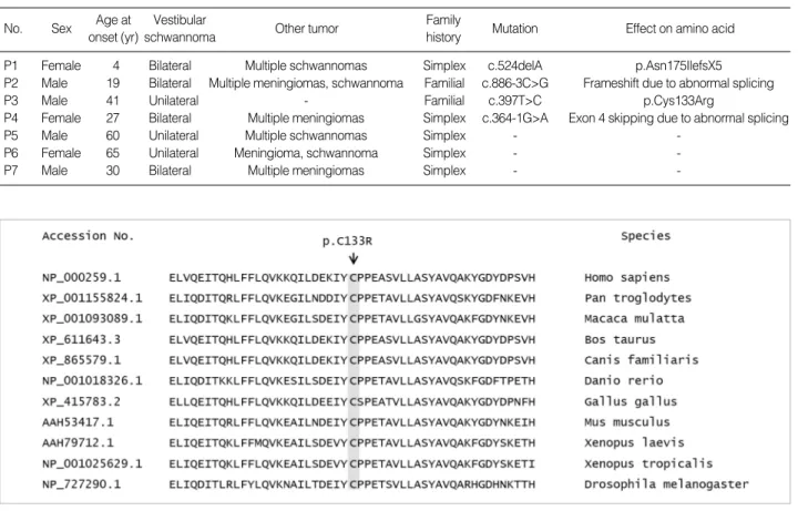
Further, we investigated the pathogenicity of the novel missense mutation c.397T>C (p.Cys133Arg). The patient had a family history of vestibular schwannoma, but the mutational status of the family members could not be deter- mined because their samples were not available. This muta- tion was not detected in 95 control subjects. The cysteine encoded by codon 133 is located on the central structural domain of the Band 4.1, Ezrin, Radixin, Moesin (FERM) domain and is highly conserved (Fig. 2). The substitution of cysteine to arginine was expected to appreciably change the hydrophobicity of the protein. Consistent with the pre- dictions of the 3 in-silico software programs utilized in the study, we considered this mutation to be pathogenic.
Fig. 2. Multiple alignment and amino acid conservation for a novel missense mutation c.397T>C (p.Cys133Arg). The cysteine at codon 133 is well-conserved among various species.
No. Sex Age at onset (yr)
Vestibular
schwannoma Other tumor Family
history Mutation Effect on amino acid
P1 Female 4 Bilateral Multiple schwannomas Simplex c.524delA p.Asn175IlefsX5 P2 Male 19 Bilateral Multiple meningiomas, schwannoma Familial c.886-3C>G Frameshift due to abnormal splicing
P3 Male 41 Unilateral - Familial c.397T>C p.Cys133Arg
P4 Female 27 Bilateral Multiple meningiomas Simplex c.364-1G>A Exon 4 skipping due to abnormal splicing
P5 Male 60 Unilateral Multiple schwannomas Simplex - -
P6 Female 65 Unilateral Meningioma, schwannoma Simplex - -
P7 Male 30 Bilateral Multiple meningiomas Simplex - -
Table 3. Clinical features and molecular findings of the 7 patients who participated in this study
DISCUSSION
The present investigation revealed that the total NF2 mutation-detection rate was 57% (100% in familial cases and 40% in simplex cases). This detection rate is similar to those in other populations, but it is much higher than that reported previously in this population (7%) [4]. However, the previous study had several limitations. In addition to the cases satisfying the NIH criteria, which correspond to the first 2 statements (criteria A and B, Table 1) of the Man- chester criteria, some extremely unsuitable cases may have been included in that study [4]. Moreover, the previous study used single-strand conformation polymorphism analysis instead of direct sequencing for mutation scanning, and the single-strand conformation polymorphism analysis did not include all the exons. The findings of our study show that the NF2gene is also an important genetic factor for the incidence of NF2 in this population. Therefore, genetic testing for NF2should be considered for molecular diag- nosis, especially in familial or BVS cases.
All mutations identified in this study were novel and res- tricted to a single patient or family. Therefore, the muta- tion spectrum of the NF2gene in this population is likely to be highly heterogeneous. Among the constitutional NF2 mutations other than large rearrangements, the following distribution could be determined on the basis of the pre- dicted effects: nonsense, 39%; frameshift, 27%; splice site, 25%; and nontruncating and other mutations, 7% [8]. Even in our study, truncating mutations such as splice-site mu- tations and nonsense mutations were more common than nontruncating mutations, although our data were obtained from a sample of limited size.
Genotype-phenotype correlations for NF2 have been well established [9, 10]. In general, nonsense or frameshift mutations are associated with severe NF2, and missense or in-frame deletions are associated with mild NF2. For 2 patients (P1 and P2) with frameshift mutations in our study, the ages at disease onset were 4 and 19 yr, respectively, and both individuals exhibited multiple schwannomas or meningiomas as well as BVS. In contrast, patient P3, who
harbored a missense mutation, was 41 yr old at disease onset and developed UVS without other tumors.
In summary, our results provide insights on the muta- tional spectrum of NF2in Korean subjects, which will be helpful for establishing a genetic screening strategy for the Korean population.
REFERENCES
1. Evans DG, Huson SM, Donnai D, Neary W, Blair V, Newton V, et al.
A clinical study of type 2 neurofibromatosis. Q J Med 1992;84:603-18.
2. Wallace AJ, Watson CJ, Oward E, Evans DG, Elles RG. Mutation scanning of the NF2 gene: an improved service based on meta-PCR/
sequencing, dosage analysis, and loss of heterozygosity analysis.
Genet Test 2004;8:368-80.
3. Kluwe L, Nygren AO, Errami A, Heinrich B, Matthies C, Tatagiba M, et al. Screening for large mutations of the NF2 gene. Genes Chro- mosomes Cancer 2005;42:384-91.
4. Yang HJ, Won YJ, Park KJ, Jung HW, Choi KS, Park JG. Germline mutations of the NF2 gene in Korean neurofibromatosis 2 patient. J Korean Cancer Assoc 1998;30:790-9.
5. Ferrer-Costa C, Orozco M, de la Cruz X. Sequence-based prediction of pathological mutations. Proteins 2004;57:811-9.
6. Ng PC and Henikoff S. Predicting deleterious amino acid substitu- tions. Genome Res 2001;11:863-74.
7. Ramensky V, Bork P, Sunyaev S. Human non-synonymous SNPs:
server and survey. Nucleic Acids Res 2002;30:3894-900.
8. Ahronowitz I, Xin W, Kiely R, Sims K, MacCollin M, Nunes FP.
Mutational spectrum of the NF2 gene: a meta-analysis of 12 years of research and diagnostic laboratory findings. Hum Mutat 2007;28:
1-12.
9. Evans DG, Trueman L, Wallace A, Collins S, Strachan T. Genotype/
phenotype correlations in type 2 neurofibromatosis (NF2): evidence for more severe disease associated with truncating mutations. J Med Genet 1998;35:450-5.
10. Baser ME, Kuramoto L, Joe H, Friedman JM, Wallace AJ, Gillespie JE, et al. Genotype-phenotype correlations for nervous system tumors in neurofibromatosis 2: a population-based study. Am J Hum Genet 2004;75:231-9.