Individual Variation in Growth Factor Concentrations in Platelet-rich Plasma and Its Influence on Human Mesenchymal Stem Cells
Hee Soon Cho, M.D.1, In Hwan Song, M.D.2, So-Young Park, M.D.3, Min Cheol Sung, M.D.4, Myun-Whan Ahn, M.D.4, and Kyung Eun Song, M.D.5
Departments of Laboratory Medicine1, Anatomy2, Physiology3, and Orthopedic Surgery4, College of Medicine, Yeungnam University, Daegu;
Department of Clinical Pathology5, Kyungpook National University School of Medicine, Daegu, Korea
Background: The objective of this study was to explore whether individual variations in the concentration of growth factors (GFs) influence the biologic effects of platelet-rich plasma (PRP) on human mesenchymal stem cells (HMSCs).
Methods: The concentrations of 7 representative GFs in activated PRP (aPRP) were measured using ELISA. The effects of PRP on the proliferation and alkaline phosphatase (ALP) activity of HMSCs were examined using several concentrations of aPRP from 3 donors;
the relationships between the GF levels and these biologic effects were then evaluated using 10% aPRP from 5 subgroups derived from 39 total donors. HMSCs were cultured in DMEM with the addition of aPRP for 4 or 12 days; then, DNA content and ALP activity were measured.
Results: The quantity of DNA increased significantly at a 10% concentration of aPRP, but the ALP activity was suppressed at this concentration of aPRP. The GF concentrations varied among donors, and 5 subgroups of characteristic GF release patterns were identified via cluster analysis. DNA levels differed significantly between groups and tended to be higher in groups with higher con- centrations of transforming growth factor-beta1 (TGF-β1) and platelet-derived growth factors (PDGFs). DNA quantity was positive- ly correlated with TGF-β1 concentration, and was negatively correlated with donor age. ALP activity was negatively correlated with PDGF-BB concentration.
Conclusions: The varying GF concentrations may result in different biologic effects; thus, individual differences in GF levels should be considered for reliable interpretation of the biologic functions and standardized application of PRP.
Key Words: Platelet-rich plasma, Mesenchymal stem cells, Growth factors, Transforming growth factor-β1, Platelet-derived growth factor-BB
Received: November 22, 2010 Manuscript No: KJLM-10-162 Revision received: April 27, 2011
Accepted: May 12, 2011
Corresponding author: Myun-Whan Ahn, M.D.
Department of Orthopedic Surgery, Yeungnam University College of Medicine, 317-1 Daemyeong-dong, Nam-gu, Daegu 705-717, Korea
Tel: +82-53-620-3643, Fax: +82-53-628-4020, E-mail: [email protected] ISSN 1598-6535 © The Korean Society for Laboratory Medicine.
This is an Open Access article distributed under the terms of the Creative Commons Attribution Non-Commercial License (http://creativecommons.org/licenses/by-nc/3.0) which permits unrestricted non-commercial use, distribution, and reproduction in any medium, provided the original work is properly cited.
INTRODUCTION
Platelets, the fundamental component of primary hemo- stasis, are also known to be reservoirs for many growth fac- tors (GFs), which they store in their α-granules. Platelet ag- gregation and activation, after vascular damage, result in
the release of several GFs that may affect the chemotaxis, proliferation, and differentiation of mesenchymal stem cells (MSCs) or other committed cells during the process of tis- sue repair and healing [1]. The GFs released from platelets include platelet-derived growth factors (PDGFs), trans- forming growth factor-beta (TGF-β), vascular endothelial growth factor (VEGF), epidermal growth factor (EGF), fi- broblast growth factor (FGF) and insulin-like growth fac- tors (IGFs) [2, 3].
To explore the possibility that platelet-rich plasma (PRP) could provide an autologous source of these essential GFs that benefit bone and soft tissue healing [4], many clinical and experimental studies dealing with the effects of PRP have been conducted [5-7]. However, the benefit of PRP on bone formation is a controversial subject. While a report suggested a stimulatory effect with the addition of PRP [5], others have observed no improvement [6] or have detected
even inhibitory effects [7]. Although the lack of standard- ization in application across these studies, including differ- ences in the preparation method or dosage of PRP, bioma- terials, species, implantation sites, and cell types, may have contributed to the inconsistent results [8], we hypothesized that the differences in GF composition among PRPs could lead to this discrepancy. GF quantities vary significantly be- tween individuals [9]. The potential efficacy of PRP depen- ds on the levels of GFs released from platelets; however, no study has yet addressed the relationship between the bio- logic effects and GF concentrations in PRP. In this study, we attempted to explore the variations in GF release patterns in PRP in order to determine whether differences in GF con- centrations influence biologic effects such as proliferation and alkaline phosphatase (ALP) activity of human MSCs (HMSCs) and to identify the factors associated with each of these biologic effects.
MATERIALS AND METHODS
This study was reviewed and approved by the Institutional Review Board of Yeungnam University College of Medicine.
Prior to drawing blood or bone marrow, informed consent was obtained from each donor.
1. Preparation of PRP and measurement of GF concentrations First, PRPs were obtained from 3 healthy male volunteers (27 yr, 39 yr, and 43 yr old) to explore the effects of PRP con- centration on the proliferation and ALP activity of HMSCs.
Then, we obtained donated PRPs from 39 other healthy vol- unteers to assess the relationship between the effects and GF concentrations. The mean donor age was 42 yr (range, 24- 68 yr), distributed at 6-8 individuals per decade. We limited our subjects to those who had not taken anti-platelet medi- cations within 1 week of donation; further, to minimize the possible influences of hormonal variation on the prolifera- tion of HMSCs, we included only male donors.
PRPs were prepared via double centrifugation of blood.
In brief, 9 mL of venous blood was drawn into a polypropyl- ene tube containing 1 mL of anticoagulant, acid citrate dex- trose (ACD). The blood was centrifuged at 1,500 rpm for 10 min at 25˚C to separate the platelet-containing plasma from the red cells. The platelet-containing plasma was then cen- trifuged at 3,000 rpm for an additional 10 min at 25˚C, and platelet-poor plasma (PPP) was separated out. Platelet pel- lets were resuspended in 1 mL of plasma and were pooled for PRP. To activate the platelets, 1 part bovine thrombin stock solution (1,000 U/mL; Sigma, St. Louis, MO, USA) was added to 9 parts PRP or PPP to yield a final thrombin
concentration of 100 U/mL. Each sample was incubated for 1 hr at 37˚C [10]. The resulting supernatants from the clot preparation were referred to as activated PRP (aPRP) and activated PPP (aPPP). Both aPRPs and aPPPs were stored at -80˚C until use. The concentrations of PDGF-AA, PDGF- AB, PDGF-BB, TGF-β1, FGF-basic (FGF-b), IGF-1, and VEGF were measured with commercially available Quanti- kine colorimetric sandwich ELISA kits (R&D Systems, Min- neapolis, MN, USA) according to the manufacturer’s in- structions.
2. Cell isolation
HMSCs were cultured using established protocols [11].
Bone marrow aspirate was obtained with consent from a 50-yr-old female patient who had undergone an autologous bone graft for the treatment of degenerative spondylolithe- sis. Mononuclear cells were isolated using a density gradient and were cultured in DMEM (Sigma, St Louis, MO, USA) with 10% fetal bovine serum (FBS), penicillin, and strepto- mycin (Gibco-BRL, Rockville, MD, USA). The cells were incubated in a humidified atmosphere of 5% CO2 at 37˚C.
Non-adherent cells were discarded after 24 hr, and the me- dium was exchanged every 3 days. First passage (P1) cells were utilized in the study.
3. The effects of aPRP and aPPP on the DNA content and ALP activity of HMSCs
The cells were seeded at a density of 1.5×10⁴ cells/well in 12-well plates (35 mm diameter) and were allowed to attach for 24 hr. After this adhesion period, the medium was re- moved and was replaced with DMEM with 10% FBS (as control) or variable volume concentrations of aPRP and aPPP (1%, 3%, 10%, and 30%) from each of the 3 donors.
Each experiment was conducted in triplicate. Media was exchanged every 4 days. The level of DNA and ALP activity were measured in quadruplicate on the fourth and twelfth days. The DNA level was measured via Hoechst DNA stain, and ALP activity was determined by quantifying the p-ni- trophenol released from p-nitrophenol phosphate [12].
4. Relationships between GF concentrations, DNA content, and ALP activity of HMSCs
In order to evaluate the effects of aPRP GF concentrations on the proliferation or ALP activity of HMSCs, the HMSCs were cultured under the same conditions as described above, in DMEM with 10% aPRPs from each of the 39 donors. Af- ter culturing for 12 days, the DNA contents and ALP activity were measured.
5. Statistical analysis
Statistical analysis was conducted using the Predict Ana- lytics Software program (PASW) version 18.0 (SPSS Inc., Chicago, IL, USA). The Shapiro-Wilk test was used to ana- lyze the normal distribution of the data. The effects of aPRP and aPPP concentrations on the DNA content and ALP ac- tivity of HMSCs were evaluated via ANOVA, or the Krus- kal-Wallis (K-W) test with Bonferroni correction. The dif- ferences in GF concentrations between aPRPs and aPPPs were examined using the paired t-test. Pearson’s correlation analysis was used to identify correlations between GF levels and age, platelet count, or mean platelet volume. The aPRP release pattern of GFs was grouped via cluster analysis using the K-means method. The relationships between GF con- centrations and the biologic effects between these groups was analyzed using ANOVA or K-W test with Bonferroni correction. Stepwise multiple linear regression analysis was employed to show which GFs were associated with DNA content or ALP activity. Significant differences were estab- lished at P<0.05; the Bonferroni corrected P value thresh- old (0.05/number of comparison) was employed for post- hoc analysis of the K-W test.
RESULTS
1. Effects of PRP and PPP on the DNA content and ALP activity of HMSCs
On the fourth day after cultivation, DNA contents were unaffected by the addition of aPRP (P =0.043 by K-W test, but multiple comparisons with Bonferroni correction did not reveal significant differences). The addition of aPPP did not affect DNA content at any concentration (P =0.408 by ANOVA). However, by the twelfth day, DNA levels differed significantly according to the aPRP and aPPP concentra- tions. Both aPRP and aPPP, at a concentration of 10% re-
sulted in increased DNA levels. However, DNA levels were reduced by aPPP at a concentration of 30% (P <0.001 by ANOVA, post-hoc analysis using Scheffe method; Fig. 1A).
The stimulatory effects of aPRP were significantly greater than those of aPPP (P =0.006 by paired t-test). The ALP ac- tivity of cultured HMSCs was reduced with the addition of aPRP (P <0.001 by ANOVA), but the addition of aPPP did not affect the cellular ALP activity (P =0.564 for the fourth day; P =0.094 for the twelfth day; both by ANOVA; Fig. 1B).
2. Characteristics and concentrations of GFs in aPRP and aPPP Plasma samples from 39 individuals were evaluated in this portion of the study. Platelet counts in native whole blood (WB) varied by individual, ranging from 117×109/L to 337×109/L (mean, 192±50×109/L). The platelet count was significantly higher in the PRP samples than in native blood (P <0.001 by paired t-test), with an average 6-fold increase;
we noted a relatively high degree of variability among indi- viduals (mean, 1,166±402×109/L; range, from 400×109/L to 2,505×109/L; Table 1). The Pearson’s correlation between the WB and PRP platelet count was 0.673 (P <0.001). The platelet counts were not correlated with donor age (P =0.222 for WB platelet count; P =0.685 for PRP platelet count).
The GF concentrations, with the exception of IGF-1, were significantly higher in aPRPs than in aPPPs (Fig. 2). The concentration of GFs also displayed considerable variation across donors, and the concentrations of TGF-β1, PDGFs,
Fig. 1. The DNA concentration (A) and ALP activity (B) of human mesenchymal stem cells after the addition of 1%, 3%, 10%, and 30% activated PRP or activated PPP. Each value is the mean of 3 donors, with the error bar showing one standard deviation (*P <0.05; **P <0.01 compared with control).
Abbreviations: ALP, alkaline phosphatase; PRP, platelet-rich plasma; PPP, platelet-poor plasma.
4.00 3.00 2.00 1.00 0.00
μg/well
DNA
PRP 4 day PPP 4 day PRP 12 day PPP 12 day
Control 1%
3%
10%
30%
**
**
**
A
Control 1%
3%
10%
30%
B 25
20 15 10 5 0
nM pNP/min/μg DNA
ALP/DNA
PRP 4 day PPP 4 day PRP 12 day PPP 12 day
**
*
****
**
**
****
Table 1. Donor characteristics (N=39) PLT in WB
(×109/L) PLT in PRP
(×109/L) PLT in PPP
(×109/L) Recovery rate
of PLT (%) Age (yr)
192±50 1,166±402 27±17 61±17 42.4±13.5
(117-337) (400-2,505) (2-76) (27-97) (24-68)
Values are mean±standard deviation (minimum-maximum).
Abbreviations: PLT, platelet count; WB, whole blood.
and FGF-b were correlated with the platelet count in both WB and PRP. The concentrations of PDGF-BB and IGF-1 displayed weak negative correlation with age (Table 2).
3. Cluster analysis of 7 GFs and between-group differences in DNA levels or ALP activity in HMSCs
We performed a cluster analysis using the K-means me- thod to identify the variations in GF release patterns and their influences on the biologic effects. Five characteristic re-
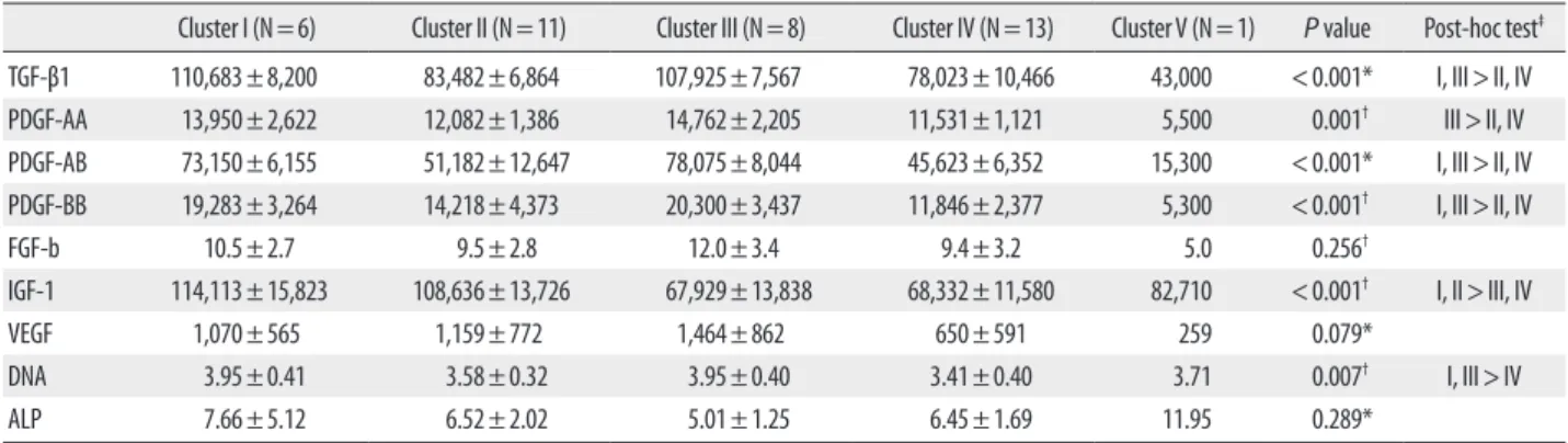
lease patterns of aPRP GFs were identified and significant differences in PDGF-AA, PDGF-AB, PDGF-BB, TGF-β1 and IGF-1 were noted between the groups. Groups 1 and 3 showed higher concentrations of TGF-β1 and PDGFs, wher- eas groups 2 and 4 exhibited lower concentrations of TGF-β1 and PDGFs. Groups 1 and 2 showed higher concentrations of IGF-1, whereas groups 3 and 4 showed lower concentra- tions of IGF-1. Group 5 exhibited substantially lower concen- trations of TGF-β1, PDGFs, and FGF-b; however, the group was excluded from ANOVA because its sample size was only 1. The DNA levels differed significantly between groups (P =0.007 by ANOVA) and were higher in groups 1 and 3 than in group 4. The differences in ALP activity between cluster groups were not significant (P =0.289 by K-W test;
Table 3).
Table 2. Pearson’s correlation coefficients for age, platelet count, and growth factor levels.
Age PLT in WB PLT in PRP Recovery rate MPV
TGF-β1 -0.307 0.444† 0.604† 0.391* -0.162
PDGF-AA -0.046 0.524† 0.707† 0.383* -0.205
PDGF-AB -0.122 0.516† 0.647† 0.318* -0.207
PDGF-BB -0.322* 0.476† 0.644† 0.371* -0.278
FGF-b 0.315 0.416† 0.419† 0.056 -0.162
VEGF 0.158 0.110 0.283 0.312 -0.172
IGF-1 -0.393* 0.227 0.110 -0.035 0.156
*p<0.05; †p<0.01.
Abbreviations: TGF-β1, transforming growth factor-β1; PDGF, platelet-derived growth factor; FGF-b, fibroblast growth factor-basic; VEGF, vascular endothelial growth fac- tor; IGF-1, insulin-like growth factor-1; PLT, platelet count; WB, whole blood; MPV, mean platelet volume.
Fig. 2. Quantification of growth factors in activated PRP and activated PPP (P <0.05 between PRP and PPP, except for IGF-1).
Abbreviations: TGF-β1, transforming growth factor-β1; PDGF, platelet-de- rived growth factor; IGF-1, insulin-like growth factor-1; FGF-b, fibroblast growth factor-basic; VEGF, vascular endothelial growth factor.
2,000 1,600 1,200 800 400 0
pg/mL
VEGF
15 12 9 6 3 0
pg/mL
FGF-b 120,000
100,000 80,000 60,000 40,000 20,000 0
pg/mL
TGF-β1 PDGF-AA PDGF-AB PDGF-BB IGF-1 PRP PPP
Table 3. Cluster characteristics of growth factors and biologic effects
Cluster I (N=6) Cluster II (N=11) Cluster III (N=8) Cluster IV (N=13) Cluster V (N=1) P value Post-hoc test‡
TGF-β1 110,683±8,200 83,482±6,864 107,925±7,567 78,023±10,466 43,000 <0.001* I, III>II, IV
PDGF-AA 13,950±2,622 12,082±1,386 14,762±2,205 11,531±1,121 5,500 0.001† III>II, IV
PDGF-AB 73,150±6,155 51,182±12,647 78,075±8,044 45,623±6,352 15,300 <0.001* I, III>II, IV
PDGF-BB 19,283±3,264 14,218±4,373 20,300±3,437 11,846±2,377 5,300 <0.001† I, III>II, IV
FGF-b 10.5±2.7 9.5±2.8 12.0±3.4 9.4±3.2 5.0 0.256†
IGF-1 114,113±15,823 108,636±13,726 67,929±13,838 68,332±11,580 82,710 <0.001† I, II>III, IV
VEGF 1,070±565 1,159±772 1,464±862 650±591 259 0.079*
DNA 3.95±0.41 3.58±0.32 3.95±0.40 3.41±0.40 3.71 0.007† I, III>IV
ALP 7.66±5.12 6.52±2.02 5.01±1.25 6.45±1.69 11.95 0.289*
Values are mean±standard deviation.
Units: growth factors, pg/mL; DNA, μg/well; ALP, nM pNP/min/μgDNA.
P values were determined by *Kruskal-Wallis test, or by †one-way ANOVA, ‡Post-hoc tests were conducted using the Bonferroni correction for the Kruskal-Wallis test or the Scheffe method for ANOVA.
Abbreviations: TGF-β1, transforming growth factor-β1; PDGF, platelet-derived growth factor; FGF-b, fibroblast growth factor-basic; IGF-1, insulin-like growth factor-1; VEGF, vascu- lar endothelial growth factor; ALP, alkaline phosphatase.
4. Relationship between DNA content or ALP activity and aPRP GF concentrations
The amount of DNA obtained from HMSCs that were cultured for 12 days with 10% aPRPs varied by donor, rang- ing from 2.59 to 4.59 μg/well (mean, 3.66±0.43 μg/well).
The ALP activity per cell also showed considerable individ- ual variations, ranging from 2.83 to 17.96 nM pNP/min/μg DNA (mean, 6.50±2.68 nM pNP/min/μg DNA).
Stepwise multiple regression analysis was employed to identify the independent variables that affected DNA levels and ALP activity. The variables included were donor age, the WB and PRP platelet count, the platelet recovery rate, and aPRP concentrations of the 7 GFs. The amount of DNA was correlated positively with the TGF-β1 concentration (Beta =0.500, P <0.001) and negatively with donor age (Beta=-0.324, P =0.017; Table 4). The ALP activity (ALP/
DNA) was correlated negatively with the concentration of PDGF-BB (Beta=-0.418, P =0.008; Table 4).
DISCUSSION
Despite 2 decades of clinical study of PRP, little basic data is available regarding the effects of variations in GF levels on biologic function. Because PRP displays the unique charac- teristics of a mixture of multiple GFs in various concentra- tions, and because the regenerative potency of PRP proba- bly depends on its GF levels, knowledge of the relationships between GF levels and biologic effects is required to ensure the reliable and reproducible use of PRP [8]. Therefore, in this study, we attempted to explore the relationship between GF levels and biologic effects.
Obtaining a sufficiently large volume of blood from the
single donor in order to analyze the various effects was dif- ficult; thus, we employed DNA analysis for proliferation markers and ALP activity for osteogenic markers of HM- SCs. First, we cultured HMSCs at several aPRP concentra- tions (1%, 3%, 10%, and 30%) from 3 donors to evaluate its effects on proliferation and ALP activity, as well as to deter- mine the optimal concentration in our experimental set- ting. Simultaneously, HMSCs were cultured with the same concentrations of aPPPs in an effort to demonstrate that the effects were attributable to the presence of platelets. Then, HMSCs were cultured with 10% aPRPs from another 39 donors to assess the relationship between the biologic ef- fects and GF concentrations.
Our findings indicated that aPRP induced HMSC prolif- eration in a dose-dependent manner. The addition of 10%
aPRP to the culture medium induced marked cell prolifera- tion in vitro; this result was consistent with the findings of previous studies [13, 14]. Because higher concentration (30%) of aPRP did not promote proliferation, as compared to controls, 10% aPRP may be optimal for the experimental ex vivo expansion of HMSCs. Some studies also showed that a high concentration of PRP did not promote or that it even suppressed cell proliferation; optimal concentration varied with the cell type or preparation method [15-17]. Few stud- ies have suggested the presence of negative regulators in PRP, such as thrombospondin [17], but the reason for the antip- roliferative effect of high-concentration-PRP is not clear. Al- though aPPP could promote cell proliferation when it is acti- vated, the proliferative effect of aPPP was significantly lower than that of aPRP in this and some other studies. Non-acti- vated PPP cannot induce cell proliferation; therefore, the proliferative effect of aPPP also might be attributed to plate- let-derived factors [16, 18]. However, why higher concentra- tions of aPPP suppress HMSCs is still unclear.
In the present study, aPRP suppressed the ALP activity of MSCs. Some studies have reported similar results in that PRP increased migration and proliferation, but reduced the osteogenic differentiation of bone marrow-derived MSCs in vitro [14, 19]. However, in this study, aPPP did not suppress ALP activity at any concentration; therefore, these inhibi- tory effects may be associated with substances that are de- rived from platelets.
Substantial variations in the PRP and WB platelet counts were noted. The PRP platelet count was correlated with both donor WB platelet counts and recovery rates. The GF con- centrations also showed considerable between-donor varia- tions. The aPRP GF concentrations, except for IGF-1, were significantly higher than the aPPP GF concentrations. This suggested that TGF-β1, PDGFs, FGF-b, and VEGF were de- sults
Variables
Unstandardized
coefficients Standardized
coefficients Significance B Standard Error Beta
The amount of DNA
(constant) 3,025.6 371.8 0.000
TGF-β1 11.9 3.1 0.500 0.000
Age -10.2 4.1 -0.324 0.017
ALP/DNA
(constant) 0.010 0.001 0.000
PDGF-BB <0.001 <0.001 -0.418 0.008
R=0.674, R2=0.454 for the amount of DNA.
R=0.418, R2=0.175 for ALP/DNA.
Abbreviations: TGF-β1, transforming growth factor-β1; PDGF, platelet-derived growth factor.
rived from platelets, but that IGF-1 did not originate from platelets [20]. The concentrations of TGF-β1, PDGFs, and FGF-b were correlated with the WB and PRP platelet counts, more strongly with the PRP than WB platelet counts. Weib- rich et al. [9] noted that the GFs contents were not well cor- related with either the WB or PRP platelet counts; however, this result may be attributed to the use of non-activated PRP in their study. In our study, PDGF-BB and IGF-1 concentra- tion were negatively correlated with age, indicating that GFs vary between individuals according to the differences in platelet count and donor age.
Since the efficacy of PRP is based on the production and release of multiple GFs when platelets are activated, we hy- pothesized that differing GF concentrations might result in different effects on MSC proliferation or osteogenic differ- entiation. To verify this hypothesis, HMSCs from 39 donors were cultured with 10% aPRP, and the relationships between GF concentrations and DNA contents or ALP activity were evaluated. The individual variations in PRP GF release pat- terns were noted, and could be divided into 5 groups based on cluster analysis.
The levels of TGF-β1, PDGFs, and IGF-1 differed between the groups. VEGF and FGF-b displayed tendencies similar to those of TGF-β1 and PDGFs, but these differences were not significant. IGF-1 showed a different tendency, suggest- ing that IGF-1 originated from a different source than the other GFs. The DNA content of HMSCs is significantly higher in groups with higher TGF-β1 and PDGF contents.
These results indicated that individual characteristic release patterns of aPRP GFs may be attributable to GFs derived from platelets and IGF-1 and may influence the different proliferative effects.
We then performed multiple regression analysis to evalu- ate independent factors that could affect DNA content or ALP activity. Multiple regression analysis results showed that the quantity of DNA was positively correlated with the concentration of TGF-β1 and was negatively correlated with donor age. This finding indicates that TGF-β1 is the most mitogenic of the aPRP GFs. TGF-β1 has been shown to stimulate the proliferation of undifferentiated MSCs, and its effects vary with the amount applied [21]. Previous re- ports have suggested that PDGF-BB and FGF could stimu- late the proliferation of MSCs or osteoblastic cells at a con- centration of 5 ng/mL [22, 23] and that the concentrations of these GFs in 10% aPRP were substantially lower than 5 ng/mL. The lack of a detectable effect of PDGF-BB and FGF on MSC proliferation in this study might have been the re- sult of low concentrations of PDGF-BB and FGF that might be present in 10% aPRP.
Although the cellular ALP activity did not differ among groups derived from cluster analysis, aPRP inhibited the ALP activity separately from aPPP. Thus, an unknown sub- stance released from the platelets has been hypothesized to suppress osteogenic differentiation. Multiple regression analysis showed that the ALP activity was negatively corre- lated with the concentration of PDGF-BB in our study.
Tokunaga et al. [24] noted that the depletion of PDGFR-β in MSCs attenuated mitogenic and migratory responses, but enhanced osteogenic differentiation. Additionally, Ranly re- ported that PDGF-BB inhibited demineralized bone matrix- induced intramuscular cartilage and bone formation in a dose-dependent manner [7]. These findings, coupled with the results of our study, indicate that the suppression of ALP activity by aPRP is attributable to PDGF-BB. However, the inhibitory effects of PRP on osteogenesis remain somewhat unclear. The principal disadvantage of this study was that only ALP activity was used as an osteogenic marker, primar- ily because of the limited available volume of PRP. Kanno et al. [25] demonstrated that ALP activity was suppressed dur- ing cell growth, but was enhanced when the cells achieved confluence. Further studies for optimal duration and the ef- fect on other osteogenic markers, such as osteopontin or RUNX2, should be conducted to demonstrate the osteo- genic effects of PRP.
In conclusion, aPRPs stimulate MSC proliferation in a dose dependent manner, and they suppress ALP activity during cell proliferation. The GF concentrations reveal indi- vidual variations that may result in different biologic effects of aPRP. MSC proliferation was positively correlated with TGF-β1 concentration, and ALP activity was negatively correlated with PDGF-BB concentration in aPRP. These re- sults indicate that individual differences in GFs should be taken into consideration in order to ensure reliable inter- pretation of the biologic function and standardized applica- tion of PRP.
Authors’ Disclosures of Potential Conflicts of Interest No potential conflict of interest relevant to this article was reported.
Acknowledgement
This work was supported by the Basic Research Program of the Korea Science & Engineering Foundation [grant num- ber R01-2005-000-10803-0].
REFERENCES
1. Anitua E, Andia I, Ardanza B, Nurden P, Nurden AT. Autologous
Thromb Haemost 2004;91:4-15.
2. Eppley BL, Woodell JE, Higgins J. Platelet quantification and growth factor analysis from platelet-rich plasma: implications for wound healing. Plast Reconstr Surg 2004;114:1502-8.
3. Alsousou J, Thompson M, Hulley P, Noble A, Willett K. The biol- ogy of platelet-rich plasma and its application in trauma and or- thopaedic surgery: a review of the literature. J Bone Joint Surg Br 2009;91:987-96.
4. Marx RE, Carlson ER, Eichstaedt RM, Schimmele SR, Strauss JE, Georgeff KR. Platelet-rich plasma: Growth factor enhancement for bone grafts. Oral Surg Oral Med Oral Pathol Oral Radiol En- dod 1998;85:638-46.
5. Lucarelli E, Fini M, Beccheroni A, Giavaresi G, Di Bella C, Aldini NN, et al. Stromal stem cells and platelet-rich plasma improve bone allograft integration. Clin Orthop Relat Res 2005;435:62-8.
6. Froum SJ, Wallace SS, Tarnow DP, Cho SC. Effect of platelet-rich plasma on bone growth and osseointegration in human maxillary sinus grafts: three bilateral case reports. Int J Periodontics Restor- ative Dent 2002;22:45-53.
7. Ranly DM, McMillan J, Keller T, Lohmann CH, Meunch T, Co- chran DL, et al. Platelet-derived growth factor inhibits demineral- ized bone matrix-induced intramuscular cartilage and bone for- mation. A study of immunocompromised mice. J Bone Joint Surg Am 2005;87:2052-64.
8. Foster TE, Puskas BL, Mandelbaum BR, Gerhardt MB, Rodeo SA.
Platelet-rich plasma: from basic science to clinical applications.
Am J Sports Med 2009;37:2259-72.
9. Weibrich G, Kleis WK, Hafner G, Hitzler WE. Growth factor levels in platelet-rich plasma and correlations with donor age, sex, and platelet count. J Craniomaxillofac Surg 2002;30:97-102.
10. Roussy Y, Bertrand Duchesne MP, Gagnon G. Activation of hu- man platelet-rich plasmas: effect on growth factors release, cell di- vision and in vivo bone formation. Clin Oral Implants Res 2007;
18:639-48.
11. Lennon DP, Haynesworth SE, Arm DM, Baber MA, Caplan AI.
Dilution of human mesenchymal stem cells with dermal fibro- blasts and the effects on in vitro and in vivo osteochondrogenesis.
Dev Dyn 2000;219:50-62.
12. Song IH, Caplan AI, Dennis JE. In vitro dexamethasone pretreat- ment enhances bone formation of human mesenchymal stem cells in vivo. J Orthop Res 2009;27:916-21.
13. Mishra A, Tummala P, King A, Lee B, Kraus M, Tse V, et al. Buff- ered platelet-rich plasma enhances mesenchymal stem cell prolif- eration and chondrogenic differentiation. Tissue Eng Part C Me-
14. Gruber R, Karreth F, Kandler B, Fuerst G, Rot A, Fischer MB, et al.
Platelet-released supernatants increase migration and prolifera- tion, and decrease osteogenic differentiation of bone marrow-de- rived mesenchymal progenitor cells under in vitro conditions.
Platelets 2004;15:29-35.
15. Graziani F, Ivanovski S, Cei S, Ducci F, Tonetti M, Gabriele M. The in vitro effect of different PRP concentrations on osteoblasts and fibroblasts. Clin Oral Implants Res 2006;17:212-9.
16. Kakudo N, Minakata T, Mitsui T, Kushida S, Notodihardjo FZ, Ku- sumoto K. Proliferation-promoting effect of platelet-rich plasma on human adipose-derived stem cells and human dermal fibro- blasts. Plast Reconstr Surg 2008;122:1352-60.
17. Hsu CW, Yuan K, Tseng CC. The negative effect of platelet-rich plasma on the growth of human cells is associated with secreted thrombospondin-1. Oral Surg Oral Med Oral Pathol Oral Radiol Endod 2009;107:185-92.
18. Anitua E, Andia I, Sanchez M, Azofra J, del Mar Zalduendo M, de la Fuente M, et al. Autologous preparations rich in growth factors promote proliferation and induce VEGF and HGF production by human tendon cells in culture. J Orthop Res 2005;23:281-6.
19. Arpornmaeklong P, Kochel M, Depprich R, Kübler NR, Würzler KK. Influence of platelet-rich plasma (PRP) on osteogenic differ- entiation of rat bone marrow stromal cells. An in vitro study. Int J Oral Maxillofac Surg 2004;33:60-70.
20. Schmidmaier G, Herrmann S, Green J, Weber T, Scharfenberger A, Haas NP, et al. Quantitative assessment of growth factors in ream- ing aspirate, iliac crest, and platelet preparation. Bone 2006;39:
1156-63.
21. Devescovi V, Leonardi E, Ciapetti G, Cenni E. Growth factors in bone repair. Chir Organi Mov 2008;92:161-8.
22. Cassiede P, Dennis JE, Ma F, Caplan AI. Osteochondrogenic po- tential of marrow mesenchymal progenitor cells exposed to TGF- beta 1 or PDGF-BB as assayed in vivo and in vitro. J Bone Miner Res 1996;11:1264-73.
23. Kim SJ, Kim SY, Kwon CH, Kim YK. Differential effect of FGF and PDGF on cell proliferation and migration in osteoblastic cell. Gro- wth Factors 2007;25:77-86.
24. Tokunaga A, Oya T, Ishii Y, Motomura H, Nakamura C, Ishizawa S, et al. PDGF receptor beta is a potent regulator of mesenchymal stromal cell function. J Bone Miner Res 2008;23:1519-28.
25. Kanno T, Takahashi T, Tsujisawa T, Ariyoshi W, Nishihara T. Plate- let-rich plasma enhances human osteoblast-like cell proliferation and differentiation. J Oral Maxillofac Surg 2005;63:362-9.