대한방사선의학호|지 1996;35(1): 131- 135
영아에서의 기관지폐 이형성증의 고해상전산화 단층 촬영소견 : 예비보고1
정윤호 · 이영석 · 김지혜 · 한 헌 · 정효선 · 차유미 · 김영채 · 김상회 2
목 적 :영아의 기관지페 이형성증 (Bronchopulmonary dysplasia, BPD)의 고해상 전산화 단층촬영 (High-Resolution Computed Tomography, HRCT) 소견에 관하여 알아보고자 하였다.
대상 및 방법 :입상 및 밤사선학적으로 기관지폐 이형성증의 판정기준에 맞는 13 명의 영아를 대상으 로 하였으며 나이는 1개월에서 12개월 사이에 있었고 미숙아 11 명, 만삭아가 2명이었으며 출생 체중은 0.97 kg에서 3.88kg으로 평균 2.03kg이었다.
검사로는 나선형 CT의 고해상도 기술로 ultra high bone algorithm을 이용하여 1mm으| 두께와 5에서 8mm의 간격으로 시행하였고 스캔시간은 0.7초로 호홉의 멈춤없이 시행하였다. 세영의 방사선과 전문 의가두번에 걸쳐서 HRCT 소견을분석 하였다.
결 과:기관지폐 이형성증으1 HRCT소견은 실질띠 (parenchymal bands) 13예( 100%), 소엽간 격막 비후 (interlobular septal thickening) 12여 1(92%) , 그리고 다발성 과도통기 (multifocal hyperaeration) 가 11 예 (85%) 였는데 이중 엽상( lobar) 또는 분절성 분포(segmen tal distribution) 를 하는것이 7예 (54%) 였고 소엽상 (Iobular) 또는 유소낭성 병변 (small cyst like lesion) 으로 보인 다발성 과도통기가 4여 1(31%) 였다. 중심소엽 결절 (centrilobular nodule)OI 7여 1(31%) , 경화 (consolidation) 또는 무기폐 (atelectasis) 가 7예 (54%) 있었으며 기관지 혈관속 비후 (bronchovascular bundle thicken ing)7
f
6예 (46%) 였다.결 론·실질띠와 소엽간 격막비후, 그리고 다발성 과도통기가 대부분의 증례에서 보이고 있어 주소 견 (major findings) 으로 분류 하였고 그외에 부소견(minor findings) 으로 소엽중심 결절, 경화 또는 무 기폐, 그리고 기관지혈관속 비후가 있었다. 이 소견들은 향후 기관지폐 이형성증을 평가하는데 있어서 의 기초자료로 이용될수 있으리라 생각된다.
서
로
」기관지폐 이형성증은 호흡곤란 증후군을 가지는 환아들 이 신생아 중환아실에서 치료를 받는 도중에 드물지 않게 발생하는 질환이다(1, 2). 기관지폐 이형성증의 진단 및 추 적 관찰에 단순 흉부 촬영이 주로 이용되어져 왔는데 이것 은 CT보다 폐실질의 병변을 감지 하는데 럴 예민하다고 알려져 있다 (3). 그래서 저자들은 병변의 범위 및 특성을 보다 정확히 알아보기 위해 CT소견에 관한 연구를 시행 하였다. 특히 영아에서의 기관지폐 이형성증에 관한 HRCT소견의 연구는 보고된 바 없어 일차적으로 영아에 서 경험한 13예에 관하여 예비보고를하고자한다.
1중앙길병원 진단방사선과 2중앙길병원 소아과
이 논문은 1995년 12월 23일 접수하여 1996 년 3월 12일에 채택되었음
대상및방법
1993년 4월부터 1995년 2월까지 127H 월 동안에 HRCT 를 시행하였고 기관지폐 이형성증의 판정기준을 만족시키 는 13명의 영아를 대상으로 하였다. 기관지폐 이형성증의 판정기준 (3-5) 으로써 1)생후 1 일에 기계적 통기법과 산 소공급을 필요로 하였고, 2) P02 60 torr 미만의 저산소증 과 PC02 45torr 초과의 고이산화탄소증이 있었으며, 3) 생후 28 일에도 계속적인 산소의존증이 있고,
4)
단순흉부 촬영 소견상 이상소견이 있었던 경우로 하였다. 단순 흉부 촬영 상의 이 상소견으로는 과팽 창 (hyperexpansion) , 비 정 상적 과투시성 영역 (abnormal hyperlucent area), 선상 혼탁( linear opaci ties) 이 있는 경우를 포함하였다 (3, 4). 나이는 1 개월에서 127H 월 사이로 평균 6개월이었고 성별 로서 열명의 남아와세명의 여아가 있었다.제태기간은 28 주에서 39주로 평균 32주 5일 이였으며 그중 미숙아는 11명 이었고 만삭아는 2명 이었다. 출생체중은 0.97kg. 에서
131 -
대 한 방사 선 의 학회 지 1996 ; 35( 1) : 131- 135
3.88kg으로 평균 2.03kg 이였다. 산소치료를 받은 기간은 30일에서 120일로 평균 49일 이었으며 인공호흡기를 사용 한기간은 14일에서 120일로평균 40일이었다.
방법은 나선형 CT(Somatom plus : Siemens, Erlangen, Germany) 의 고해상도 기술로써 ultra high bone algor ithm을 이용하였는데 폐첨에서 폐저까지 열에서 열두개 의 스캔을 시행하였으며 1mm의 두께와 5에서 8mm의 간 격으로 시행하였다. 스캔시간은 0.7초였고 137kVp와 195 mAs 를 이용하였다. 관심 영역 (Field of view) 은 9에서 14 cm였고 호흡의 멈춤 없이 시행하였다. chloral hydra- te (Pocral, 0.6ccd/kg) 를 1-3회, 경구로 투여하여 진정시 킨후 앙와위 자세로 촬영 하였으며 촬영시 담당의사가 입 회하였다.
세명의 방사선과 전문의가 2회에 걸쳐서 HRCT스캔을 보면서 소견들을 정리 하였고 모든예에서 합의에 도달하
U
였다. CT소견의 분석은 실질띠, 소엽간 격막비후, 다발성 과도통기, 중심소엽 결절, 경화 또는 무기폐. 그리고 기관 지혈관속 버후 등을 알아 보았다. 실질띠는 한개이상의 이 차성 소엽의 범위를 벗어나는 병변을 기준으로 했으며 다 발성 과도통기는 엽성 또는 분절성 분포를 하는 것과 소엽 기종또는유소낭성 병변을보이는경우로구분하고,폐경 화는 일부 동반된 무기폐와의 구분이 되지않은 경우가 많 아서 무기폐 동반 유무에 관계없이 폐경화에 포함시켰다.
결 과
영아에서 기관지폐 이형성증의 HRCT 소견과 빈도는 Table 1 에 요약되 어 있다. 그 소견을 보면 13예 의 환아중 실질띠 13예(1 00%) 소엽간 격막 비후 12예 (92%) 그리고 다발성 과도통기가 11 예 (85%) 였는데 이중 엽성 또는 분
a b
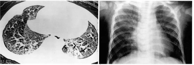
Fig. 1. HRCT scan 01 2-month-old inlant shows linear parenchymal bands(l ong arrows), consolidation with atelectasis(short ar- row), and multilocal hyperaerations in both lungs
b. Radiograph shows linear opacity in left lower lung(arrow) and multilical hyperaerations in both lungs
a
Fig. 2. a. HRCT scan 01 2.month-old inlant shows lobular or small cyst like multilocal hyperaeratiions(short arrows) and interlobular septal thickenings(l ong arrows)
b. Radiograph shows multilocal linear opacities and multilocal hyperlucent areas in both lungs.
132
정윤호 외 영아에서의 기관지폐 이형성증의 고해상 전산화 단층 촬영
、‘
a
Fig. 3. a. HRCT scan of 9-month-old infant shows multiple centrilobular nodules(short arrows) in posterobasal segment of both lower lobes and parenchymal band in left lower lobe(long arrow)
b. Radiograph shows hyperaeration of both lungs and linear opacities in both lower lungs
Table 1. HRCT findings of BPD (n=13) Findings
Parenchymal bands
Interlobular septal thickening Multifocal hyperaeration
Lobar or segmental Lobular or small cyst like Centrilobular nodules
Consolidation and/or atelectasis Bronchovascular bundle thickening
No. (%) 13 (100%)
12 (92%) 11 (85%) 7 (54%) 4 (31%) 7 (54%) 7 (54%) 6 (46%)
절성 분포가 7예 (54%) 였고 소엽성 또는 유소낭성 병변으 로 보인 다발성 과도 통기가 4예 (31%) 였다. 중심소엽 결 절이 7예 (54%) 였으며 경화 또는 무기폐가 7예 (54%) 였고 기관지 혈관속 비후가 6예 (46%) 였다. 이중 실질띠와 소엽 간 격막 비후, 그리고 다발성 과도통기는 대부분의 증례에 서 보이고 있어 주소견으로 분류하였고, 중심소엽결절, 경 화 또는 무기폐, 기관지속 혈관 비후는 부소견으로 분류하 였다 (Fig. 1
,
2,
3).고 찰
기 관지 폐 이 형성증은 1967년 Northway 등에 의 해 호홉 곤란증후군환아에서 인공호흉기와고농도의 산소치료를 받은영아들에서 발생하는만성 폐질환으로처음기술되었 다(1). 그는 병리학적 방사선학적 특정을 네가지 단계 (Stage) 로 분류 하였다. 제 l단계는 처음 2-3일에 얼어나 는데 이때에는 심한 호흡곤란 증후군과 구분할 수 없으며
‘급성 호흡곤란 증후군의 기간’이라고도 한다. 흉부 방사선 소견은 전반적 인 과립상의 양상 (generalized granular pattern)을 보이고 넓게 퍼진 폐포의 무기폐( widespread alveolar atelectasis) 와 일치하는 폐밀도의 증가를 보인다,
제 2단계는 4-10일에 일어나는데 폐의 혼탁이 두드러지며 경계가 잘그려지는 공기 -기관 음영 (air-bronchogram) 양상을 보인다. 이때는 ‘재생 (regeneration) 의 기간’이라 고도 하는데 조직학적으로 폐포상피의 괴사와 수선 (re
pair) 이 있게된다. 제 3단계는 10-20 일 사이에 일어 나는 데 ‘만성 폐질환으로의 이행기’라고도 한다. 이때는 폐의 완전 혼탁이 보이 며 공기 -기 관 음영 이 양쪽 폐 에 전체적 으로 퍼져있는 수포 (bulla) 와 비슷한 작고 퉁근 저음영으 로 보이는 특정적언 소견을 나타내며 여기에 더하여 간질 폐기종 (interstitial emphysema) 이 나타나 무기폐와 폐기 종의 복합적 양상으로 나타난다. 조직학적으로는 기관지 와 세 기 관지 의 점 막에 이 형 성 (metaplasia) 이 나타난다.
제 4단계는 ‘만성 질환의 기간’ 이라고도 하며 1개월 이상 생존한 경우에 나타난다. 이전의 수포들은 점점 크지며 기 종 (pneumatocele)이 될정도로 자란다. 조직학적으로 기관 지 주변 평활근의 두드러진 비대가 나타난다(1, 2, 6, 7).
기관지폐 이형성증 발생의 주요 병언론으로는 폐의 미 숙성, 고 농도의 산소독성, 인공호흡기에 의한 압력상해 (barotrauma) 등이 알려져 있으며 (2, 8) , 그 이외에 폐부 종, 증가된 기도저항등이 알려져 있다 (8) . 폐의 미숙성은 중요한 역활을 하는데 저체중 환아에서 많이 나타난다. 고 농도의 산소 또한 폐 에 손상을 주며 특히 미숙아에서는 실 내공기보다 약간더 높은 산소 농도에서도 손상을 줄수 있 다 (2). 인공호흡기의 사용에서 간헐적인 양압은 폐신장 (Iung stretch) 이 일어나 말단 기도에 허혈증 또는 괴사가 일어난다 (2, 9, 8).
기관지폐 이형성증의 CT 소견에 대한 보고는 적으며 Oppenheim등은 2개 월 에 서 13세 사이 의 소아에 서 ( 평 균
4.1
세) 기관지폐 이형성증의 후유증에서의 HRCT와 Elec-
tron-beam CT소견으로 다발성 과도통기, 경계가 분명한
선상 혼탁, 삼각 늑막하 흔탁(Triangular subpleural opa- cities), 분절성 또는 엽성 무기폐등을 보고 하였다(3).
- 133 -
대 한 방 사 선 의 학 회 지 1996 : 35( 1) : 131- 135
저자들은 1개월에서 127~ 월 사이의 영아만을 대상으로 하여 기관지폐 이형성증의
HRCT
소견을 분석하였는데 실질띠, 다발성 과도통기가 주소견으로 보였던 Oppen heim 등 (3) 의 보고와 일치하지만, 본 연구에서는 지금까 지HRCT
소견으로 언급이 없었던 소엽간 격막비후, 중심 소엽 결절, 경화 및 무기폐, 기관지 혈관속 비후 등이 관찰 되었다.이와같이 다른소견이 추가로보언 것은본연구에 서는 영아를 대상으로 소견을 분석 하였고, 영아에서는 영 아기 이후의 소아보다는 급성 폐질환, 즉 폐렴, 폐부종, 무 기폐등에 더 쉽게 영향을받기 때문이 아닌가생각된다.이 연구의 제한점으로 CT소견과병리 소견의 상관관계 를 규명하지 못한 점이 있으나 본 연구의 결과는 향후 기 관지폐 이형성증 연구의 기초 자료로 이용가능하리라 생 각된다.
~J C그 고 ï r E그 헌
1. Northway WH Jr, Rosan RC , Porter DY. Pulmonary disease lollowing respiratory therapy 01 hyaline membrane disease.
N Engl J Med 1967; 276: 357-368
2. Silverman FN, Kuhn JP, Berdon WE, et al. Calley’s pediatrics X-ray diagnosis. 9th ed. Philadelphia: Mosby , 1993: 601-603 3. Oppenheim C, Mamou-Mani T, Sayegh N, De Blic JD‘ Sche-
inmann P, Lallemand D. Bronchop비 monary dysplasia :Value 01 CT in identilying p 비 monary sequelae. AJR 1994;163 169-172
4. Griscom NT, Wheeler WB, Sweezey NB, Klm YC, Lindsey JC,
Wohl MEB. Bronchopulmonary dysplasia: Radiographic ap- pearance in middle childhood. Radiology 1989; 171 :811-814 5. Davis JM, Rosenleld WN, Chronic lung disease. In Avery GB,
Fletcher MA , MacDonald MG , eds. Neonatology: Pathophysi- ology and management 01 the newborn. 4th ed. Philadelphia:
J. B. Li ppincott company , 1994: 453-477
6. Harrod JR, L’Heureux P, Wangensteen OD, Hunt CE. Long- term lollow-up 01 severe respiratory distress syndrome treated with IPPB. J Pediatr 1974; 84 ‘ 277-286
7. Stocker JT. Pathologic leatures 01 long-standing “Healed"
bronchopulmonary dysplasia: A study 01 28 3-to 40-month-old inlants. Hum Patho/1986; 17: 943-961
8. Bancalari E, Gerhardt T. Bronchopulmonary dysplasia Pediatr Clin North Am 1986; 33: 1-23
9. De Kleine MJ, Roos CM, Vorn WJ, Jansen HM, Koppe JG Lung lunction 8-18 years alter intermittent positive pressure ventilation lor hyaline membrane disease. Thorax 1990; 45 941-9946
m m
정윤호 외 영아에서의 기관지폐 이형성증의 고해상 전산화 단층 촬영
Journal of the Korean Radiological Society 1996; 35( 1) ; 131-135
High-Resolution CT Findings in Infants with Bronchopulmonary Dysplasia: Preliminary Report'
Yoon Ho Chung, M.D., Young Seok Lee, M.D., Ji Hye Kim, M.D., Heon Han, M.D., Hyo Sun Chung, M.D., Yoo Mi Cha, M.D., Young Chae Kim, M.D., Sang Hee Kim, M.D.2
1 Department o( Diagnostic Radiology, Chung-Ang Gil Hospital
2Department o( Pediatrics, Chung-Ang Gil Hospital
Purpose: To evalute high resolution CT(HRCT) findings in infants with bronchopulmonary dysplasia(BPD).
Materials and Methods: In 13 infants(age range, 1 -12 months; 11 premature babies, two full-term babies; birth weight, 0.97 -3.88kg ; mean 2,03 kg) with clinico-radiologically suggested BPD, HRCT findings of the lung were reviewed retrospectively.
Spiral CT using ultra high bone algorithm, 1 mm collimation with 5-8mm interval, and 0.7sec scan time was perfomed without regard to breathing-control of infants. Three radiologists each analysed the HRCT findings twice.
Results: HRCT findings of BPD were as follows; parenchymal bands(n=13), interlobular septal thickenings (n=12), multifocal hyperaeration involving lobar or segmental distribution(n=7), and involving lobular distri- bution or small cyst-like lesion(n=4), centrilobular nodules(n=7), consolidation and/or atelectasis(n=7), and bronchovascular bundle thickening(n=6).
Conclusion : Parenchymal bands, interlobular septal thickenings, and multifocal hyperaerations were the major findings in cases of bronchopulmonary dysplasia whereas, centrilobular nodules, consolidation and/or atelectasis, and bronchovascular bundle thickenings were the minor findings. These findings may be used as basic data in the evaluation of BPD in future studies.
Index Words: Infants, respiratory system
Computed tomography(CT), high-resolution Lung, diseases
Address reprint requests to; Yoon Ho Chung, Department of Diagnostic Radiology, Chung-Ang Gil Hospital, # 1198,
Kuwol 깅 ong, Namdong-Ku, 405-220 Korea. Tel. 82-32-460-3060 Fax.82-32-467-9302
π
μ
국제 학술대회 일정표[ II J
• Sandwichcursus Uroradiology (1997/02/11-12) venue: De Jaarbeurs Utrecht, The Netherlands
contact: Mrs. F.E. Blommendaal, NVvRd, P.O. Box 8171, 3505 RD Utrecht. The Netherlands
(tel: 31 - 30 -2474294; fax: 31 - 30 - 2474439)
• Medical Imaging (1997/02/16 -20)
venue: New Port BeachMarriott Hl Newport Beach, CA, USA‘
contact‘ Ms. Roberta Hart, Spie Bellingham,
P.O. Box 10, Bellingham, 매IA 98227-0010, USA (tel: 1-360-6763290; fax: 1-360-6471445)
• Seminar on Cross-Sectional Imaging (1997/02/24 -28)
venue: Hyatt Regenc St. John R‘ Virgin Islands, USA.
contact: Janice Ford, Hosp. of the Univ. of PA,
3400 Spruce Street, Philadelphia, PA 19104, USA‘ (tel: 1 - 215 -6627825; fax: 1 -215 - 3495925)
• 7th European Symposium on Radiopharmacy & Rad- iopharmaceuticals (1997/03/02 -05)
venue: MECC Maastricht, The Netherlands
contact: Congress Bureau, Van Namen & Westerlaken, P.O. Box 1558, 6501 NB Nijmegen, The Netherlands (tel:31-24-3234471; fax:31-24-3601159)
• 10th European Congress of Radiology (1997/03/02 -07)
venue ‘ Austria Centre Vienna, Austria contact: MR. P. Baierl, ECR-office,
Neutorgasse 9/2A, A-IOIO Vienna, Austria (tel: 43 - 1 - 5334064; fax: 43 - 1 - 5334064 -9)
• 20th Annual Skeletal Symposium: Musculoskeletal MRI (1997/03/03-08)
venue: The peaks (Hotel & Spa) Telluride, Colorado, USA contact: Janice Ford, Hosp. of the Univ. of PA,
3400 Spruce Street, Philadelphia, PA 19104, USA‘ (tel: 1- 215-6626904; fax: 1-215-3495925)
• Hospimedica '97: INT. Hospital, Pharmaceutical &
Medical Equipm. & Supplies EXH. (1997/03/05 -08) venue: Singapore Int. Conv./Exh. Ct Singapore, singapore contact: Ms. S. Lim/Ms. J. Yip, East-West Exhibitions Pte,
No. 5 Temasek Boulevard, # 05-05 Suntec City Tower, Singapore 038985
(tel ‘ 65 - 3329670; fax: 65 - 3370366)
• 22nd Annual Meeting Soc. of Cardiovascular and Interventional Radiology (1997/03/08-13)
venue: Washington, DC, USA.
contact: SCVIR, 10201 Lee Highway, Suite 500, Fairfax, VA 22030, USA.
(tel: 1-703 -6911805; fax: 1 -703 - 6911855)
• 3rd International Conference on Magnetic Reson- ance Imaging (1997/03/12-16)
venue: The Regent Hotel Melbourne, Australia
contact κ1s. Probati κ1ilton , B. Sc., Convention Professionals, P.O. Box 4031, Balwyn East, Victoria 3129, Australia (tel: 61 - 3 -98990368; fax: 61-3 -98990368)
• Annua
’
Meeting American Institute of Ultrasound in Medicine (1997/03/23-26)venue: San Diego, USA‘
contact: Jenny Clark, AIUM, Suite 100,
14750 Sweitzer Lane, Laurel, MD 20707-5906, USA.
(tel: 1 - 301 -4984100; fax: 1 -301 -4984450)
• Second Congress Asian & Ocenian Soc. of Neuro-/ Head & Neck Radiology (1997/03/24-27)
venue: Taipei Veterans Gen. Hosp Taipei, Taiwan, R. O. C contact: Dept. of Radiology, Veterans Gεn. Hospital, 201,
Sec. 2, Shih-Pai Rd., Taipei, Taiwan 11217, R. O. C (tel: 886 - 2 - 8757357; fax ’ 886 -2 -8733643)
• Course: Neuroradiology Update 1997 (1997/03/24 -28)
venue ‘ undetermined, USA‘
contact‘ Janice Ford, Hosp. of the Univ. of PA,
3400 Spruce Street, Philadelphia, PA 19104, USA (tel: 1 -215 -6626904; fax: 1 -215 - 3495925)
• Course: Radiology for MRCP (1997/04/00- 00) venue 씨lolfson Conference Centre London. United
Kingdom
contact: wolfson Conference Centre, Hammersmith Hospital, Du Cane Road, London WI2 ONN, United Kingdom (tel :44- 181-7403245; fax: 44- 181 -7404950)
• Course on Radiation Protection (1997/04/00-00) venue: Wolfson Conference Centre London, United
Kingdom
contact: Wolfson Conference Centre, Hammersmith Hospital, Du Cane Road, London WI2 ONN, United Kingdom.
(tel:44-181-7403245; fax:44-181-7404950)
• 8th Annual Meeting European Society of Pediatric Urology (1997/04/03 -05)
venue: Pontificia Univ. Urban. Rome, ltaly
contact: ESPU Meetings, Central Secretariat, 42 Devonshire Road, Cambridge CBl 2BL, United Kingdom (tel‘44-1223 - 323437; fax: 44- 1223 - 460396)
• 56th Soc. Assembly of Japan Radiological Soc./53rd Soc. Assembly of JSRT (1997/04/03-06)
venue: Pacifico Yokohama Yokohama city, Japan contact: JMCP, Kitaotemachi Bldg., 1-7-6,
Chiyoda-ku, Tokyo 101, Japan.
(tel:81-3- 52810456; fax:81-3-52810457)
• 3rd International Conference of Nuclear Cardiology (1997/04/06 -09)
venue: Florence, Italy
contact: OIC, Via A. La Marmora 24, Firenze, Italy 50121
(tel: ; fax: 39 - 55 - 570227)
• 3rd Asian-Pacific Congress of Cardiovascular and Interventional Radiology (1997/04/06-11)
venue: World Congress Centre Melbourne, Australia contact: Mrs .. Davies, APCCVIR, Radiology Department,
The Royal Melbourne Hosp., Parkville, Victoria 3050, Australia. (tel: 61 - 3 -93427293; fax: 61 - 3 - 93428369)