DOI:10.4078/jkra.2009.16.2.133
<접수일:2009년 5월 26일, 수정일:2009년 6월 2일, 심사통과일:2009년 6월 2일>
※통신저자:김 호 연
서울시 서초구 반포동 505번지
가톨릭대학교 의과대학 류마티스연구센터
Tel:02) 2258-7471, Fax:02) 599-4287, E-mail:[email protected]
이 논문은 2009년도 정부(교육과학기술부)의 재원으로 한국과학재단의 지원을 받아 수행된 연구임(R11-2002- 098-05001-0), (No.M10870060005-08N7006-00510).
인간의 말초혈액에서 염증성 사이토카인에 의한 Th17 세포의 분화에 미치는 영향
가톨릭대학교 의과학연구원 류마티스연구센터
허유정ㆍ박미경ㆍ주지현ㆍ박경수ㆍ조미라ㆍ김호연
= Abstract =
The Effect of Inflammatory Cytokines on the Differentiation of Th17 Cells in Human Peripheral Blood
Yu-Jung Heo, Mi-kyung Park, Ji-Hyeon Ju, Kyung-Su Park, Mi-La Cho, Ho-Youn Kim
The Rheumatism Research Center, Catholic Research Institute of Medical Science, The Catholic University of Korea, Seoul, Korea
Objective: IL-17-producing T cells (Th17 cells) have been identified as a distinct lineage of CD4+ T helper cells in mice. Since this discovery, many efforts have been made to investigate the characteristics and the role of human Th17 cells and the factors involved in their differentiation. This study was undertaken to assess the effects of cytokines and stimulatory conditions on the differentiation of human CD4+ T cells into Th17 cells.
Methods: Peripheral blood CD4+ T cells were isolated from healthy humans and then these cells were cultured with using various stimulatory conditions. The Th17 cells and regulatory T (Treg) cells were detected by flow cytometry (FACs). The related gene expressions of cytokines, transcription factors and chemokine receptors were determined by ELISA and RT-PCR.
Results: In the presence of inflammatory cytokines, TNFa and IL-1b, the human CD4+ T cells rapidly produced IL-17 in response to anti-CD3/anti-CD28 stimulation, whereas, with anti-CD3/
anti-CD28 stimulation alone, the CD4+ T cells expressed low levels of IL-17. TNFa and IL-1b were also important inducers of IL-22 production. IL-6 and IL-23 up-regulated the RORgammat, CCR4 and CCR6 expressions in the human CD4+ T cells. In response to TGF-b and IL-2, the human CD4+ T cells were rapidly induced to express FoxP3, IL-10 and CCR7, as compared
with anti-CD3/anti-CD28 stimulation alone.
Conclusion: The effect of inflammatory cytokines on the differentiation of human Th17 cells may help us to understand their pathogenic role. Moreover, the differential expression of chemokine receptors and transcription factors of the subsets of CD4+ T cells with the different features of Th17 and Treg, may raise new issues concerning the pathogenesis of autoimmune inflammatory diseases.
Key Words: Human CD4+ T cells, Th17 cells, Treg cells, RORγt, FoxP3
서 론
T세포는 특정 사이토카인들과 전사인자의 발현을 통해 몇가지 계통의 helper T세포(Th 세포)로 분화 발달 하는 것으로 알려졌다. Th1세포는 interleukin-12 (IL-12) 신호전달을 통해 T-bet 전사인자가 활성화되 고 IFN-γ를 생성하는 T세포군이며, Th2세포는 IL-10 의 신호전달을 통해 GATA binding protein 3 (GATA3) 전사인자를 발현하고 IL-4를 생성하는 T세포군이다 (1). 이들 세포군과 길항역할을 할 수 있는 세포로 알려진 면역조절세포(regulatory T cells, Treg세포)는 여러가지 환경에서 분화 발달 할 수 있고 Forkhead box P3 (FxoP3) 전사인자를 발현하며 사이토카인 transforming growth factor-β (TGF-β)와 IL-10을 생 성하고 이들 사이토카인과 또는 세포표면에 발현하 는 여러 수용체 또는 리간드를 통해 다른 면역세포 활성을 억제하는 것으로 알려졌다 (2).
최근에 알려진 IL-17을 생산하는 T세포(Th17세포) 는 IL-6와 TGF-β에 의해서 분화되며, 전사인자인 re- tinoic acid receptor-related orphan nuclear receptor gamma t (ROR)γt, RORα를 발현한다 (3,4). Th17세포는 생 쥐나 인간에서 공통적으로 IL-17 구성원 중 IL-17A 와 IL-17F를 분비하고, 호중구의 활성과 염증성 사 이토카인과 케모카인의 생성에 관여한다 (5). 염증성 자가면역질환에서 Th17세포는 기존 염증관련 세포 로 알려진 Th1세포보다 질환 발병과 밀접한 관련이 있는 중요한 세포로서 최근 그 연구가 활발히 진행 되고 있다.
생쥐에서 Th17세포의 분화 발달에는 TGF-β, IL-6 와 IL-23가 중요한 역할을 하며, IL-6는 IL-21의 분비 를 촉진하고, IL-21은 자가분비를 통해 Th17세포의 분화를 돕는다 (6). 생쥐에서 Th17세포는 IL-21 뿐만
아니라 IL-22와 같은 새로운 사이토카인도 생산할 수 있다고 알려졌으며, 몇몇 경우에 IL-10과 IL-22가 동시에 분비된다는 보고도 있다 (7,8). 하지만 아직 까지 세포활성을 일으키는 조건과 이들 세포의 기능 에 대해 더 많은 연구가 요구된다고 생각되고 있다.
인간세포를 대상으로 in vitro에서 Th17세포를 분 화시키기 위해서는 생쥐세포에서 보다 더 많은 자극 이 요구된다. IL-6, TGF-β와 IL-23 뿐만 아니라 tu- mor necrosis factor-alpha (TNF-α)나 IL-1β같은 염증 성 사이토카인이 동반되어야 IL-17생성이 증가하며, 이때 Th1과 Th2세포 발달에 관여하는 IL-12와 IL-4 를 중성화시키는 항체를 넣어주면 Th17세포 발달이 잘 일어나는 것으로 알려져 있다 (9). TGF-β와 IL-6 는 Th17세포의 분화에 필수적인 요소이며, IL-1β와 TNF-α는 Th17세포의 확장에 관여하고 IL-23는 Th17 세포의 표현형을 결정하는 것으로 알려져 있다 (10- 12). 면역질환동물모델과 환자에서 IL-17의 활성이 중요한 것으로 밝혀지고 있으며 현재도 많은 연구가 진행 중이다.
TGF-β가 Th17세포 분화에 부정적인 작용을 한다 는 반대의 보고도 있어 주요 논쟁의 요소가 되고 있 다 (13,14). TGF-β는 Treg세포의 주요 전사인자인 FoxP3 활성을 유도하기 위해 mothers against decap- entaplegic (MAD)와 the Caenorhabditis elegans protein SMA (SMAD)를 직접적으로 활성화시켜 Treg세포를 유도한다 (15). Treg세포는 자기와 비자기 항원에 대 하여 발생하는 면역반응을 조절하는 대표적인 세포 로, 생쥐와 인간에서 전체 CD4+ T세포 중 5∼10%정 도를 차지한다 (16,17). Treg세포는 자가면역 질환의 세포치료에 활용되기도 하는 중요한 세포이다.
이러한 Treg세포와 Th17세포의 특성분석은 생쥐모 델에서 매우 활발하게 이루어졌고 인간세포에서 조 사는 아직 미흡한 실정이다. 본 연구에서는 정상인
의 말초혈액 세포에서 염증성 사이토카인에 의한 Th17세포 활성 관련 조건을 조사하고 이들의 특성 을 분석하였다. 그리고 이렇게 분환 된 Th17세포에 서 발현되는 인자들이 Treg세포에서는 어떻게 발현 되는지 조사하고자 한다.
대상 및 방법 1. 대 상
정상인 10명을 대상으로 하였으며, 대상자의 성별 은 여성이 8명, 남성이 2명으로 평균나이는 28±2세 였다. 본 연구는 가톨릭중앙의료원 임상연구관리 규 정을 준수하여 실험하였다.
2. 시 약
사용한 자극제로는 anti-human CD3 항체, anti-human CD28 항체(BD Pharmingen, San Diego, CA)을 사용 하였고 사이토카인으로는 IL-12, IL-4, IL-2, TNF-α, IL-6, IL-23 (R&D, Minneapolis, MN), IL-1β (Endogen, Rockford, IL), TGF-β (Peprotech, Rocky Hill, NJ), Phorbol 12-Myristate 13-Acetate (PMA), Ionomycine (Sigma Addich, St. Louis, MO)을 사용하였다. 중화 항체로는 anti-IFN-γ, anti-IL-4 (R&D)를 사용하였다.
3. CD4+ T세포 분리
정상인으로부터 헤파린 처리한 주사기를 이용하여 혈액을 채취한 후 말초혈액 단핵세포(peripheral blood mononuclear cells, PBMC)를 Ficoll-PaqueTM (Amersham Biosciences, burkinghamshire, England)를 이용하여 cen- trifuge gradient법으로 분리하였다. 분리한 PBMC를 anti-human CD4+ T cell Biotin-Ab Cocktail (Milteny biotec, Bergisch Gladbach, Germany)과 4oC, 15분간 반응시킨 후 anti-human CD4 biotin Microbead (Milteny biotec)와 4oC, 15분 반응시켰고, MACs buffer로 세척 한 뒤 autoMACs (Milteny biotec)를 사용하여 음성 분획으로 CD4+ T세포를 분리하였다. 분리된 CD4+ T 세포를 PBS로 세척하고 55oC에서 30분 동안 불활성 화된 10% 우태아 혈청, Penicillin (100 U/mL), Strepto- mycine (100 g/mL)이 포함된 세포 배양액 (RPMI 1640, Gibco BRL, USA)에 배양하였다.
4. Th세포로의 분화 유도
분리된 CD4+ T세포(Th0) 5×10⁵와 5000 rad에서 조 사한 항원공여세포 5×10⁵을 anti-CD3가 1 μg/mL로 coating 처리된 24 well plate (NUNC, Denmark)에 분 주하고, anti-CD28 항체 1 μg/mL를 넣고, 여기에 여 러 사이토카인들을 처리하여 Th1, Th2, Th17, Treg세 포로 분화를 유도하였다. Th1세포의 분화를 위해서 anti-IL-4와 IL-12 10 ng/mL을 넣어 주었고, Th2세포 의 분화를 위해서는 anti-IFN-γ와 IL-4 10 ng/mL을 넣었다. Th17세포의 분화를 위해서는 IL-6 10 ng/mL, TGF-β 2 ng/mL, IL-1β 10 ng/mL, TNF-α 5 ng/mL 로 자극하고 3일 후 IL-23 10 ng/mL로 재자극 해주 어 3일 배양 하였다. Treg 세포로의 분화를 위해서 는 TGF-β 10 ng/mL과 IL-2 10 ng/mL로 자극하였다.
중화항체(anti-IFN-γ 10 μg/mL, anti-IL-4 10 μg/mL) 는 30분 전처리 하였다.
5. 유세포 분석
각 계통으로 분화시킨 세포들의 세포표면항원의 발현과 함께 세포의 공조 수용체의 발현 정도를 유 세포 분석을 통해서 알아보았다. 모든 항체는(e-bio- science, San Diego, CA) 제품을 사용하였다. 먼저 6 일 동안 자극한 뒤 수집된 세포의 표면항원을 분석 하기 위해서 PMA 25 ng/mL, Ionomycine 250 ng/mL 과 Golgistop (BD Phamingen, San Diego, CA)을 5시 간 전처리 하였다. 먼저 수집한 세포를 Peridinin chlo- rophyll protein (PerCP)-anti-human CD4항체와 Allophy- cocyanin (APC)-anti-human CD25항체와 4oC에서 30분 간 암시야에서 반응시킨 뒤 FACs buffer (0.002% so- dium azide, 0.2% BSA/PBS)로 세척하고, Cytofix/perm 으로 4oC에서 20분간 암시야에서 반응시킨 뒤, Fluo- rescein isothiocanate (FITC)-anti-human Foxp3 항체, Phcoe- rythrin (PE)-anti-human IL-17항체와 4oC에서 30분간 암시야에서 반응시켰다. 마지막으로 이 세포들을 Perm- wash buffer로 두 차례 세척 한 후 FACs buffer에 재 부유하여 유세포 분석기(FACs Calibur; Becton Dic- kinson, San Diego, CA)로 분석하였다. 분석 프로그 램으로는 Flow jo (Version 4.5, Treestar, Ashland, OR)를 사용하였다. 림프구 그룹은 전체세포를 for- ward/side scatter 특징에 따라 나눈 그룹 중에 선별하
Table 1. Primers used in RT-PCR
Primers (5'-3')
Template Annealing
Sense Anti-sense
(bp) oC Transcription factor
T-bet CACTACAGGATGTTTGTGGACGTGC CTAAAGCTGACAAACAACAAGGGG 204 62 GATA3 AACTGTCAGACCACCACAACCACAC CCATGACTATGAAGAAGGAAGGACATCC 221 60
RORc AGTCGGAAGGCAAGATCAGA CAAGAGAGGTTCTGGGCAAG 192 58
Chemokine receptor
CXCR3 CAACGCCACCCACTGCCAATACAA CAGGCGCAAGAGCAGCATCCACA 415 60 CCR4 AAGAAGAACAAGGCGGTGAAGATG AGGCCCCTGCAGGTTTTGAAG 269 57 CCR6 CCTGGGGAATATTCTGGTGGTGA CATCGCTGCCTTGGGTGTTGTAT 404 57 CCR7 GTGCCCGCGTCCTTCTCATCAG GGCCAGGACCACCCCATTGTAG 352 60 β-actin GGACTTCGAGCAAGAGATGG TGTGTTGGCGTACAGGTCTTTG 198 60 여 분석하였다.
6. 사이토카인 측정
배양된 상층액을 모아 IL-17, IL-22와 IL-10을 sand- wich ELISA를 이용하여 농도를 측정하였다. sand- wich ELISA용 96 well plate (NUNC, Denmark)에 단 클론성 항체 IL-17 항체, IL-10 항체(R&D) 4 μg/mL 을 50 μl/well씩 넣고, 4oC에서 밤새 반응시킨 다음 차단용액(1% BSA/PBST)을 200 μl/well씩 넣고 실온 에서 2시간 반응시켰다. Standard로는 재조합 IL-17, IL-10 (R&D)을 이용하여 10 ng/mL∼15.6 pg/mL 농 도로 사용하였다. 표준시료와 함께 측정할 세포배양 상층액을 50 μl/well씩 넣고 실온에서 2시간 반응시 켰다. 반응용기를 세척용액(0.05% Tween 20/PBS)으 로 4회 세척하고 biotinylated goat-anti-human IL-17, IL-10을 100 ng/mL 희석하여 50 μl/well씩 넣어 실온 에서 2시간 반응시킨 후 세척용액으로 4회 세척 하 였다. 마지막으로는 ExtraAvidin-Alkaline Phosphatase conjugate (SIGMA)를 1:2,000으로 희석하여 50 μl/
well씩 넣고 실온에서 2시간 반응시키고 세척 후 PNPP (Fluka, Phosphate Disodium Salt Hexahydrate)/DEA 용 액(Diehanolamine 97 mL, NaN3 0.2 g, MgCl2HO 0.1 g, 1차 증류수 800 mL)에 1 mg/mL농도로 녹여 50 μl/well씩 넣어 20∼30분 후 0.2 N NaOH로 반응을 멈추고 405 nm 파장에서 흡광도로 측정하였다. IL- 22는 peprotech kit 제품을 사용하였으며, kit에서의 절차를 사용하였다.
7. RNA 분리와 Th17 분화와 관련된 역 전사 중합 효소반응(RT-PCR)
3일 동안 배양한 세포로부터 총 RNA를 RNA zol BTM (TEL TEST, Friendswood, TX)을 이용하여 추 출하였다. 추출한 총 RNA를 주형으로 cDNA를 합성 하기 위하여 0.5 μg random ninemer (TakaRA, Shiga, JAPAN)와 70oC에서 5분 반응시킨 뒤 4oC에서 급냉 시킨 다음 10 mM dNTP mix (Invitrogen, Carlsbad, Cali- fornia) 1 μl, 역전사 효소 M-MULV (MBI Fermentas, Hanover, MD) 1 unit, 5x M-MULV 희석용액(MBI Fermentas) 4 μl, RNase Imhibitor (MBI Fermentas) 0.5 μl를 가하고 전체를 nuclease free water (Promega, Madison, WI) 20 μl로 맞춘 뒤, 25oC 10분, 42oC에서 60분, 72oC에서 10분간 반응시켰다. 이렇게 생성된 cDNA 산물을 이용하여 중합효소 연쇄반응을 시행 하였다. 본 실험에서 사용한 β-actin, T-bet, GATA3, RORc, Foxp3, CXCR3, CCR6, CCR4와 CCR7의 시발 체는 모두 Genotec Co (Seoul, Korea)에서 제작하였 고, 염기서열은 아래 표 1에 표시하였다. 증폭 과정 중 각각 서로 다른 양으로 역 전사된 cDNA와 증폭 수를 변경하면서 RT-PCR을 시행하여 대수적으로 증 가하는 cDNA양과 증폭수를 결정하였다. RT-PCR를 위한 반응 화합물은 총 25 μl가 되도록 하였고, 용 액의 조성은 2.5 μl의 10x reaction buffer (10 mM Tris-HCl, pH 8.3:15 mM MgCl2:50 mM KCl;
Takara, Shiga, Japan), 0.5 mM의 dNTP (Takara, Shiga,
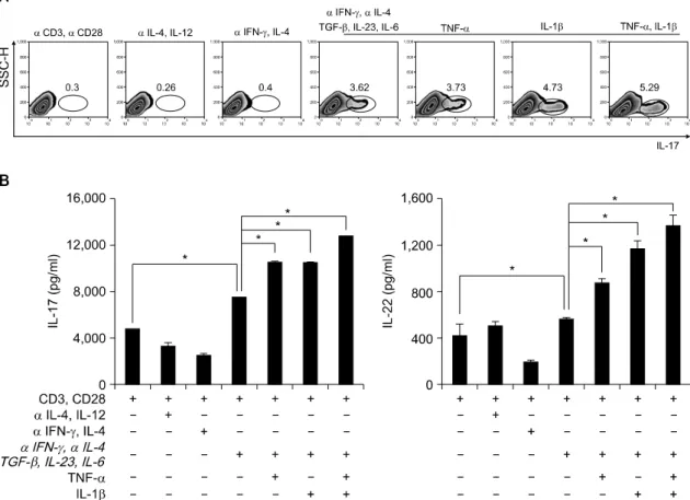
Fig. 1. Th17 cell differentiation in human CD4+ T cells. (A) Flow cytometry of intracellular human IL-17 producing CD4+ T cells from peripheral blood. CD4+ T cells cultured with plate bound anti-CD3, anti-CD28 and various cytokines in vitro for 6d. Neutralize antibodies used anti-IFN-γ and anti-IL-4. Data are representative of ten independent experiments. (B) ELISA quantification of IL-22 and IL-17 in culture supernatants of A. Data are representative of ten independent experiments. Values are mean±SD. *p<0.01.
Japan)를 사용하였다. 증폭을 위해 Dual-bay Thermal cycler system (MJ Research)를 사용하였다. 음성 대 조군으로 추출한 cDNA 대신 증류수를 사용하여 PCR 산물이 관찰되지 않는 것을 통해 PCR 오염이 없음을 확인하였다.
8. 통계적 유의성의 검증
실험 결과는 평균±표준 오차로 표현하였으며, 통 계적 유의성은 Student’s t-test를 실시하였고 p값이 0.05이하 일 때 통계적으로 유의하다고 분석하였다.
결 과
1. 정상인의 CD4+ T세포에서 염증성 사이토 카인 에 의한 Th17세포 분화의 최적조건 조사
Th17세포의 기본적인 특성을 조사하기 위해 정상 인의 혈액으로부터 Th1, Th2, Th17 세포를 유도하였 다. 기존 보고와 마찬가지로 Th1세포에서는 IFN-γ 생성이, Th2에서는 IL-4의 생성이 특이적으로 증가 되었다(결과 제시하지 않음). Th17세포의 분화는 기 존 보고와 같이 Th1과 Th2계통을 anti-IFN-γ와 anti- IL-4를 사용하여 차단한 후, TGF-β, IL-23와 IL-6로 자극하여 분화를 유도하였다. 한편 분화된 Th17세포
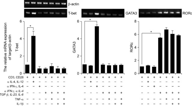
Fig. 2. Expression of transcription factors in human CD4+ Th17 cells. RT-PCR analysis of T-bet, GATA3 and RORc expression in CD4+ T cells. CD4+ T cells cultured with plate bound anti-CD3, anti-CD28 and various cytokines in vitro for 3d. β-actin was used as a loading control. Results were normalized for β-actin expression. Statistical data repeat of three independent experiments. Values are mean±SD. *p<0.01.
에서는 IL-17의 생성이 다른 Th세포에 비하여 증가 되었으며, 추가적으로 염증성 사이토카인인 TNF-α 또는 IL-1β를 처리하여, IL-17의 생성이 증가되는 지를 관찰하였다(그림 1A). 또한, Th17세포에서 TGF-β, IL-23와 IL-6로 자극한 조건보다 TNF-α 또 는 IL-1β를 처리하였을 때, IL-22의 분비가 통계적 으로 유의하게 증가 되었다(그림 1B).
2. 염증성 사이토카인에 의한 Th세포 분화에서 전 사인자와 케모카인 수용체 발현조사
각 계통의 특이 전사인자 발현 조사와 더불어 케 모카인 수용체의 발현을 조사하였다. 정상인의 혈액 으로부터 CD4+ T세포를 분리하여 중화항체를 처리 한 뒤 방사선 조사를 한 항원제시 세포와 1:1의 비 율로 3일 동안 공조 배양 하였다. 기존 보고와 마찬 가지로, anti-IL-4와 IL-12를 처리한 Th1세포에서는 T-bet의 발현이 증가되었으며, anti-IFN-γ와 IL-4를 처리한, Th2세포는 GATA3의 발현이 증가됨을 관찰 하였다. Anti-IFN-γ, anti-IL-4를 처리한 후, TGF-β,
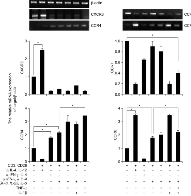
IL-23와 IL-6를 처리하여 분화를 유도한 Th17세포에 서는 RORc의 발현이 통계적으로 유의하게 증가되었 다. 특히, 이러한 RORc의 발현 증가는 TGF-β, IL-23와 IL-6를 처리한 조건보다 TNF-α 또는 IL-1β 를 처리한 조건에서도 Th0, Th1과 Th2 세포에 비해 서 현저히 증가되었다(그림 2). 다음으로, T세포의 아형을 아는데 도움이 되는 케모카인 수용체(CXCR3, CCR7, CCR4와 CCR6)의 발현을 Th세포 형태별로 조사하였다 (18,19). Th1세포에서는 CXCR3와 CCR6 의 발현이 증가 되었으며, Th2세포는 CCR4의 발현 이 증가되었다. Th17세포에서는 CCR4와 CCR6의 발 현이 증가되었으며, 일반적으로 Treg세포에서 증가 되어 있는 CCR7의 발현에는 변화가 없음을 관찰하 였다(그림 3).
3. 정상인의 CD4+ T 세포에서 Foxp3를 발현하는 Treg세포와 염증성 사이토카인에 의해 분화된 Th17세포의 활성 표지자 비교 조사
앞에 결과를 통해 TNF-α와 IL-1β에 의해 유도된
Fig. 3. Expression of chemokines receptors in human CD4+ Th17 cell. RT-PCR analysis of CXCR3, CCR7, CCR4, and CCR6 expression in CD4+ T cells. CD4+ T cells cultured with plate bound anti-CD3, anti-CD28 and various cytokines in vitro for 3d. Results were normalized for β-actin expression. Statistical data repeat of three independent experiments. Values are mean±SD. *p<0.01.
Th17세포의 CCR7 발현이 통계적으로 유의하게 감 소되어있었다(그림 3). 정상인의 CD4+ T세포와 방사 선 조사를 한 항원제시세포를 1:1의 비율로 6일 동 안 공조 배양한 후 Th0, Th17과 Treg세포로의 분화 를 유도하여 Th17세포와 Treg세포 내 특이적인 전사 인자와 사이토카인의 발현을 동시에 조사하였다.
Th17세포로의 분화를 위해서는 TGF-β, IL-23, IL-6, IL-1β와 TNF-α를 모두 사용하였으며, Treg세포로 의 분화를 위해서는 TGF-β와 IL-2를 처리하여 분화 를 유도하였다. Th17세포와 Treg세포로 분화시킨 뒤 Foxp3의 발현은 Treg세포에서 증가되어 있었으며, Th17세포에서는 현저히 저하되어 있었다(그림 4A).
Fig. 4. Th17 versus Treg lineages are differential expression of cytokine and chemokines receptors in human CD4+ T cells. (A) Flow cytometry of intracellular human Foxp3 expression CD4+ CD25+ T cells from peripheral blood. CD4+ T cells cultured with plate bound anti-CD3, anti-CD28 and various cytokines in vitro for 3d (Th0:
anti-CD3 and anti-CD28, Th17: anti-IFN-γ, anti-IL-4, TGF-β, IL-6, IL-23, IL-1β and TNF-α, Treg: anti-IFN-γ, anti-IL-4, TGF-β and IL-2). Data are representative of ten independent experiments. (B) ELISA quantification of IL-22, IL-17 and IL-10 in culture supernatants of A. Data are representative of ten independent experiments.
Values are mean±SD. (C) RT-PCR analysis of CXCR3, CCR4, CCR6, and CCR7 expression in CD4+ T cells.
CD4+ T cells cultured with plate bound anti-CD3, anti-CD28 and various cytokines in vitro for 3d. β-actin was used as a loading control. Results were normalized for β-actin expression. Statistical data repeat of three independent experiments. Values are mean±SD. *p<0.01 versus Th0 or Th17.
이들이 분비하는 사이토카인과 케모카인 수용체 역 시 상반된 조절을 보였다. Th17세포에서 분비되는 IL-17과 IL-22의 분비는 Treg세포에서 감소되어 있었 으며, Treg세포에서 분비되는 IL-10의 생산은 Th17세 포에서 통계적으로 유의하게 감소되어 있었다(그림 4B). 또한, Th17세포에서 증가되어있는 CCR4와 CCR6 의 발현은 Treg세포에서 감소되어 있었으며, Treg세
포에서 증가되어 있는 CCR7의 발현은 Th17세포 에 서 의미있게 감소되어 있었다(그림 4C).
고 찰
자가면역질환을 비롯한 많은 질병관련 동물모델에 서 염증성 사이토카인 유래 Th17세포가 핵심적인 병
인반응을 주도하는 것이 밝혀지면서 Th17세포 연구 가 활발하게 진행되고있다 (20-23). 하지만 인간에서 의 Th17세포연구는 세포 사용의 한계와 인간 개개 인의 편차가 심하여 균일화된 생쥐에서처럼 활발한 연구가 어려운 문제점이 있다. 최근 많은 연구자들 이 인간 Th17세포 연구를 시작하였으며 Th세포의 발생과 발달 그리고 기능에 관하여 많은 연구가 진 행 중이다.
본 연구에서는 정상인 CD4+ T세포에서 Th17세포 의 활성조건을 조사하고 동시에 표지자와 활성분자 발현을 이들 세포와 대조적인 기능을 가진 것으로 알려진 Treg세포와 비교 조사하였다.
지금까지 생쥐에서 Th17세포는 TGF-β, IL-6와 IL- 21에 의해 분화 발달 되고 RORc, signal transducer and activator of transcription 3 (STAT3), interferon regu- latory factor 4 (IRF4)와 같은 전사인자가 활성화 되 어 IL-23 세포내 신호전달을 통해 IL-17A, IL-17F와 IL-21를 분비한다고 보고되었다 (24,25). TGF-β의 역할에 대해서 인간에서는 Th17세포의 분화는 생쥐 와는 달리 상반된 보고가 있다 (13,14). TGF-β에 의 해 분화가 억제된다는 보고도 있고 생쥐와 동일하게 분화를 유도한다는 보고도 있어 추가연구가 필요한 실정이다. 이러한 TGF-β와 함께 IL-6와 IL-23에 의 해 분화가 일어나고 전사인자 RORc가 활성화되어 IL-17A, IL-17F, IL-22, IL-26와 같은 사이토카인을 분비하는 Th17세포가 발달하는 것으로 알려져 있다 (26,27).
본 연구에서는 정상인의 말초혈액 세포로부터 분 리한 CD4+ T세포에 TGF-β, IL-6와 IL-23를 처리하 여 Th17세포의 분화를 유도하였으며 이때 염증성 사이토카인인 IL-1β와 TNF-α에 의해서 IL-17의 발 현이 더욱 증가되는 것을 관찰하였다. 또한 인간 Th17세포에서 IL-17과 동시에 IL-22 생성이 증가되 는 것을 관찰하였다. 생쥐와 달리 인간세포에서는 Th17세포의 활성에 IL-1β와 TNFα와 같은 염증성 사이토카인이 중요하며, 이로 보아 염증성 자가면역 질환에서 Th17세포가 활성되는 기전에 이러한 염증 성 사이토카인들이 관여함을 짐작할 수 있다. 실제 로 자가면역질환 환자군에서 TNF-α를 수용성 수용 체 또는 항체로 차단하면 Th17세포 활성이 감소된 다고 알려졌다 (28).
본 연구에서는 Th세포 발달에 중요한 전사인자 발현을 조사하였는데 Th1세포에서는 T-bet의 발현이, Th2에서는 GATA3의 발현이 증가되었고, Th17세포 에서는 RORc의 발현이 증가되었다. RORc가 과발현 되는 Th17세포에서는 T-bet과 GATA3의 발현이 억제 되어 있는 것을 확인하였다. 케모카인 수용체 역시 Th1에서는 CXCR3, CCR6가, Th2에서는 CCR4의 발 현이 증가되었으며, Th17세포에서는 CCR4, CCR6의 발현이 증가되었다. 즉 TNF-α나 IL-1β의 자극이 없 이도 Th17세포의 활성조건에 의해서 이미 특정 전 사인자나 케모카인 수용체 발현이 유의하게 나타남 을 관찰하였다. 인간 Th세포 종류에 따른 케모카인 수용체의 발현 차이는 매우 흥미로운 결과이며, 생 쥐에서도 Th세포 종류에 따라서 케모카인 수용체 발현이 다르게 나타난다고 보고되었다 (18,19). 이는 이들과 접촉하는 세포나 표적조직이 다르다는 것을 의미한다. 이러한 결과는 세포의 기능연구에 매우 중요한 단서가 될 수 있다.
이런 의미에서 Th17세포는, 특히 염증이 있는 상 태에 있는 자가면역질환 동물모델에서 핵심적인 병 인 T세포로 주목을 받게 된다. Th17세포는 IFN-γ를 생산하는 Th1세포 또는 IL-4를 생산하는 Th2세포에 의해서 활성이 억제되며 특히, Foxp3를 발현하는 Treg세포에 의해 억제된다 (29,30). 이런 결과들은 Th17 세포는 다른 Th세포와 길항작용이 있음을 암시한다.
하지만, 인간에서 Th17세포와 Treg세포 사이에서 상 호작용에 대해서는 아직 연구가 필요한 상태이다.
TGF-β는 Th17세포 발달을 촉진하고 동시에 면역 조절능을 가지고 있는 Treg세포의 발달에도 관여하 는 것으로 알려져 있다 (31). 이러한 과정에서 TGF- β가 Th17세포와 Treg세포 사이에서 경쟁적으로 발 달과 활성을 조절할 것으로 생각되는 결과들이 동물 연구를 통해서 발표되고있다 (30). Treg세포의 종류 는 FoxP3를 발현하는 CD4+CD25+ T세포, IL-10을 생 성하는 Tr1세포, TGF-β를 생성하는 Th3 세포등이 알려져 있다 (32). 이러한 세포들은 면역질환의 병인 반응을 억제 조절하여 자가면역질환을 효과적으로 제어한다는 보고도 있어 질환제어를 위한 세포로 주 목받고 있다. 인간 T세포 연구에서 이들 Treg세포와 Th17세포의 특징을 비교 조사하는 것은 매우 중요 하며 이러한 연구 결과는 자가면역질환 활성 조절에
도 활용될 수 있을 것이다.
본 연구는 인간세포에서 염증성 사이토카인에의해 Th17세포의 분화가 촉진되며 Treg세포에서 증가되어 있는 Foxp3와 IL-10의 발현이 Th17세포에서는 현저 히 감소되어 있었다. 그리고 Th17세포에서 증가되어 있는 IL-22와 IL-17의 분비는 Treg세포에서는 감소되 어 있었다. 케모카인 수용체 역시 Treg세포에서는 CCR7의 발현이 현저하게 증가된 반면에 Th17세포 에서는 감소되어 있었으며, Th17세포에서 증가 되어 있는 CCR6와 CCR4의 발현이 Treg세포에서는 저하 되어 있었다.
결 론
본 연구에서는 인간에서 Th17세포의 분화는 IL-1 β와 TNF-α와 같은 염증환경 유발 사이토카인 의 해 증가되며, 분화된 Th17세포는 RORc, CCR6와 CCR4를 발현하고 IL-22와 IL-17을 분비하는 특성을 가진다. 이러한 Th17세포의 전사인자, 케모카인 수 용체, 사이토카인 발현은 Treg세포와는 대조를 보였 다. 이러한 결과는 인간 Th17세포와 Treg세포 조절 기전을 연구하는데 중요한 기초 자료가 될 수 있다 고 생각된다.
참고문헌
1) Mosmann TR, Coffman RL. TH1 and TH2 cells:
different patterns of lymphokine secretion lead to different functional properties. Annu Rev Immunol 1989;7:145-73.
2) Hori S, Nomura T, Sakaguchi S. Control of regulatory T cell development by the transcription factor Foxp3.
Science 2003;14:1030-1.
3) Ivanov II, McKenzie BS, Zhou L, Tadokoro CE, Lepelley A, Lafaille JJ, et al. The orphan nuclear receptor RORgammat directs the differentiation program of proinflammatory IL-17+ T helper cells.
Cell 2006;126:1121-33.
4) Harrington LE, Mangan PR, Weaver CT. Expanding the effector CD4 T-cell repertoire: the Th17 lineage.
Curr Opin Immunol 2006;18:349-56.
5) Weaver CT, Hatton RD, Mangan PR, Harrington LE.
IL-17 family cytokines and the expanding diversity of effector T cell lineages. Annu Rev Immunol. 2007;
25:821-52.
6) Wei L, Laurence A, Elias KM, O'Shea JJ. IL-21 is produced by Th17 cells and drives IL-17 production in a STAT3-dependent manner. J Biol Chem 2007;
282:34605-10.
7) McGeachy MJ, Bak-Jensen KS, Chen Y, Tato CM, Blumenschein W, McClanahan T, et al. TGF-beta and IL-6 drive the production of IL-17 and IL-10 by T cells and restrain T(H)-17 cell-mediated pathology.
Nat Immunol 2007;8:1281-3.
8) Liang SC, Tan XY, Luxenberg DP, Karim R, Dunussi-Joannopoulos K, Collins M, et al. Interleukin (IL)-22 and IL-17 are coexpressed by Th17 cells and cooperatively enhance expression of antimicrobial peptides. J Exp Med 2006;203:2271-9.
9) Chen W, Jin W, Hardegen N, Lei KJ, Li L, Marinos N, et al. T(H)-17 differentiation: of mice and men.
Nat Immunol 2007;8:903-5.
10) Elisabetta V, Nicolas St, Raphael Zr, Sofia IB, Phi- lippe H, Emmanuel B, et al. A critical function for ransforming growth factor-b, interleukin 23 and proinflammatory cytokines in driving and modulating human Th-17 responses. Nat Immunol 2008;9:650-7.
11) Eva AR, Laura R, Jens G, David J, Marco G, Antonio L, et al. Surface phenotype and antigenic specificity of human interleukin 17-producing T helper memory cells. Nat Immunol 2007;8:639-46.
12) Estelle B, Mohamed O, Vijay KK. Th-17 cells in the circle of immunity and autoimmunity. Nat Immunol 2007;8:345-50.
13) Acosta-Rodriguez EV, Napolitani G, Lanzavecchia A, Sallusto F. Interleukins 1beta and 6 but not rans- forming growth factor-beta are essential for the differentiation of interleukin 17-producing human T helper cells. Nat Immunol 2007;8:903-5.
14) Wilson NJ, Boniface K, Chan JR, McKenzie BS, Blumenschein WM, Mattson JD, et al. Development, cytokine profile and function of human interleukin 17-producing helper T cells. Nat Immunol 2007;8:
950-7.
15) Sakaguchi S, Sakaguchi N, Asano M, Itoh M, Toda M. Conversion of peripheral CD4+CD25- naive T cells to CD4+CD25+ regulatory T cells by TGF-beta nduction of transcription factor Foxp3. J Exp Med 2003;15:1875-86.
16) Sakaguchi S, Sakaguchi N, Shimizu J, Yamazaki S, Sakihama T, Itoh M, et al. Immunologic self-tolerance maintained by activated T cells expressing IL-2 receptor alpha chains (CD25). Breakdown of a single
mechanism of self-tolerance causes various auto- immune diseases. J Immunol 1995;1:1151-64.
17) Shevach EM, Piccirillo CA, Thornton AM, McHugh RS. Immunologic tolerance maintained by CD25+
CD4+ regulatory T cells: their common role in con- trolling autoimmunity, tumor immunity, and rans- plantation tolerance. Immunol Rev 2001;182:18-32.
18) Matsui T, Akahoshi T, Namai R, Hashimoto A, Kurihara Y, Rana M, et al. Selective recruitment of CCR6-expressing cells by increased production of MIP-3 alpha in rheumatoid arthritis. Clin Exp Im- munol 2001;125:155-61.
19) Ruth JH, Shahrara S, Park CC, Morel JC, Kumar P, Qin S, et al. Role of macrophage inflammatory pro- tein-3alpha and its ligand CCR6 in rheumatoid arthritis. Lab Invest 2003;83:579-88.
20) Brennan FM, Feldmann M. Cytokines in auto- immunity. Curr Opin Immunol 1996;8:872-7.
21) Miossec P. Interleukin-17 in rheumatoid arthritis: if T cells were to contribute to inflammation and des- truction through synergy. Arthritis Rheum 2003;48:
594-601.
22) Nakae S, Komiyama Y, Nambu A, Sudo K, Iwase M, Homma I, et al. Antigen-specific T cell sensitization is impaired in IL-17-deficient mice, causing sup- pression of allergic cellular and humoral responses.
Immunity 2002;17:375-87.
23) Nakae S, Nambu A, Sudo K, Iwakura Y. Suppression of immune induction of collagen-induced arthritis in IL-17-deficient mice. J Immunol 2003;171:6173-7.
24) Elias KM, Laurence A, Davidson TS, Stephens G, Kanno Y, Shevach EM, et al. Retinoic acid inhibits Th17 polarization and enhances FoxP3 expression through a Stat-3/Stat-5 independent signaling path-
way. Blood 2008;111:1013-20.
25) Anne B, Sylvia H, Magdalena H, Christine R, Christine S, Philipp Y, et al. The development of inflammatory Th-17 cells requires interferon-regulatory factor 4. Nat Immunol 2007;8:958-66.
26) Mangan PR, Harrington LE, Quinn DB, Helms WS, Bullard DC, Elson CO, et al. Transforming growth factor-beta induces development of the T(H)17 lineage.
Nature 2006;441:231-4.
27) Chung Y, Yang X, Chang SH, Ma L, Tian Q, Dong C. Expression and regulation of IL-22 in the IL- 17-producing CD4+ T lymphocytes. Cell Res 2006;
16:902-7.
28) Macias I, Garcia-Perez S, Ruiz-Tudela M, Medina F, Chozas N, Giron-Gonzalez JA. Modification of pro- and antiinflammatory cytokines and vascular-related molecules by tumor necrosis factor-a blockade in patients with rheumatoid arthritis. J Rheumatol 2005;
32:2102-8.
29) Aarvak T, Chabaud M, Miossec P, Natvig JB. IL-17 is produced by some proinflammatory Th1/Th0 cells but not by Th2 cells. J Immunol 1999;162:1246-51.
30) Bettelli E, Carrier Y, Gao W, Korn T, Strom TB, Oukka M, et al. Reciprocal developmental pathways for the generation of pathogenic effector TH17 and regulatory T cells. Nature 2006;441:235-8.
31) Afzali B, Lombardi G, Lechler RI, Lord GM.
Department of Nephrology and Transplantation, The role of T helper 17 (Th17) and regulatory T cells (Treg) human organ transplantation and autoimmune disease. Clin Exp Immunol 2007;148:32-46.
32) Shevach EM. CD4+ CD25+ suppressor T cells: more questions than answers. Nat Rev Immunol Review 2002;2:389-400.