www.jpis.org
pISSN 2093-2278 eISSN 2093-2286 Copyright © 2013 Korean Academy of PeriodontologyThis is an Open Access article distributed under the terms of the Creative Commons Attribution Non-Commercial License (http://creativecommons.org/licenses/by-nc/3.0/).
Bone apposition on implants coated with calcium phosphate by ion beam assisted deposition in oversized drilled sockets: a histologic and histometric analysis in dogs
Min-Soo Kim1,†, Ui-Won Jung1,†, Sungtae Kim2, Jung-Seok Lee1, In-Seop Lee3, Seong-Ho Choi1,*
1Department of Periodontology, Research Institute for Periodontal Regeneration, Yonsei University College of Dentistry, Seoul, Korea
2Department of Periodontology, Seoul National University School of Dentistry, Seoul, Korea
3Institute of Physics and Applied Physics, Atomic-Scale Surface Science Research Center, Yonsei University, Seoul, Korea
Purpose: The purpose of this study was to evaluate the osseointegration of calcium phosphate (CaP)-coated implants by ion beam assisted deposition with a lack of primary stability.
Methods: A total of 20 CaP-coated implants were bilaterally placed in the mandible of five dogs. In the rotational implant group, the implants were inserted in oversized drilled sockets without mechanical engagement, while the conventional surgi- cal protocol was followed in the control group. Each group was allowed to heal for 4 and 8 weeks. The bone-to-implant contact (BIC, %) was measured by a histometric analysis.
Results: All of the implants were well-maintained and healing was uneventful. In the histologic observation, all of the im- plants tested were successfully osseointegrated with a high level of BIC at both observation intervals. There was no significant difference in BIC among any of the groups.
Conclusions: Within the limitation of this study, successful osseointegration of CaP-coated implants could be achieved in un- favorable conditions without primary stability.
Keywords: Biocompatible coated materials, Calcium phosphates, Dental Implants, Osseointegration.
INTRODUCTION
For many years, dental implants have been widely used to restore missing dentition [1]. The success of the implant is dependent on the nature of osseointegration, the firm bond- ing between natural bone and the implant [2]. Several studies have established that the surfaces of the implants play an im- portant role in the bone apposition around implants [3-6].
Many studies have concluded that implants with a rough
surface showed superior bone formation compared to im- plants with a smooth surface [7,8]. Various methods for in- creasing the roughness of the implants’ surface have been introduced, including acid etching of the titanium surface, blasting with other solid materials, and coating of biocom- patible materials [9]. Calcium phosphate (CaP) is used as a material for modifying the surface in order to improve os- seointegration of titanium implants. CaP is known to have characteristics such as enhancing rapid fixation and direct Received: Nov. 28, 2012; Accepted: Jan. 18, 2013
*Correspondence: Seong-Ho Choi
Department of Periodontology, Research Institute for Periodontal Regeneration, Yonsei University College of Dentistry, 50 Yonsei-ro, Seodaemun-gu, Seoul 120-752, Korea
E-mail: [email protected], Tel: +82-2-2228-3189, Fax: +82-2-392-0398
†Min-Soo Kim and Ui-Won Jung contributed equally to this study.
implants is still controversial because of the coating layer’s potential separation from the base material and interference with successful osseointegration by surface degradation.
Therefore, various methods for CaP coating have been used, such as plasma spraying, flame spraying, and dip coating [11].
Notably, ion beam assisted deposition (IBAD) has been shown to produce a superior bonding strength between tita- nium and CaP [12,13]. Primary stability is achieved with initial rigid fixation of implants and is considered essential for a successful treatment outcome. The slightest ability for move- ment of the implant in the initial stage of healing is regarded to be hazardous. Nevertheless, in some cases within normal surgical protocol, clinicians fail to achieve primary stability due to deficient bone quality or quantity. To overcome this, biocompatible and osteoconductive implants are essential.
To evaluate the osteoconductivity of implants with various surface structures, a surgically created circumferential gap defect model has been used [14,15]. Osteoconductivity is eval- uated by histologic and histometric analyses of the bone for- mation around the implants. There are two different pro- cesses of bone formation around implant sites, contact os- teogenesis and distant osteogenesis. Contact osteogenesis is where new bone forms in direct contact with the implant surface and distance osteogenesis is when new bone forms on the surfaces of the parent bone [16,17]. Both processes par- ticipate in closing the defects between the implant and na- tive bone. Specifically for the gap defect model, investigators can discover both processes through histologic observation.
In the present study, a histologic and histometric analyses of bone apposition on the surfaces was conducted to evaluate the osseointegration of CaP-coated implants in dogs with or without primary stability.
MATERIALS AND METHODS
Implant surface preparation
Titanium implants (grade IV) were sandblasted using alu- mina particles and then acid etched by hydrochloric acid. CaP thin film (–500 nm) was deposited on sandblasted, large-grit, acid-etched titanium by an electron-beam deposition system.
An electron beam evaporator (Telemark, Battle Ground, WA, USA) at 7.5 kV and 0.13 A, and an end-Hall type ion gun (Com- monwealth Scientific, Alexandria, VA, USA) at 90 V and 2.0 A were employed for deposition. Heat treatment after the de- position was conducted at 450°C in the vacuum of 3 Torr, mmHg. The thickness of the deposited CaP layer was mea- sured by a surface profiler (Model P-10, Tencor, Santa Clara, CA, USA). Evaporants of CaP were prepared by sintering the mixed powder of hydroxyapatite (Alfa Aesar, Johnson Mat-
Louis, MO, USA) at 1,000°C for 2 hours.
Animals
Five male mongrel dogs, 18 to 24 months old and weighing about 30 kg, were chosen. All of the animals had intact denti- tion and healthy periodontium. Animal selection, manage- ment, preparation, and surgical procedures followed a proto- col approved by the Animal Care and Use Committee, Yonsei University Health System, Seoul, Korea.
Experimental design
The implants were classified into two groups by existence of initial stability: rotational implants (RI) and control (C).
Each group was then classified into different healing periods of 4 or 8 weeks. To evaluate the effectiveness of plasma pro- cessing, bone-to-implant contact (BIC) and bone density (BD) in histologic samples were measured.
Surgical protocol
All surgical procedures were performed under general an- esthesia in a sterile operating room. The animals received an intravenous injection of atropine (0.05 mg/kg; Kwangmyung Pharmaceutical, Seoul, Korea) and an intramuscular injection of xylazine (2 mg/kg; Rompun, Bayer Korea, Seoul, Korea) and ketamine hydrochloride (10 mg/kg; Ketalar, Yuhan, Seoul, Ko- rea). Local infiltration anesthesia was also performed using 2% lidocaine hydrochloride (Lidocaine, Kwangmyung Phar- maceutical) followed by inhalation anesthesia using 2% en- flurane (Gerolan, Choongwae Pharmaceutical, Seoul, Korea).
All mandibular premolars and the first molar were extracted and allowed to heal for 8 weeks. The implants were then placed under the same edentulous conditions as extraction of teeth. A midcrestal incision was performed to make a muco- periosteal flap on the left side of the mandible. The implant site was prepared using surgical drills in both groups (RI, C).
We used the same CaP-coated implants in all of the groups, but the method for preparing the site differed in each group.
The final drill used in the RI group was 3.4 mm in diameter, the same as that of the implant, which allows for relatively free movement of implants in surgical sites after surgery. On the other hand, in the C group, we gave rigid fixation of im- plants using smaller final drills with diameters of 2.85 mm.
All mucoperiosteal flaps were sutured with glyconate mono- filament (Monosyn 4.0, B.Braun, Tuttlingen, Germany), and the implants were maintained in a submerged state for the whole healing period. To generate a different healing time, the same procedures were performed on the right side of the mandible 4 weeks later. All of the animals were sacrificed with an anesthesia drug overdose 8 weeks after the first sur-
ered formalin for 10 days.
Specimen preparation
Specimens were dehydrated in ethanol, then embedded in methacrylate, and sectioned in the buccolingual plane using a diamond saw (Exakt, Apparatebau, Norderstedt, Germany).
From all of the block sections, the central section was reduced to a final thickness of about 20 µm and processed by hema- toxylin-eosin staining. Histometric analysis using a stereomi- croscope (MZFLIII, Leica, Wetzlar, Germany) and microscope (DM-LB, Leica) was done followed by general histologic ob- servation. An automated image-analysis system (Image-Pro Plus, Media Cybernetics, Silver Spring, MD, USA) was used for histometric analysis.
RESULTS
Clinical findings
During the experimental period, all of the implants were well-maintained and healing was uneventful. There were no active signs of inflammation or complications including wound dehiscence, swelling, or bleeding in the mucosa adja- cent to the implants.
Histologic findings Four-week group
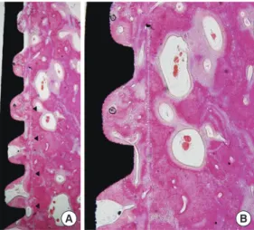
Newly formed bone was observed along the threads of the implants. In the RI group, reversal lines indicating implant ostectomy were observed away from the threads (Fig. 1). Most of the gap areas between the threads were filled with new bone. There was no specific histologic difference between
Eight-week group
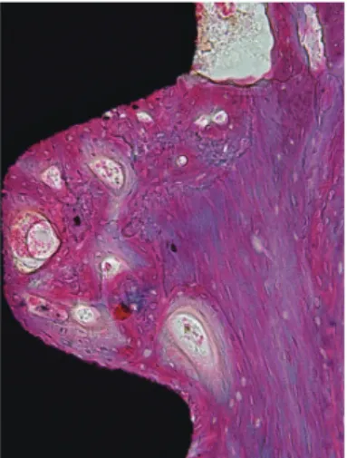
In the 8-week group, woven bone along the threads of the implants was replaced by mature lamellar bone and well-or- ganized osteons were observed. Many primary and second- ary osteons were seen within the thread area when compared with the 4-week group. The bone marrow contained adipo- cytes, vessels, collagen fibers, and some mononuclear leuko- cytes. A thin rim of newly formed bone apparently covered most of the rough surface in the bone marrow compartment (Figs. 3 and 4).
Histometric analysis
The histometric analysis results of all 20 implants are pre- sented in Figs. 5 and 6. The mean BIC and BD showed a slight
Figure 1. Rotational implants group, 4-week healing period (H&E:
A, ×40; B, ×100). Arrowheads: reversal line representing implant ostectomy.
A B
Figure 2. Control group, 4-week healing period (H&E: A, ×40; B,
×100).
A B
Figure 3. Rotational implants group, 8-week healing period (H&E,
×200). Primary and secondary osteons within the thread area. Note that the reversal line still remains.
increase in the RI group, and a tendency toward a decrease in the control group over time. However, there were no statisti- cally significant differences in BIC and BD between the two groups.
DISCUSSION
A dental implant is an optimal treatment option for various clinical conditions. Following an extraction of a tooth, the al- veolar bone undergoes complicated stages of healing [18].
This creates a challenging situation for clinicians to obtain primary stability of the implant. Several options have been utilized to address this issue, including alveolar ridge split- ting, modifying the drilling method, and use of wide im- plants. As osteoconductivity of the implants is essential for the success of these procedures, a variety of surface modifi- cations to the implant have been developed and adjusted. In a systematic review of newly developed and marketed oral implants, Junker et al. [7] reported that modifying the proce- dures for deposition on the surface of the implants can affect the chemical composition of oral implants. They concluded that sufficient proof of a safe and predictable implant-to- bone response is related to surface roughness. A review of experimental surface alterations revealed that CaP-coated implants can improve osseointegration compared to non- coated titanium implants. Despite promising clinical results, a relatively long treatment time frame is required for dental implants when compared to conventional dental prostheses including fixed crowns and removable dentures. To shorten the time spent restoring missing dentition, the concept of an immediate implant was introduced [19], but the primary sta- bility in the immediate implant is much harder to achieve because of discrepancies arising between the dimensions of
the natural teeth and implants. To solve this clinical problem, several studies have suggested methods that evaluate the de- gree of osseointegration using different methods of analysis.
Stadlinger et al. [20] reported that majority of in vivo studies used histological and histomorphometric methods although the location of the implant and animals used differed among the studies. Results of the histometric analysis tend to in- crease with time while results of the radiographic assessment decreased. In the present study, an oversized-drilled model was used to evaluate the osteoconductivity of implants coat- ed with CaP using IBAD. Despite the different healing peri- ods and different methods of implant site preparation, all implants were successfully osseointegrated. The results of this study are in agreement with a study performed by Chae et al. [21]. They obtained a rapid formation of new bone that was in contact with the implant by coating the implants with nano-sized CaP. Previous studies have revealed that implants installed without mobility exhibited a higher BIC value in the early healing phase, and maintained it. On the other hand, the mean BIC value in the rotationally mobile implants in- creased gradually according to time [22]. Song et al. [23] com- pared two different modified surfaces of implants using gap defect models and Um et al. [24] compared different post- coating heat treatment methods [24]. CaP layers coated using the IBAD technique provided high bond strength between implants and natural teeth. In addition, cell adhesion to the implant surface improved in a histologic observation [23]. A postcoating heat treatment can also affect the cell attach- ment to implant surfaces by increasing the crystallinity of the coated surface [24]. These advantageous characteristics lead Figure 5. Mean bone-to-implant contact (%) in the six most coronal threads. RI: rotational implants.
4-Week 8-Week
0 20 40 60 80 100
78.09 69.52 64.43
77.38
Figure 6. Mean bone density (%) in the six most coronal threads.
RI: rotational implants.
4-Week 8-Week
0 10 20 30 40 50 60 70
64.62 57.98 42.63
58.54
Control RI
Figure 4. Control group, 8-week healing period (H&E, ×200). Cell- rich bone marrow zone was seen.
might play an important role in achieving consistency of os- seointegration. Further clinical studies in humans evaluating osseointegration with a standardized protocol could provide a theoretical background that could be applied to clinical practice.
In conclusion, within the limitation of this study, successful osseointegration of CaP-coated implants could be achieved under unfavorable conditions without primary stability. CaP coating using IBAD could be an effective method for the sur- face treatment of dental implants.
CONFLICT OF INTEREST
No potential conflict of interest relevant to this article was reported.
ACKNOWLEDGEMENTS
This work was supported by a grant (code #: 2011K000191) from the Center for Nanostructured Materials Technology under the 21st Century Frontier R&D Program of the Minis- try of Education, Science and Technology, Korea.
REFERENCES
1. Albrektsson T, Jansson T, Lekholm U. Osseointegrated dental implants. Dent Clin North Am 1986;30:151-74.
2. Branemark PI. Osseointegration and its experimental background. J Prosthet Dent 1983;50:399-410.
3. Cochran DL. A comparison of endosseous dental implant surfaces. J Periodontol 1999;70:1523-39.
4. Botticelli D, Berglundh T, Buser D, Lindhe J. The jumping distance revisited: an experimental study in the dog. Clin Oral Implants Res 2003;14:35-42.
5. Botticelli D, Berglundh T, Lindhe J. Resolution of bone de- fects of varying dimension and configuration in the mar- ginal portion of the peri-implant bone. An experimental study in the dog. J Clin Periodontol 2004;31:309-17.
6. Botticelli D, Berglundh T, Persson LG, Lindhe J. Bone re- generation at implants with turned or rough surfaces in self-contained defects: an experimental study in the dog. J Clin Periodontol 2005;32:448-55.
7. Junker R, Dimakis A, Thoneick M, Jansen JA. Effects of implant surface coatings and composition on bone inte- gration: a systematic review. Clin Oral Implants Res 2009;
20 Suppl 4:185-206.
8. Shalabi MM, Gortemaker A, Van’t Hof MA, Jansen JA, Creugers NH. Implant surface roughness and bone heal- ing: a systematic review. J Dent Res 2006;85:496-500.
SH, et al. Surface characteristics of a novel hydroxyapa- tite-coated dental implant. J Periodontal Implant Sci 2012;
42:59-63.
10. Lacefield WR. Hydroxyapatite coatings. Ann N Y Acad Sci 1988;523:72-80.
11. Hayashi K, Inadome T, Mashima T, Sugioka Y. Compari- son of bone-implant interface shear strength of solid hy- droxyapatite and hydroxyapatite-coated titanium implants.
J Biomed Mater Res 1993;27:557-63.
12. van Dijk K, Schaeken HG, Wolke JC, Maree CH, Habraken FH, Verhoeven J, et al. Influence of discharge power level on the properties of hydroxyapatite films deposited on Ti6A14V with RF magnetron sputtering. J Biomed Mater Res 1995;29:269-76.
13. Lee IS, Zhao B, Lee GH, Choi SH, Chung SM. Industrial application of ion beam assisted deposition on medical implants. Surf Coat Technol 2007;201:5132-7.
14. Wikesjo UM, Susin C, Qahash M, Polimeni G, Leknes KN, Shanaman RH, et al. The critical-size supraalveolar peri- implant defect model: characteristics and use. J Clin Peri- odontol 2006;33:846-54.
15. Jung UW, Kim CS, Choi SH, Cho KS, Inoue T, Kim CK.
Healing of surgically created circumferential gap around non-submerged-type implants in dogs: a histomorpho- metric study. Clin Oral Implants Res 2007;18:171-8.
16. Davies JE. Understanding peri-implant endosseous heal- ing. J Dent Educ 2003;67:932-49.
17. Sivolella S, Bressan E, Salata LA, Urrutia ZA, Lang NP, Bot- ticelli D. Osteogenesis at implants without primary bone contact: an experimental study in dogs. Clin Oral Implants Res 2012;23:542-9.
18. Araujo MG, Lindhe J. Dimensional ridge alterations fol- lowing tooth extraction. An experimental study in the dog.
J Clin Periodontol 2005;32:212-8.
19. Lazzara RJ. Immediate implant placement into extraction sites: surgical and restorative advantages. Int J Periodon- tics Restorative Dent 1989;9:332-43.
20. Stadlinger B, Pourmand P, Locher MC, Schulz MC. Sys- tematic review of animal models for the study of implant integration, assessing the influence of material, surface and design. J Clin Periodontol 2012;39 Suppl 12:28-36.
21. Chae GJ, Jung UW, Jung SM, Lee IS, Cho KS, Kim CK, et al.
Healing of surgically created circumferential gap around nano-coating surface dental implants in dogs. Surf Inter- face Anal 2008;40:184-7.
22. Jung UW, Kim S, Kim YH, Cha JK, Lee IS, Choi SH. Osseo- integration of dental implants installed without mechani- cal engagement: a histometric analysis in dogs. Clin Oral Implants Res 2012;23:1297-301.
et al. The effect of a multi-treated implant surface on gap defect healing in dogs. Thin Solid Films 2009;517:5352-6.
24. Um YJ, Song JE, Chae GJ, Jung UW, Chung SM, Lee IS, et
coated implants on the healing of circumferential coronal defects in dogs. Thin Solid Films 2009;517:5375-9.