CASE REPORT
알코올 남용 및 만성 간질환의 과거력이 없는 환자의 다발성 과혈관 과형성 간결절 1예
도병주, 박인영, 이소연, 송진경, 장명국, 조성진1, 남은숙1, 윤은주2
한림대학교 의과대학 강동성심병원 내과학교실, 병리학교실1, 영상의학교실2
A Case of Multiple Hypervascular Hyperplastic Liver Nodules in a Patient with No History of Alcohol Abuse or Chronic Liver Diseases
Byoung Joo Do, In Young Park, So Yon Rhee, Jin Kyung Song, Myoung Kuk Jang, Seong Jin Cho1, Eun Sook Nam1, and Eun Joo Yun2 Departments of Internal Medicine, Pathology1, and Radiology2, Kangdong Sacred Heart Hospital, Hallym University College of Medicine, Seoul, Korea
Up-to-date imaging modalities such as three-dimensional dynamic contrast-enhanced CT (3D CT) and MRI may contribute to detection of hypervascular nodules in the liver. Nevertheless, distinguishing a malignancy such as hepatocellular carcinoma from benign hypervascular hyperplastic nodules (HHN) based on the radiological findings is sometimes difficult. Multiple incidental liver masses were detected via abdominal ultrasonography (US) in a 65-year-old male patient. He had no history of alcohol intake and no remarkable past medical history or relevant family history, and his physical examination results and laboratory findings were normal. 3D CT and MRI showed numerous enhanced nodules with hypervascularity during the arterial phase.
After US guided liver biopsy, the pathological diagnosis was HHN. To date, several cases of HHN have been reported in patients with chronic alcoholic liver disease or cirrhosis. Herein, we report on a case of HHN in a patient with no history of alcoholic liver disease or cirrhosis. (Korean J Gastroenterol 2015;65:321-325)
Key Words: Liver; Nodule; Hyperplasia; Hypervascular
Received August 3, 2014. Revised October 16, 2014. Accepted October 17, 2014.
CC This is an open access article distributed under the terms of the Creative Commons Attribution Non-Commercial License (http://creativecommons.org/licenses/
by-nc/4.0) which permits unrestricted non-commercial use, distribution, and reproduction in any medium, provided the original work is properly cited.
Copyright © 2015. Korean Society of Gastroenterology.
교신저자: 윤은주, 134-701, 서울시 강동구 성안로 150, 한림대학교강동성심병원 영상의학과
Correspondence to: Eun Joo Yun, Department of Radiology, Hallym University Kangdong Sacred Heart Hospital, 150 Seongan-ro, Gangdong-gu, Seoul 134-701, Korea. Tel: +82-2-2224-2445, Fax: +82-2-2224-2647, E-mail: [email protected]
Financial support: None. Conflict of interest: None.
INTRODUCTION
With the recent progress of imaging modalities, detection of liver nodules is common. In particular, three-dimensional dynamic contrast-enhanced CT (3D CT) and MRI contribute mainly to making a differential diagnosis of nodules.
Diagnostic imaging with 3D CT and MRI, however, shows hy- pervascularity in both hepatic malignancy, such as hep- atocellular carcinoma (HCC), and benign hypervascular hep- atic nodules, such as focal nodular hyperplasia (FNH), hep-
atocellular adenoma (HCA) and, rarely, hypervascular hyper- plastic nodule (HHN).1 Therefore, distinguishing these two conditions based on the radiologic findings is sometimes difficult. In clinical situations, however, differential diagnosis to exclude malignancy is important in order to avoid needless therapeutic interventions. Therefore, in a difficult case of dif- ferentiating benign from malignant nodules based on the ra- diologic findings, there is an occasional need to perform biop- sy for histological confirmation.
To date, only a few cases of HHN have been reported, and
Fig. 1. Abdominal ultrasonography. Multiple-numerous hypoechoic nodules are seen in both lobes of the liver (arrows).
Fig. 2. Abdominal three-dimensional dynamic contrast-enhanced CT images. (A) Pre-enhancement. Multiple nodules are highly enhanced in the arterial phase (B), with decreased enhancement in the portal phase (C), followed by iso-attenuation in the delayed phase (D).
disease.
CASE REPORT
A 65-year-old man was referred from the local clinic due to multiple variable-sized liver masses detected incidentally on abdominal ultrasonography (US) for the patient’s regular health screening examination. He had no symptoms or signs on the referred area of the abdomen. He had no history of al- cohol intake, smoking, or drug intake, and no remarkable past medical history or relevant family history. His physical examination results were also normal. His height was 155.3
Fig. 3. Abdominal MRI. The hepatic nodules show iso-signal intensity on T1-weighted image (A) and T2-weighted image (B). The gadoxetate acid enhanced MR images obtained during the arterial (C) and portal phases (D) show the same findings shown on CT.
cm, body weight was 55 kg, and body mass index was 22.8 kg/m2. The laboratory findings upon admission were as fol- lows: hemoglobin 13.3 g/dL, hematocrit 39%, white blood cell count 4,300/L, platelet count 187,000/L, AST 30 IU/L, ALT 33 IU/L, total protein 7.4 g/dL, albumin 4.5 g/dL, total bilirubin 0.6 mg/dL, ALP 79 IU/L, GGT 51 IU/L, PT 95.3%, CRP 0.4 mg/dL, AFP 2.41 ng/mL, and protein in- duced by vitamin K absence II 19 mAU/mL. The patient’s HBsAg, anti-HBs, anti-HBc IgG, and anti-HCV were all negative.
Esophagogastroduodenoscopy showed no evidence of por- tal hypertension or varices.
The abdominal US showed variable-sized multiple-nume- rous nodular lesions, ranging from 5 to 15 mm in diameter, throughout the liver, appearing as hypoechoic nodules (Fig. 1).
The subsequent 3D CT showed multiple enhanced nodular lesions in the arterial and portal phase and isodense nodules in the delayed phase, which were well-matched with those found on the US (Fig. 2). The MRI showed an iso-intense sig- nal on T1- and T2-weighted images. Following gadoxetate
acid (Gd-EOB-DTPA, Primovist; Bayer Schering Pharma, Berlin, Germany) enhancement, numerous nodules were highly en- hanced in the arterial phase (Fig. 3).
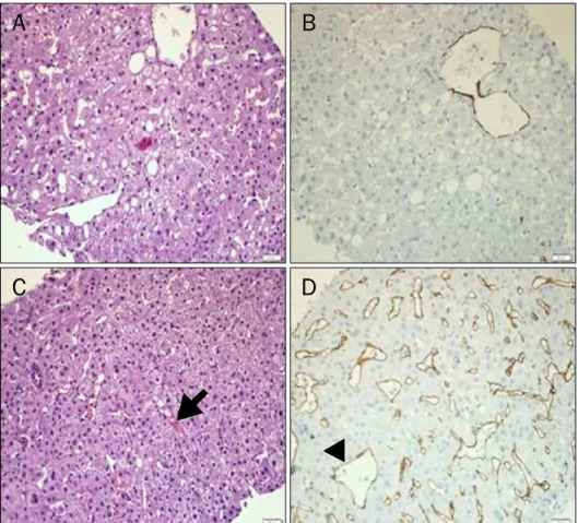
US-guided liver gun biopsy was performed six times on separate lesions to confirm the nature of the hypervascular nodules histologically. The liver biopsy showed hepatocelluar nodules with mild fatty changes and slightly increased cel- lular densities without cellular or structural atypia and mitotic figure (Fig. 4). It also showed hypervascularity of the unpaired arteries and sinusoidal dilatation with congestion. In im- munohistochemistry, the endothelial cells of the sinusoids were positive to CD34, indicating the capillarization of the si- nusoids (Fig. 4). The background liver was normal except for the findings mentioned above (Fig. 4).
In this patient with no history of chronic alcoholic liver dis- ease or cirrhosis, the diagnosis of multiple HHN was made based on the aforementioned radiologic and pathologic findings.
Fig. 4. Histopathology. (A) The non- nodular portion shows a normal parenchymal structure except for the mild fatty change (H&E stain, ×100).
(B) The non-nodular portion shows little immunoreactivity for CD34 (CD34 stain, ×100). (C) The nodules have slightly increased cellularity without cellular atypia and mitotic figures, showing sinusoidal dilatation (arrow) with mild congestion (H&E stain, ×100). (D) The nodules show abundant sinusoidal capillarization and sinusoidal expression of CD34 (arrowhead) (CD34 stain, ×100).
DISCUSSION
Hypervascular nodules in the liver include hemangioma, FNH, HCA, fibrolamellar carcinoma, HCC and metastases from primary tumors such as islet cell tumor, carcinoid, renal cell carcinoma, melanoma, and thyroid carcinoma. Angiosar- coma, infantile hemangioendothelioma, peliosis hepatis, and intrahepatic splenosis rarely present as hypervascular hepatic masses.7 Besides these, the HHN in the current case is also considered a rare entity showing hypervascularity.
The most common benign hypervascular nodule is hemangioma. Histology shows a series of vascular lakes and channels, with larger lesions developing areas of thrombosis and fibrosis.7 The second one is FNH. A well circumscribed le- sion consisting of a stellate scar or a fibrous body surrounded by multiple benign nodules appearing as hepatocytes is the characteristic feature of FNH.1 The third one is HCA, which consists of a sheet of hepatocytes and is formed as a pseudo- capsule related to compression of the adjacent hepatic parenchyma. In contrast to FNH, however, HCA does not form bile ducts.7 On the other hand, HHN shows increased cellular
density compared to that of the normal liver but less than that of HCC. In addition, it has an unpaired artery and shows in- creased sinusoidal capillarization.
International Working Party Classification8 and/or World Health Organization classification9 has been widely used of late for classification of hepatocytic nodular lesions. However, HHN is not described in either classification. One of the clin- ical concerns regarding the absence of a uniform classi- fication scheme is the inability of determining the probability of premalignancy or malignancy.
In the aspect of prognosis, HHN appears to be benign, thus requiring no specific therapeutic intervention. Eight HHNs in the liver were recently reported in seven patients with chronic alcoholic liver disease or cirrhosis.4 In six of the eight cases with follow-up CT, five nodules showed a decrease in size within 36 months, and one disappeared within eight months.
Park et al.3 reported a case of HHN in alcoholic liver cirrhosis, in which the nodules showed no change in size and number after four months from the initial diagnosis. In the case re- ported herein, 3D CT was performed after six months, and the follow-up 3D CT showed that the nodules had the same
sizes and numbers. However, due to the limited number of re- ported cases, the prognosis of HHN remains undetermined.
The pathogenesis of HHN in the liver has not yet been clearly established. A recent hypothesis considers abnormal hep- atic circulation, including changes in the portal venous or ar- terial system as the possible etiology.10
As described above, to date, reported patients with HHN had chronic liver diseases or at least a history of heavy drinking.11 Another few cases of HHN have been reported in patients with Budd-Chiari syndrome.12 In the current case, however, the patient had neither symptoms and signs nor risk factors of chronic liver disease, such as those that modify hepatic blood circulation. To the best of these authors’ knowl- edge, this is the first reported case in South Korea of multiple HHN with a normal background liver in a patient with no his- tory of alcohol abuse or chronic liver disease.
It is still not clear what kinds of surveillance protocols are needed for HHN patients. Accumulation of data will be neces- sary in order to clarify the etiologies, risk factors, prognosis, and strategy for management that is evidence-based. It is hoped that the current report will serve as an educational case for physicians listing HHN as a differential diagnosis of hypervascular liver nodule and as the first step in accumu- lation of HHN in a patient with no history of alcohol abuse or chronic liver disease so as to elucidate the etiologies, risk fac- tors, prognosis, and evaluation protocols.
REFERENCES
1. Tani J, Miyoshi H, Sasaki M, et al. Multiple hypervascular FNH-like lesions in a patient with no history of alcohol abuse or chronic liver disease. Intern Med 2013;52:2225-2230.
2. Nakashima O, Kurogi M, Yamaguchi R, et al. Unique hyper- vascular nodules in alcoholic liver cirrhosis: identical to focal nodular hyperplasia-like nodules? J Hepatol 2004;41:992-998.
3. Park JE, Kim BS, Lee CH, Choi JH, Park YC, Park KK. A case of hy- pervascular hyperplastic nodules mimicking hepatocellular car- cinoma in alcoholic liver cirrhosis. Korean J Hepatol 2009;15:
193-200.
4. Park WK, Chang JC, Kim JW, et al. Hypervascular hyperplastic nodules appearing in chronic alcoholic liver disease: benign in- trahepatic nodules mimicking hepatocellular carcinoma. J Korean Radiol Soc 2006;54:113-119.
5. Moon JH, Ahn CM, Chung HS, Ahn SH, Park YN. A case of hyper- vascular hyperplastic nodules in a patient with alcoholic liver cirrhosis. Yonsei Med J 2006;47:881-886.
6. Lee WJ. Focal nodular hyperplasia-like nodule. Korean J Hepatol 2008;14:537-540.
7. Namasivayam S, Salman K, Mittal PK, Martin D, Small WC.
Hypervascular hepatic focal lesions: spectrum of imaging features. Curr Probl Diagn Radiol 2007;36:107-123.
8. International Working Party. Terminology of nodular hep- atocellular lesions. Hepatology 1995;22:983-993.
9. Bosman FT, World Health Organization, International Agency for Research on Cancer. WHO classification of tumours of the diges- tive system. 4th ed. Lyon: IARC, 2010:196-261.
10. Kondo F. Benign nodular hepatocellular lesions caused by ab- normal hepatic circulation: etiological analysis and introduction of a new concept. J Gastroenterol Hepatol 2001;16:1319-1328.
11. Kim SR, Maekawa Y, Ninomiya T, et al. Multiple hypervascular liv- er nodules in a heavy drinker of alcohol. J Gastroenterol Hepatol 2005;20:795-799.
12. Maetani Y, Itoh K, Egawa H, et al. Benign hepatic nodules in Budd-Chiari syndrome: radiologic-pathologic correlation with emphasis on the central scar. Am J Roentgenol 2002;178:
869-875.