주의력결핍 과잉행동장애 아동에서 Osmotic-Controlled Release Oral Delivery System Methylphenidate 투여가
국소 대뇌관류에 미치는 영향
양영희1)・황준원2)・김붕년3)・강혜진4)・이재성4)・이동수4)・조수철5)
국립서울병원 소아정신과,1) 강원대학교병원 정신건강의학과,2) 서울대학교병원 정신건강의학과,3)
서울대학교병원 핵의학과,4) 국군수도병원 정신건강센터5)
A Study about Effects of Osmotic-Controlled Release Oral Delivery System Methylphenidate on Regional Cerebral Blood Flow in Korean Children
with Attention-Deficit Hyperactivity Disorder
Young-Hui Yang, M.D.1), Jun-Won Hwang, M.D., Ph.D.2), Boong-Nyun Kim, M.D., Ph.D.3), Hyejin Kang, Ph.D.4), Jae-Sung Lee, M.D., Ph.D.4),
Dong-Soo Lee, M.D., Ph.D.4), and Soo-Churl Cho, M.D., Ph.D.5)
1)Department of Child Psychiatry, Seoul National Hospital, Seoul, Korea
2)Department of Psychiatry, Kangwon National University Hospital, Chuncheon, Korea
3)Department of Neuropsychiatry, Seoul National University Hospital, Seoul, Korea
4)Department of Nuclear Medicine, Seoul National University Hospital, Seoul, Korea
5)Mental Health Center, Korean Armed Forces Capital Hospital, Seongnam, Korea
Objectives: The objective of this study was to examine the effects of osmotic-controlled release oral delivery system methyl- phenidate on changes in regional cerebral blood flow (rCBF) in children with attention-deficit hyperactivity disorder (ADHD) using single photon emission computed tomography (SPECT).
Methods: A total of 26 children with ADHD (21 boys, mean age: 9.2±2.05 years old) were recruited. Each ADHD participant was examined for changes in rCBF using technetium-99m-hexamethylpropylene amine oxime brain SPECT before and after 8 weeks methylphenidate medication. Brain SPECT images of pediatric normal controls were selected retrospectively. SPECT images of ADHD children taken before medication were compared with those of pediatric normal controls and those taken after medica- tion using statistical parametric mapping analysis on a voxel-wise basis.
Results: Before methylphenidate medication, significantly decreased rCBF in the cerebellum and increased rCBF in the right precuneus, left anterior cingulate, right postcentral gyrus, right inferior parietal lobule and right precentral gyrus were observed in ADHD children compared to pediatric normal controls (p-value<.0005, uncorrected). After medication, we observed significant hypoperfusion in the left thalamus and left cerebellum compared to pediatric normal controls (p-value<.0005, uncorrected). In the comparison between before medication and after medication, there was significant hyperperfusion in the superior frontal gyrus and middle frontal gyrus and significant hypoperfusion in the right insula, right caudate, right middle frontal gyrus, left subcallosal gyrus, left claustrum, and left superior temporal gyrus after methylphenidate medication (p-value<.0005, uncorrected).
Conclusion: This study supports dysfunctions of fronto-striatal structures and cerebellum in ADHD. We suggest that methyl- phenidate may have some effects on the frontal lobe, parietal lobe, and cerebellum in children with ADHD.
KEY WORDS: ADHDㆍMethylphenidateㆍSPECT.
Date received: October 6, 2015 / Date of revision: February 12, 2016 / Date accepted: February 16, 2016
Address for correspondence: Soo-Churl Cho, M.D., Ph.D., Mental Health Center, Korean Armed Forces Capital Hospital, 81 Saemaeul-ro 177beon-gil, Bundang-gu, Seongnam 13574, Korea
Tel: +82.31-725-6081, Fax: +82.31-706-0987, E-mail: [email protected]
본 연구는 한국얀센의 연구비 지원으로 이루어졌음(This study was supported by Janssen Korea).
서 론
아동청소년기에 진단되는 주요 정신장애 중의 하나인 주의 력결핍 과잉행동장애(attention-deficit hyperactivity disor- der, ADHD)는 주의 집중의 어려움, 충동성, 과잉행동 및 실 행기능의 저하를 특징으로 하는 신경발달학적 장애로 학습 부진과 대인관계의 어려움을 초래한다.1) 메칠페니데이트(me- thylphenidate)는 ADHD의 약물치료 중 하나로 ADHD아동 의 약 70%에서 효과를 나타내며, 나머지 30%에서는 치료효 과가 불분명하다고 알려져 있다.2) 현재까지 알려진 메칠페니 데이트의 작용 기전은 전두엽과 기저핵에 있는 도파민 또는 노르에피네프린 운반체의 작용을 억제하여 시냅스 내의 이들 신경전달물질의 농도를 증가시키는 것이다.3) ADHD의 진단 과 약물치료 효과 평가는 주로 병력과 임상 증상 변화 관찰 을 통해 이루어지고 있으며, 뇌영상이나 혈액분석 등과 같은 실험실검사는 일반적으로 이용되고 있지 않다.
최근까지 보고된 ADHD의 뇌영상 연구에서는 전전두엽 (prefrontal cortex), 배측 전대상회(dorsal anterior cingulate cortex), 미상핵(caudate), 조가비핵(putamen), 두정엽, 그리고 소뇌 등에서 구조적 또는 기능적 이상이 보고되었다.4,5) 그러 나 아직 연구 대상이나 연구 방법 등에 따라서 이상을 보이는 구체적인 뇌영역들은 연구에 따라서 일부 차이가 있다. ADHD 에서 약물치료가 뇌기능에 미치는 영향에 대한 초기 연구 중 의 하나는 단일광전자방출단층촬영(single photon emission computed tomography, SPECT)을 이용한 연구로 전두엽, 두정엽, 그리고 기저핵 등에서 나타난 혈류감소가 메칠페니데 이트 투약 하루 만에 교정됨을 보고하였다.6) 이후 연구에서7) 약물치료 이후 전두엽과 선조체(striatum), 시상(thalamus)에 서 혈류의 증가가 보고되었고, 또 다른 연구에서는8) 오른쪽 선조체와 배측 고위 시각 영역(ventral higher visual area)의 혈류 감소와 상부 전전두엽의 혈류 증가가 보고되었다. 또한 약물치료 후의 증상변화와 혈류 변화와의 관련성을 비교한 연구들로, Anderson 등9)은 약물치료 후에 ADHD 아동의 과 잉활동 정도에 따라 소뇌 충부(cerebellar vermis)에서 혈류 변화의 차이가 있다고 보고하였고, 또 다른 연구에서는10) 약물 치료 반응이 없는 ADHD 아동은 치료 반응이 있는 아동에 비해 전대상회, 왼쪽 전장(claustrum), 오른쪽 조가비핵의 혈 류 증가와 오른쪽 상부 두정엽의 혈류 감소를 보였다. 성인 ADHD를 대상으로 약물치료 전후를 비교한 연구에서는11) 약물치료 이후 소뇌 충부의 혈류 증가와 중심앞이랑(pre- central gyri), 왼쪽 미상핵, 오른쪽 전장에서의 혈류 감소가 있었고, 또 다른 연구에서는12) 과제 수행의 호전과 함께 전전
두엽의 혈류감소와 오른쪽 시상과 중심앞이랑의 혈류 증가가 보고되었다. 최근에 시행된 연구에서는 기능적 뇌자기공명영 상(functional magnetic resonance imaging, fMRI)을 이용 하여 ADHD에서 약물치료 이후에 운동피질(motor cortex), 오른쪽 하부 전두엽, 왼쪽 전대상회와 보조운동영역(supple- mentary motor area), 양측 후대상회에서 활성의 저하와13) 양측 하부 전두엽과 섬이랑(insula)에서 활성증가가14) 보고되 었다.
ADHD의 뇌의 구조적 및 기능적 이상과 약물치료 이후의 뇌기능의 변화는 지속적으로 보고되었으나 이상을 보이는 뇌영역은 연구마다 부분적으로 서로 다른 결과를 보이고 있 다. 또한 앞서 언급한 연구들 중 정상대조군을 포함한 연구 는 Lee 등8)과 Schweitzer 등12)을 비롯하여 소수에 불과하다.
이로 인해 약물치료로 인한 뇌기능의 변화가 정상대조군과 어떠한 차이를 보이는지 알아보는 데 한계가 있었다.
본 연구에서는 SPECT를 이용하여 국소 대뇌관류(regional cerebral blood flow)의 변화를 측정하여 ADHD 아동청소 년에서 약물치료 전후의 뇌기능의 변화를 확인하고자 하였다.
또한 정상대조군의 SPECT 뇌영상과 비교분석하여 ADHD에 서 약물치료로 변화를 보이는 뇌영역을 알아보고자 하였다.
방 법
1. 연구 참여 대상자
환자군은 국내의 일 대학병원의 소아정신과 외래를 방문 한 아동 중 임상적 면담과 평가를 통해 ADHD로 진단되고 연구 참여기준과 배제기준을 모두 만족한 경우의 아동으로 구성되었다. 참여기준은 정신과 전문의가 한국판 Kiddie- Schedule for Affective Disorders and Schizophrenia- Present and Lifetime Version으로15) 평가하여 ADHD로 진 단된 만 5세 이상 아동이며, 배제기준은 1) 정신지체, 2) 두부 외상, 경련성 장애, 주요 신경질환의 과거력, 3) 전반적 발달장 애, 양극성 장애, 정신병적 장애, 심한 학습장애, 뚜렛장애, 강 박장애 등을 비롯한 주요 정신장애, 4) 임신의 위험성 등이 있 는 경우이다. 대조군은 환자군과 동일한 병원에 방문한 만 5 세 이상의 아동 중 발달력을 비롯한 임상적 면담 및 평가에서 주요한 정신과적 장애를 만족시키지 않고, 정신지체가 아니면 서 진료 시 평가 과정에서 SPECT 촬영을 시행하였던 아동을 후향적으로 모집하였다. 연구에 참여한 아동과 보호자에게는 연구의 목적과 과정에 대해서 설명한 이후에 연구참여의 동 의를 받았고, 보호자와 13세 이상의 아동은 서면 동의 과정 을 거쳤다. 본 연구는 연구가 수행된 병원의 임상시험심의위 원회(Institutional Review Board)의 승인을 거쳐 시행하였다.
2. 대상군 평가 및 약물치료
환자군은 약물치료 전 초기 평가와 technetium-99m- hexamethylpropylene amine oxime(99mTC-HMPAO) brain SPECT를 시행하였고, 이후에 methylphenidate-Osmotic- Controlled Release Oral Delivery System(OROS)을 8주간 복용하였다. 처음 하루 복용 용량은 18mg으로 이후 부작용 과 효과를 관찰하면서 증량하였다. 최고 용량은 체중을 기준 으로 하여 1.0mg/kg/day를 넘지 않도록 하였다. 약물의 효과 와 부작용과 관련된 평가는 약물치료 시작 전 1회, 8주간 중 간에 1회와 8주 종결 시에 1회로 총 3번의 평가를 시행하였다.
8주간 약물치료 이후에 임상평가와 SPECT 촬영을 시행하였 다. 약물치료 이후 2차 SPECT 뇌영상을 촬영하는 날에는 영 상을 촬영하기 최소 90분 전에 약물을 복용하도록 하였다.
3. SPECT 뇌영상의 획득
환자군에서 약물치료 전과 8주간 약물치료 후 2회의 SPECT 뇌영상을 촬영하였다. 뇌영상 촬영 시에는 등을 바닥에 대고 바로 누운 자세에서 눈을 감은 채 어두운 조명의 조용한 방에 누워서 검사를 시행하였다. 555MBq 99mTC-HMPAO가 주사된 이후 촬영되었고, SPECT 영상은 저에너지, 고해상도 (low-energy, high-resolution) 평행다중구멍 조준기(parallel hole collimator)를 부착한 3중 헤드 감마 카메라(Prism 3000;
Picker International, Cleveland, OH, USA)를 사용하여 획 득하였다. 투사 영상은 140keV 광절정 중심에 15%의 대칭 에너지 창을 이용하였다. 120개의 정적 투사 영상을 step and shoot mode로 획득하였고, 각각 투사 영상획득 시간은 20초 였다. 각 투사 영상은 128×128 화소크기로, 횡단면 영상(tran- saxial image)은 Metz filter(x=1.5-2.0)를 통해 64×64matrix 로 재구성하였고, 모든 영상은 Chang’s method를 사용하여 감쇠(attenuation)를 보정하였다.16) 대뇌피질의 상부에서 소 뇌의 기저면까지 걸친 40-50개의 영상으로 재구성하였다.
4. 뇌영상의 분석과 통계분석
뇌영상분석은 매트랩(Matlab)(the MathWorks Inc., Natick, MA, USA)에서 구동된 SPM8(Statistical Parametric Map- ping, version 8; Wellcome Department of Imaging Neu- roscience, London, UK)을 이용하여 시행하였다. 모든 재구 성된 영상은 분석 형식(analyze format)으로 전환되었고, SPM8 프로그램 내의 SPECT 표준 주형(standard template) 에 정규화(normalization)하였다. 이후에 신호 대 잡음 비율 (signal to noise ratio)을 높이기 위하여 가우시안 커널(Gau- sian kernel) full-width at half maximum을 16mm로 한 smoothing 과정을 거쳐 얻어진 뇌영상을 복셀(voxel) 단위 로 비교하였다. 환자군에서 약물치료 전후의 영상을 paired t-test를 이용하여 비교하였고, 대조군의 영상을 환자군의 약물치료 전과 약물치료 후 영상과 각각 t-test를 이용하여 비교하였다. 뇌영상의 통계적 분석 시 extent threshold는 SPECT의 해상도를 고려하여 125 복셀로 하였고,17) 통계적 유의 수준을 p-value<.0005(uncorrected, voxel-level)로 하였다. 뇌영상 분석에서 얻어진 Montreal Neurological Ins- titute 좌표를 Talairach 좌표를 바꾸고, 이의 해부학적 위치 는 Talairach and Tournoux atlas를18) 이용하여 정하였다. 인 구학적 및 임상적 변수에 대한 분석은 SPSS 17.0 version (SPSS Inc., Chicago, IL, USA)을 이용하여 비모수적 통계 분석을 시행하였고, 통계적 유의성은 p-value<.05를 기준으 로 검증하였다.
결 과
1. 인구학적 및 임상적 특징
ADHD 환자군 28명과 대조군 13명이 모집되었다. 환자군 28명 중 26명의 SPECT 영상이 분석 가능하였다. 이 환자군 26명 중 남아는 21명(81%)이었고, 평균 나이는 9.2(±2.05)세, 평균 지능은 109(±13.37)였다. 환자군 26명 중 12명은 약물
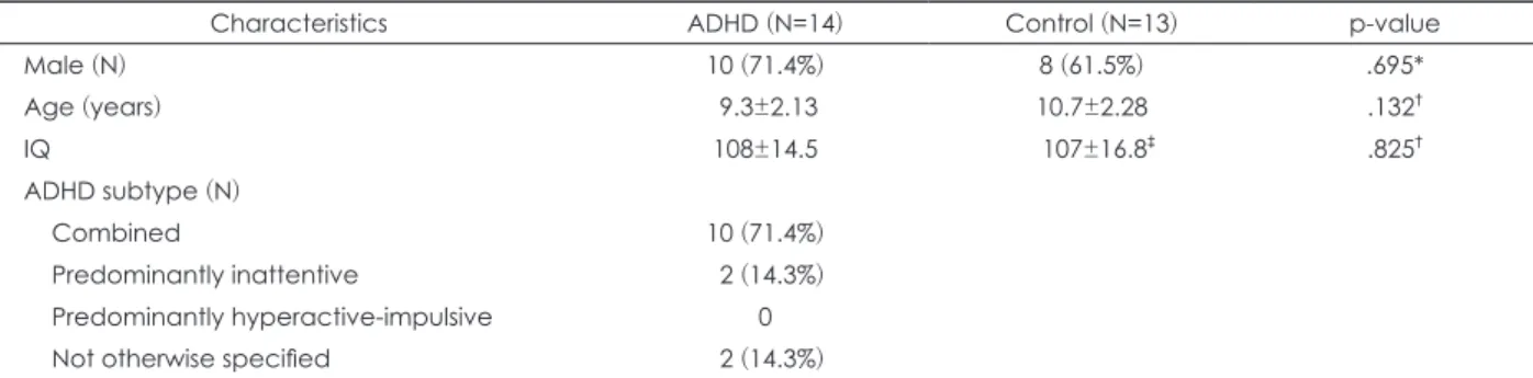
Table 1. Demographic and clinical characteristics of subjects with ADHD and pediatric normal controls
Characteristics ADHD (N=14) Control (N=13) p-value
Male (N) 10 (71.4%) 8 (61.5%) .695*
Age (years) 9.3±2.13 10.7±2.28 .132†
IQ 108±14.5 107±16.8‡ .825†
ADHD subtype (N)
Combined 10 (71.4%)
Predominantly inattentive 2 (14.3%)
Predominantly hyperactive-impulsive 0
Not otherwise specified 2 (14.3%)
*: chi-square test, †: Mann-Whitney U test, ‡: number of control in IQ: N=9. ADHD: attention-deficit hyperactivity disorder, IQ: intelli- gence quotient
치료 전 SPECT 영상만을 촬영하였고, 약물치료 전후 SPECT 영상을 촬영한 환자는 14명이었다. 약물치료 전후 영상을 촬 영한 14명 중 남아의 비율은 10명(71.4%)이었고, 대조군 13명 중에서는 8명(61.5%)이 남아였다. 평균 나이는 환자군 9.3(±
2.13)세, 대조군 10.7(±2.28)세였다. 환자군의 평균 지능은 108(±14.5)이었고, 대조군 9명의 평균 지능은 107(±16.8)이 었다. 두 그룹 간 성별과 나이에서 통계적으로 유의한 차이는 없었다. ADHD 환자군에서 복합형(combined type)은 10명 (71.4%), 주의력결핍 우세형(predominantly inattentive type) 은 2명(14.3%), ‘달리 명시되지 않는(not otherwise specified)’
에 속하는 아동은 2명(14.3%)이었다(Table 1).
2. SPECT 뇌영상 분석결과
약물치료 전 SPECT 영상이 있는 환자군 26명과 대조군 13명의 영상을 비교하였고, 환자군 26명 중 약물치료 후의 영상을 촬영한 환자 14명에 대해서 약물치료 전후를 비교하 고, 각각을 대조군과 비교 분석하였다.
1) 약물치료 전의 환자군(N=26)과 대조군(N=13)과의 비교 ADHD군은 대조군에 비해서 오른쪽 중심앞이랑(x=67, y=-10, z=32, Z-value=4.57), 오른쪽 중심뒤이랑(x=61, y=-13, z=47, Z-value=4.15), 오른쪽 하두정소엽(right, inferior pa- rietal lobule; x=50, y=-36, z=55, Z-value=3.67)의 혈류증가 와 양측 소뇌 후엽(both posterior lobes, cerebellum; x=-30,
y=-48, z=-31, Z-value=5.12; x=-20, y=-46, z=-35, Z-value=
4.87; x=16, y=-87, z=-33, Z-value=4.83)의 혈류 감소를 보 였다(p-value<.0005, uncorrected).
2) 약물치료 전후의 환자군(N=14)과 대조군(N=13)과의 비교 (Table 2, Fig. 1)
약물치료 전 ADHD군은 대조군에 비해서 오른쪽 쐐기앞 소엽(precuneus), 왼쪽 전대상회, 오른쪽 중심뒤이랑(post- central gyrus), 오른쪽 하두정소엽, 오른쪽 중심앞이랑에서 혈류 증가를 보였고, 혈류 감소는 양측 소뇌 후엽과 왼쪽 소 뇌 전엽(anterior lobe)에서 보였다(p-value<.0005, uncor- rected). 약물치료 후 ADHD군은 대조군에 비해서 혈류 증 가를 보이는 부위는 없었으며, 왼쪽 시상(thalamus), 왼쪽 소 뇌 전엽과 소뇌 후엽에서 혈류감소가 나타났다(p-value<
.0005, uncorrected).
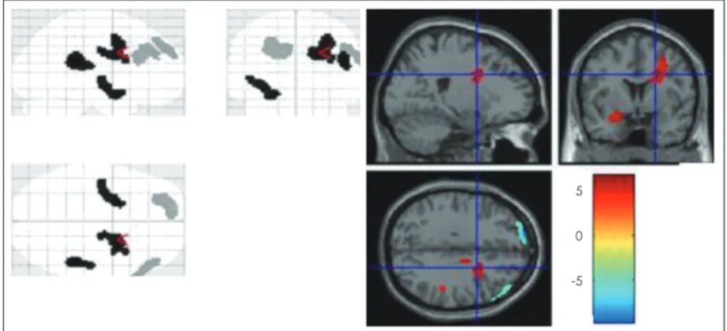
3) 환자군(N=14)의 약물치료 전후 비교(Table 3, Fig. 2) 약물치료 전후의 영상 비교 시 약물치료 전에 비해 약물치 료 이후에 왼쪽 상전두엽과 오른쪽 중간전두엽의 혈류 증가가 나타났고, 혈류 감소가 나타난 부위는 오른쪽 섬이랑, 오른쪽 미상핵, 오른쪽 중간전두엽, 왼쪽 뇌량하엽(subcallosal gyrus, frontal lobe), 왼쪽 전장, 왼쪽 상측두엽이었다(p-value<
.0005, uncorrected).
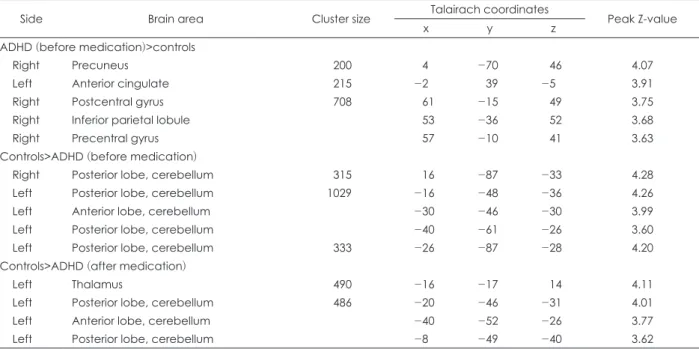
Table 2. Significant changes of rCBF in subjects with ADHD (N=14) before and after methylphenidate medication compared to pe- diatric normal controls (N=13)
Side Brain area Cluster size Talairach coordinates
Peak Z-value
x y z
ADHD (before medication)>controls
Right Precuneus 200 4 -70 46 4.07
Left Anterior cingulate 215 -2 39 -5 3.91
Right Postcentral gyrus 708 61 -15 49 3.75
Right Inferior parietal lobule 53 -36 52 3.68
Right Precentral gyrus 57 -10 41 3.63
Controls>ADHD (before medication)
Right Posterior lobe, cerebellum 315 16 -87 -33 4.28
Left Posterior lobe, cerebellum 1029 -16 -48 -36 4.26
Left Anterior lobe, cerebellum -30 -46 -30 3.99
Left Posterior lobe, cerebellum -40 -61 -26 3.60
Left Posterior lobe, cerebellum 333 -26 -87 -28 4.20
Controls>ADHD (after medication)
Left Thalamus 490 -16 -17 14 4.11
Left Posterior lobe, cerebellum 486 -20 -46 -31 4.01
Left Anterior lobe, cerebellum -40 -52 -26 3.77
Left Posterior lobe, cerebellum -8 -49 -40 3.62
ADHD: attention-deficit hyperactivity disorder, rCBF: regional cerebral blood flow
고 찰
본 연구에서는 ADHD 환자군에서 약물치료 전후의 SPECT 영상을 정상 대조군과 비교 분석하여 ADHD 아동에서의
약물치료로 인한 국소 대뇌혈류의 변화를 알아보았다. 그 결 과 ADHD군과 대조군의 비교 시 전두엽, 두정엽과 소뇌 등 에서 두드러진 변화를 보였고, ADHD 환자의 약물치료 전후 비교 시에는 전두엽, 측두엽과 피질하 구조물(subcortical structure)에 변화가 관찰되었다.
Fig. 1. Significant changes of rCBF in subjects with ADHD (N=14) before and after methylphenidate medication compared to pedi- atric normal controls (N=13). (A) Comparison between ADHD (before medication) and pediatric normal controls. red: hyperperfu- sion, blue: hypoperfusion, color bar: T-value. (B) Comparison between ADHD (after medication) and pediatric normal controls. yel- low: hypoperfusion, color bar: T-value. ADHD: attention-deficit hyperactivity disorder, rCBF: regional cerebral blood flow
A
4 2 0 -2 -4
B
4
2
0
Fig. 2. Significant changes of rCBF in subjects with ADHD after methylphenidate medication. blue: after medication>before medi- cation, red: before medication>after medication, color bar: T-value. ADHD: attention-deficit hyperactivity disorder, rCBF: regional cerebral blood flow
5 0 -5
구체적으로 보면 약물치료 전 ADHD군은 대조군에 비해 오른쪽 일차 운동영역(primary motor area)과 일차 체성감 각영역(primary somatosensory area), 하두정소엽, 오른쪽 쐐기앞소엽, 그리고 오른쪽 전대상회의 혈류 증가와 양측 소 뇌 후엽의 혈류 감소가 두드러졌다. 이러한 양상은 약물치료 전 영상만을 촬영한 ADHD 환자군 12명을 포함하여 분석 을 시행하였을 때에도 일부 영역을 제외하고 유지되었다. 약물 치료 후에는 대조군과 비교하여 혈류가 증가한 부분은 없었 고, 왼쪽 소뇌 후엽의 혈류 감소영역이 약물치료 전에 비해서 면적이 줄어들었으며, 왼쪽 시상의 혈류 감소가 관찰되었다.
약물치료 전의 일차 운동 영역의 혈류 증가는 ADHD의 과잉 행동 증상과 관련이 있는 것으로 보이며, 본 연구에는 이와 같은 혈류의 증가는 약물치료 이후에 정상화되었다. 체성감 각영역의 혈류 또한 약물 전 증가된 양상이었으나 약물치료 이후에 정상화되었다. 이는 이전 연구에서 지속적으로 보고 되고6,8) 있으며, 이 부위는 시상과 연결되어 감각 자극을 처 리한다. 혈류의 증가를 보인 하두정소엽과 쐐기앞소엽은 주 의력(attention)과 밀접한 관련이 있다고 알려져 있다.19) 하두 정소엽은 ADHD의 기능적 뇌영상연구에서 다른 뇌영역의 기능저하에 대한 보상적 역할을 수행하는 부위로 주목을 받 고 있다.20) 쐐기앞소엽은 전두엽과 연결성을 가지고 있으며, 주변의 정보를 모아 처리하고 새로운 자극으로 주의를 전환 하는 데 주요한 역할을 한다.19) 또한 혈류의 증가를 보인 전대 상회는 시상과 전전두엽을 연결하며 자극을 처리하고 적절 한 반응을 선택하여 행동을 조정하는 역할을 한다.21) 본 연구 결과에서 나타난 약물치료 전 ADHD 아동에서 보이는 중심 뒤이랑, 하두정소엽, 쐐기앞소엽, 전대상회의 활성의 증가는 휴지 상태에서 ADHD 아동이 새로운 자극에 반응하고 처리
하는 과정이 대조군 아동에 비해서 활성화되어 있음을 반영 하는 것으로 보이며, 이는 주변 자극에 쉽게 반응하여 주의 가 분산되는 ADHD 아동의 임상특징과 관련이 있을 것으로 보인다. 이러한 활성의 증가는 약물치료 이후 대조군과 차이 가 없이 정상화되는 양상을 보였다.
ADHD 아동에서 소뇌의 구조 또는 기능의 이상은 이전 연구들에서 지속적으로 보고되고 있다.9,11,22-25) 소뇌는 과거 주로 운동 기능의 조절과 관련이 있다고 알려져 있었으나, 최 근에는 전두엽과 연결되어 주의력을 비롯한 고위 인지기능의 조절에 주요한 역할을 하는 것으로 알려졌다.26) 본 연구에서 소뇌의 혈류 감소 면적이 약물치료 이후에 줄어든 결과와 소 뇌의 신경세포에 도파민 운반체가 많이 존재한다는 이전 연 구결과를27) 고려하였을 때, 소뇌가 메칠페니데이트 약물치료 와 관련이 있을 것으로 보인다. 약물치료 이후에 보인 시상의 혈류 감소는 이전 연구와7) 다른 결과이다. 본 연구에서 시상 의 혈류 감소는 약물치료 전에 시상과 밀접한 연결성을 가지 고 있는 중심뒤이랑, 전대상회, 쐐기앞소엽, 하두정소엽의 혈 류 증가가 약물치료 이후에 정상화된 것과 관련이 있을 것으 로 보인다. 이는 ADHD 아동에서 휴지 상태에 활성을 보이는 뇌기능의 연결성을 조사하는 연구로 그 관련성을 좀 더 확인 할 수 있을 것이다.
약물치료 전후의 영상 비교에서는 약물치료 이후에 왼쪽 상 전두엽과 오른쪽 중간전두엽의 혈류 증가가 관찰되었다. 약물 치료 이후의 전두엽의 혈류 증가는 이전 연구에서 비교적 일 관되게 보고되고 있다.6-8,14) 이는 메칠페니데이트가 전두엽의 노르에피네프린계와 도파민계에 영향을 주어 전두엽의 기능 을 활성화한다는 이론을 지지하는 결과이며 또한 전두엽의 주요 기능 중의 하나인 주의력과 실행 기능의 호전을 보이는
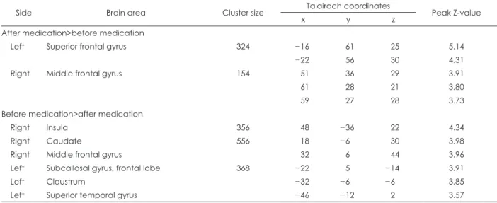
Table 3. Significant changes of rCBF in subjects with ADHD (N=14) after methylphenidate medication
Side Brain area Cluster size Talairach coordinates
Peak Z-value
x y z
After medication>before medication
Left Superior frontal gyrus 324 -16 61 25 5.14
-22 56 30 4.31
Right Middle frontal gyrus 154 51 36 29 3.91
61 28 21 3.80
59 27 28 3.73
Before medication>after medication
Right Insula 356 48 -36 22 4.34
Right Caudate 556 18 -6 30 3.98
Right Middle frontal gyrus 32 6 44 3.96
Left Subcallosal gyrus, frontal lobe 368 -22 5 -14 3.91
Left Claustrum -32 -6 -6 3.85
Left Superior temporal gyrus -46 -12 2 3.57
ADHD: attention-deficit hyperactivity disorder, rCBF: regional cerebral blood flow
임상 양상과도 관련이 있을 것이다. 반대로 섬이랑과 상측두 엽, 미상핵, 전장 등의 피질하 구조물들, 그리고 일부 전두엽 에서 혈류의 감소가 나타났다. 섬이랑의 혈류의 감소는 최근 에 fMRI를 이용하여 메칠페니데이트 효과를 조사한 연구에 서14) 혈류의 증가가 보고된 것과는 상반된 결과이며, 미상핵 의 혈류 감소는 이전 연구들과 일치하거나,8) 상반된6,7) 결과이 다. 이들 구조물들은 전두엽을 비롯한 뇌의 다른 영역들과 상호연결(reciprocal connection)을 하고 있는데, 이러한 연결 을 통해 고위 인지 및 운동 기능을 조절한다. 이러한 조절 기 능의 변화가 약물의 치료효과와 관련이 있을 것으로 생각된 다. 이는 일정한 과제가 주어진 상황에서 뇌기능의 변화를 알 아보는 연구를 통해 그 기전을 좀 더 알아볼 수 있을 것이다.
전반적으로 본 연구를 통해서 ADHD의 전두엽-선조체 및 소뇌의 이상을 다시 한 번 확인할 수 있었다.
본 연구의 제한점으로 우선 영상을 촬영하는 과정에서 참 여 대상자들에게 일정한 과제가 주어지지 않아 참여자들마 다 동일한 뇌기능을 사용하고 있다고 가정하기에 제약이 따 른다. 그러나 ADHD 아동에서 특정한 과제가 없는 상태를 평가한 연구로서 의의가 있고, 연구결과를 통해서 휴지 상태 의 ADHD 아동의 뇌 기능의 특징을 알아볼 수 있었다. 또한 본 연구에서는 연구윤리에 따라 정상대조군을 후향적으로 모집하였다. 이에 따라 대조군은 연구 참여 과정이 ADHD 아동과 차이가 있어 이에 대한 고려가 필요하다. 마지막으로 참여자의 수가 적어 약물치료 반응에 따른 비교 분석이 이루 어지지 않았다. 최근에 ADHD 아동에서 약물치료 이후의 혈 류의 변화가 노르아드레날린 체계와 관련이 있다는 보고가 있 었다. 노르에피네프린 운반체(norepinephrine transporter) 의 유전자형은 측두엽의 혈류변화28)와, α-2A-아드레날린 수 용체 유전자형은 소뇌, 렌즈핵, 그리고 대상회의 혈류변화29) 와 관련이 있었다. 향후 약물치료 반응과 유전형에 따른 뇌기 능의 변화를 측정한 연구를 통해서 ADHD 아동에서 약물치 료 반응에 대한 이해를 높일 수 있을 것으로 기대된다.
결 론
본 연구에서는 ADHD 환자에서 약물치료 전후 국소 대뇌 관류의 변화를 SPECT를 이용하여 조사하여 약물 치료와 관련된 뇌기능의 변화를 알아보았다. 그 결과 대조군과 비교 시 ADHD 환자군은 약물 치료 전 전두엽, 두정엽, 전대상회 에서 혈류의 증가를, 소뇌에서 혈류의 감소를 보였다. 약물치 료 이후에 이러한 혈류 증가는 정상화되고, 소뇌의 혈류 감소 면적은 줄어들었다. ADHD 환자에서 약물치료 전후 비교 시 약물치료 이후에 전두엽의 혈류 증가가 두드러졌으며, 섬이랑,
미상핵, 그리고 전장 등과 같은 피질하 구조물에서 혈류 감 소를 보여 이러한 부위의 기능의 변화가 약물치료와 관련이 있을 것으로 보인다. 이 연구는 ADHD 환자에서 전두엽-선 조체 연결뿐만 아니라 두정엽과 소뇌의 기능이 ADHD 환자 의 약물치료 효과와 밀접한 관련이 있음을 보여주었다. 향후 ADHD 환자에서 일정한 과제 하에서 뇌영역의 기능적 연결 과 약물치료 효과를 통합하여 분석하는 연구가 이루어진다 면 약물치료에 따른 뇌영역의 기능 변화의 기전에 대해서 더 자세히 알아볼 수 있을 것이다.
중심 단어: 주의력결핍 과잉행동장애・메칠페니데이트・
단일광자 단층촬영(SPECT).
Conflicts of Interest
The authors have no financial conflicts of interest.
References
1) American Psychiatric Association. Diagnostic and statistical man- ual of mental disorders: DSM-5. 5th ed. Arlington, VA: American Psychiatric Publishing;2013.
2) Greenhill L, Beyer DH, Finkleson J, Shaffer D, Biederman J, Con- ners CK, et al. Guidelines and algorithms for the use of methylphe- nidate in children with attention-deficit/hyperactivity disorder. J Atten Disord 2002;6 Suppl 1:S89-S100.
3) Engert V, Pruessner JC. Dopaminergic and noradrenergic contri- butions to functionality in ADHD: the role of methylphenidate.
Curr Neuropharmacol 2008;6:322-328.
4) Bush G, Valera EM, Seidman LJ. Functional neuroimaging of at- tention-deficit/hyperactivity disorder: a review and suggested fu- ture directions. Biol Psychiatry 2005;57:1273-1284.
5) Seidman LJ, Valera EM, Makris N. Structural brain imaging of at- tention-deficit/hyperactivity disorder. Biol Psychiatry 2005;57:1263- 1272.
6) Lou HC, Henriksen L, Bruhn P. Focal cerebral hypoperfusion in children with dysphasia and/or attention deficit disorder. Arch Neurol 1984;41:825-829.
7) Kim BN, Lee JS, Cho SC, Lee DS. Methylphenidate increased re- gional cerebral blood flow in subjects with attention deficit/hy- peractivity disorder. Yonsei Med J 2001;42:19-29.
8) Lee JS, Kim BN, Kang E, Lee DS, Kim YK, Chung JK, et al. Re- gional cerebral blood flow in children with attention deficit hy- peractivity disorder: comparison before and after methylphenidate treatment. Hum Brain Mapp 2005;24:157-164.
9) Anderson CM, Polcari A, Lowen SB, Renshaw PF, Teicher MH.
Effects of methylphenidate on functional magnetic resonance re- laxometry of the cerebellar vermis in boys with ADHD. Am J Psychiatry 2002;159:1322-1328.
10) Cho SC, Hwang JW, Kim BN, Lee HY, Kim HW, Lee JS, et al. The relationship between regional cerebral blood flow and response to methylphenidate in children with attention-deficit hyperactivity disorder: comparison between non-responders to methylphenidate and responders. J Psychiatr Res 2007;41:459-465.
11) Schweitzer JB, Lee DO, Hanford RB, Tagamets MA, Hoffman JM, Grafton ST, et al. A positron emission tomography study of meth- ylphenidate in adults with ADHD: alterations in resting blood flow and predicting treatment response. Neuropsychopharmacol- ogy 2003;28:967-973.
12) Schweitzer JB, Lee DO, Hanford RB, Zink CF, Ely TD, Tagamets MA, et al. Effect of methylphenidate on executive functioning in
adults with attention-deficit/hyperactivity disorder: normalization of behavior but not related brain activity. Biol Psychiatry 2004;56:597- 13) Schulz KP, Fan J, Bédard AC, Clerkin SM, Ivanov I, Tang CY, et 606.
al. Common and unique therapeutic mechanisms of stimulant and nonstimulant treatments for attention-deficit/hyperactivity disor- der. Arch Gen Psychiatry 2012;69:952-961.
14) Rubia K, Alegria AA, Cubillo AI, Smith AB, Brammer MJ, Radua J. Effects of stimulants on brain function in attention-deficit/hy- peractivity disorder: a systematic review and meta-analysis. Biol Psychiatry 2014;76:616-628.
15) Kim YS, Cheon KA, Kim BN, Chang SA, Yoo HJ, Kim JW, et al.
The reliability and validity of Kiddie-Schedule for Affective Dis- orders and Schizophrenia-Present and Lifetime version- Korean version (K-SADS-PL-K). Yonsei Med J 2004;45:81-89.
16) Chang LT. A method for attenuation correction in radionuclide computed tomography. IEEE Trans Nucl Sci 1978;25:638-643.
17) Varghese GI, Purcaro MJ, Motelow JE, Enev M, McNally KA, Levin AR, et al. Clinical use of ictal SPECT in secondarily gener- alized tonic-clonic seizures. Brain 2009;132:2102-2113.
18) Talairach J, Tournoux P. Co-planar stereotaxic atlas of the human brain: 3-dimensional proportional system: an approach to cerebral imaging. Stuttgart: Georg Thieme;1988.
19) Clark DL, Boutros NN, Mendez MF. The brain and behavior: an introduction to behavioral neuroanatomy. 3rd ed. New York, NY:
Cambridge University Press;2010.
20) Cherkasova MV, Hechtman L. Neuroimaging in attention-deficit hyperactivity disorder: beyond the frontostriatal circuitry. Can J Psychiatry 2009;54:651-664.
21) Haznedar MM, Buchsbaum MS, Hazlett EA, Shihabuddin L, New A, Siever LJ. Cingulate gyrus volume and metabolism in the schizo- phrenia spectrum. Schizophr Res 2004;71:249-262.
22) Castellanos FX, Giedd JN, Berquin PC, Walter JM, Sharp W, Tran T, et al. Quantitative brain magnetic resonance imaging in girls with attention-deficit/hyperactivity disorder. Arch Gen Psychiatry 2001;58:289-295.
23) Kim BN, Lee JS, Shin MS, Cho SC, Lee DS. Regional cerebral per- fusion abnormalities in attention deficit/hyperactivity disorder.
Statistical parametric mapping analysis. Eur Arch Psychiatry Clin Neurosci 2002;252:219-225.
24) Hill DE, Yeo RA, Campbell RA, Hart B, Vigil J, Brooks W. Mag- netic resonance imaging correlates of attention-deficit/hyperac- tivity disorder in children. Neuropsychology 2003;17:496-506.
25) Bledsoe JC, Semrud-Clikeman M, Pliszka SR. Neuroanatomical and neuropsychological correlates of the cerebellum in children with attention-deficit/hyperactivity disorder--combined type. J Am Acad Child Adolesc Psychiatry 2011;50:593-601.
26) Middleton FA, Strick PL. Cerebellar projections to the prefrontal cortex of the primate. J Neurosci 2001;21:700-712.
27) Melchitzky DS, Lewis DA. Tyrosine hydroxylase- and dopamine transporter-immunoreactive axons in the primate cerebellum. Ev- idence for a lobular- and laminar-specific dopamine innervation.
Neuropsychopharmacology 2000;22:466-472.
28) Park MH, Kim JW, Yang YH, Hong SB, Park S, Kang H, et al. Re- gional brain perfusion before and after treatment with methylphe- nidate may be associated with the G1287A polymorphism of the norepinephrine transporter gene in children with attention-deficit/
hyperactivity disorder. Neurosci Lett 2012;514:159-163.
29) Park S, Bae JH, Kim JW, Yang YH, Oh S, Hong SB, et al. Regional brain perfusion before and after treatment with methylphenidate according to the MspI polymorphism of the alpha-2A adrenergic receptor gene in children with attention-deficit hyperactivity dis- order. J Korean Acad Child Adolesc Psychiatry 2013;24:21-27.